Overview of Pulmonary Hemorrhage in Infants
Pulmonary hemorrhage in infants is a severe condition characterized by bloody discharge from the upper respiratory tract or endotracheal tube, often associated with prematurity, lung complications, infections, or trauma. The etiology, pathophysiology, clinical manifestations, and diagnosis of pulmonary hemorrhage are discussed, highlighting potential risk factors and symptoms to watch for in affected infants.

Download Presentation

Please find below an Image/Link to download the presentation.
The content on the website is provided AS IS for your information and personal use only. It may not be sold, licensed, or shared on other websites without obtaining consent from the author. Download presentation by click this link. If you encounter any issues during the download, it is possible that the publisher has removed the file from their server.
E N D
Presentation Transcript
Pulmonary Hemorrhage Gorup 8
Definition Pulmonary hemorrhage is an acute, catastrophic event characterized by discharge of bloody fluid from the upper respiratory tract or the ETT. It is a form of fulminant lung edema with leakage of red blood cells and capillary filtrate into the lungs.
Etiology 1. Prematurity, RDS, and exogenous surfactant therapy 2. Preterm infants with echocardiographic evidence of a large left-to-right shunt across a PDA and a high pulmonary blood flow have a high incidence of pulmonary hemorrhage 3. Infants who are small for gestational age 4. Lung complications. PIE and pneumothorax. 5. Infections. Bacterial, viral, or fungal infections 6. Meconium aspiration syndrome 7. Inherited coagulation disorder 8. Trauma
Pathophsyiology The underlying mechanisms of pulmonary hemorrhage remain uncertain. The pulmonary effluate has very high protein content, as well as a large number of cellular elements from the blood. Thus the hemorrhage may be a consequence of increased transcapillary pore size. A series of interrelated factors may lead to an eventual bleeding episode
Some experts consider pulmonary hemorrhage as a manifestation of an exaggerated hemorrhagic pulmonary edema brought about by an acute increase in pulmonary blood flow. The latter can occur from multiple, interrelated causes, including the normal postnatal drop in the pulmonary vascular resistance, improved pulmonary compliance after surfactant therapy, and normal postnatal absorption of lung fluid. These changes may lead to an acute increase in pulmonary blood flow and hemorrhagic pulmonary edema
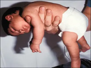
Clinical Manifestaions sudden deterioration of the infant with pallor, cyanosis, bradycardia, or apnea. Pink or red frothy liquid drains from the mouth hypotensive and is often limp and unresponsive a tachycardia and the murmur of a PDA is often heard hepatosplenomegaly, peripheral edema, and a triple rhythm
Diagnosis Haematological: although the haematocrit of the oedema fluid is usually less than 10%,9 considerable quantities of blood can be lost and the baby can become severely anaemic. Secondary DIC can develop. Biochemical: infants with PH usually have the same problems as those with severe RDS, namely hypoglycaemia, hypocalcaemia, hypoalbuminaemia and renal failure, and these should be sought and remedied.
Chest X-ray: infants with a massive PH show a virtual white out with just an air bronchogram visible. As the con- dition improves with IPPV, the changes may clear or merge into those of bronchopulmonary dysplasia (BPD). Rarely, a lobar pat- tern of consolidation is found, suggesting that the haemorrhage has just occurred in a part of the lung.
Blood gases: all components of the blood gas deteriorate rapidly with severe hypoxia, hypercarbia and metabolic acidosis. Septic screen: the possibility of infection should be considered and the infant should have a blood culture taken and be com- menced on antibiotics.
Management Resuscitation Initial resuscitation is the priority. The airway should be cleared with suction, and the infant should be intubated and ventilated and/or the ventilator pressures increased. The circulatory volume should be restored with boluses of colloid 20 ml/kg, a combination of fresh frozen plasma, blood, and platelets, with regular reassessment.
Ventilation All babies with massive pulmonary hemorrhage should be intubated and ventilated. They usually have severe lung disease, and a PIP above 30 cm H2O may be required. A ventilation strategy of high PEEP (up to 6 to 7 cm H2O) is used with a long Ti (0.4 to 0.5 seconds). Although in experimental studies this does not reduce the total lung water, it redistributes it back into the interstitial space, improving oxygenation and ventilation perfusion balance. Frequent suctioning may be required to keep the ETT clear.
Surfactant Paradoxically, although surfactant may precipitate pulmonary hemorrhage, after stabilizing the baby on the ventilator a single dose of surfactant has been suggested to improve oxygenation.
Circulation Once the initial circulating volume is restored, the infant must be reassessed for signs of left ventricular failure and pulmonary edema. In such cases, furosemide (1 mg/kg) should be given as soon as possible after the hemorrhage and repeated as necessary, to treat fluid overload.
Antibiotics Sepsis is a recognized cause of PH, thus broad-spectrum antibiot- ics should be started after taking cultures.