Case Report: Submassive Bilateral Pulmonary Embolism in a 69-Year-Old Female

Anne Knisely, a 69-year-old female with a history of endometrial cancer, presented with left leg pain, pleuritic chest pain, and shortness of breath. Imaging revealed extensive bilateral pulmonary emboli, right heart strain, and lower extremity deep vein thrombi. She was managed with anticoagulation, IVC filter placement, and antibiotic therapy for an ESBL-producing UTI. Follow-up included apixaban, vascular medicine, and plans for IVC filter removal.

Download Presentation

Please find below an Image/Link to download the presentation.
The content on the website is provided AS IS for your information and personal use only. It may not be sold, licensed, or shared on other websites without obtaining consent from the author. Download presentation by click this link. If you encounter any issues during the download, it is possible that the publisher has removed the file from their server.
E N D
Presentation Transcript
Case Report Anne Knisely, MS4 Diagnostic Radiology elective
SK, 69 y.o. F Initially admitted with heavy vaginal bleeding and severe anemia Transfused, started on Megace (megestrol) 5 cm mass on TVUS, grade 1 endometrioid adenocarcinoma on EMBx Readmitted for surgery 1 week later (robotic TLH/BSO, pelvic LND) Required 1U pRBCs, d/c ed on PSD#2 with Foley (removed PSD#11) Presented to ED on PSD#13 with left leg pain and edema, R-sided pleuritic chest pain, mild SOB CXR, CTPA, b/l LE Doppler US, TTE
Hospital Course TTE: RV severely dilated with reduced EF, evidence of severe pulmonary artery systolic pressure elevation Troponin peak of 0.03 Admitted to MICU with telemetry monitoring Heparin drip enoxaparin IVC filter placed by IR HDS since admission ESBL-producing E. coli UTI Bactrim x 7 d. Discharged on apixaban with Vascular medicine f/u in 1 mo., TTE and IVC filter removal in 3 mo.
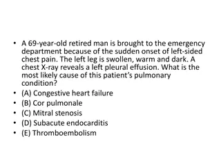
Diagnosis Submassive bilateral PE Extensive bilateral pulmonary emboli with evidence of right heart strain (CT with RV:LV ratio of 1.26, TTE) Bilateral lower extremity deep vein thrombi
Radiographic Features and Ddx Pulmonary artery intraluminal filling defect(s) Thromboembolism Mass compression/tumor emboli Respiratory motion artifact Flow-related artifact/vascular bifurcation Primary pulmonary artery sarcoma Non-compressible lower extremity vein(s) Acute thrombus Chronic thrombus Proximal obstructions caused by extrinsic masses Venous distension 2/2 CHF
References Wittram C et al. (2004) CT Angiography of Pulmonary Embolism: Diagnostic Criteria and Causes of Misdiagnosis. RSNA RadioGraphics 24:5. DiVittorio R, Bluth E, and Sullivan M. (2002) Deep Vein Thrombosis: Diagnosis of a Common Clinical Problem. Ochsner Journal. 4(1):14-17.