X-ray Image-Based Navigation for Hip Osteotomy Experiment
X-ray Image Based Navigation
for Hip Osteotomy
Experiment with a cadaver specimen to evaluate
(1) feasibility of BB implantation and
(2) visibility on X-ray images
Team Members
:
Michael Van Maele & Jesse Hamilton
Mentors
:
Dr. Mehran Armand, Dr. Yoshito Otake, & Ryan Murphy
Date:
April 10, 2012
Location:
International Center for Orthopaedic Advancement,
Bayview Campus, Johns Hopkins University
Summary
•
Date
: April 10, 2012
•
Time
: 11:30AM – 1:30 PM
•
Place
: International Center for
Orthopaedic Advancement, Bayview
Medical Center, Alpha Center
•
Materials/Devices
–
Cadaver specimen (extracted femur)
–
C-arm (make/model: ***?)
–
Beads (McMaster-Carr, stainless steel)
–
Super glue (Loctite Ultra Gel Control)
–
Drill with small diameter bit
–
Forceps, scissors, scalpel
Femur with different size BBs
attached with super glue
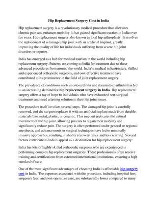
Methods
1.
Cleared overlying tissue using scissors and scalpel to expose bone
2.
Drilled three very shallow burrs in distal end of femur (closely spaced,
~1-2 cm apart)
3.
Applied super glue to burrs
4.
Placed BB in each burr using forceps
5.
Generously applied super glue over the BBs
Repeated steps 1-5 for four different size BBs, using three BBs of each size.
6.
Let glue dry for at least 10 minutes.
7.
Acquired two C-arm images at different view angles.
8.
Attempted to dislodge BBs from bone by
a.
Hitting bone against hand
b.
Striking bone with mallet
c.
Picking BBs off with forceps
9.
Evaluated images for
a.
BB visibility (image contrast between BBs and surrounding bone/tissue)
b.
Ability to distinguish between BB sizes
Methods
burr
After step 1
Step 2
Distal end of femur
Step 5
Step 8b
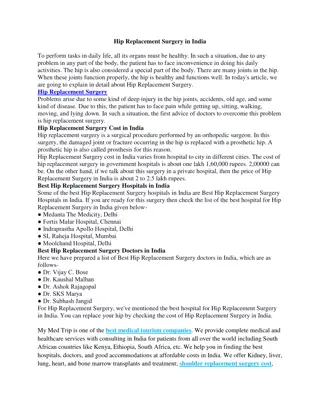
Results
X-ray
shot #1
X-ray
shot #2
X-ray setting:
**kVp, **mA
X-ray setting:
**kVp, **mA
femur
table
~**cm (approximate
distance)
~**cm
C-arm setup
What was the C-arm setup?
What were the x-ray settings?
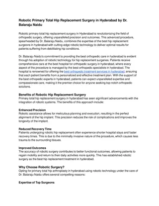
Results (Zoom In)
Discussion – BB Fixation
•
No BBs dislodged after hitting bone
against hand and striking bone with
mallet
–
Similar to mock surgical conditions
•
Could pick off BBs with forceps
–
Not similar to mock surgical conditions
•
Conclusion
: We predict that our
method should hold BBs in place
during a cadaver surgery.
•
BBs visible on x-ray. Good
image contrast versus
background tissue.
•
Radial symmetry algorithm
could automatically
distinguish between large
and small BBs.
–
Conclusion:
Should be
possible to use different size
BBs as confidence and
fragment points.
Discussion – Image Analysis
Controls detection
sensitivity (higher
value
fewer BBs
detected)
Experiment conducted using a cadaver specimen to assess the feasibility of BB implantation and visibility on X-ray images. The team cleared tissue, drilled burrs in the femur, attached BBs, and evaluated visibility and sizing on C-arm images. Results showed successful BB fixation. Predicted method feasibility for cadaver surgeries.

Download Presentation

Please find below an Image/Link to download the presentation.
The content on the website is provided AS IS for your information and personal use only. It may not be sold, licensed, or shared on other websites without obtaining consent from the author.If you encounter any issues during the download, it is possible that the publisher has removed the file from their server.
You are allowed to download the files provided on this website for personal or commercial use, subject to the condition that they are used lawfully. All files are the property of their respective owners.
The content on the website is provided AS IS for your information and personal use only. It may not be sold, licensed, or shared on other websites without obtaining consent from the author.
E N D
Presentation Transcript
X-ray Image Based Navigation for Hip Osteotomy Experiment with a cadaver specimen to evaluate (1) feasibility of BB implantation and (2) visibility on X-ray images Team Members: Michael Van Maele & Jesse Hamilton Mentors: Dr. Mehran Armand, Dr. Yoshito Otake, & Ryan Murphy Date: April 10, 2012 Location: International Center for Orthopaedic Advancement, Bayview Campus, Johns Hopkins University
Summary Date: April 10, 2012 Time: 11:30AM 1:30 PM Place: International Center for Orthopaedic Advancement, Bayview Medical Center, Alpha Center Materials/Devices Cadaver specimen (extracted femur) C-arm (make/model: ***?) Beads (McMaster-Carr, stainless steel) Super glue (Loctite Ultra Gel Control) Drill with small diameter bit Forceps, scissors, scalpel Femur with different size BBs attached with super glue
Methods 1. 2. Cleared overlying tissue using scissors and scalpel to expose bone Drilled three very shallow burrs in distal end of femur (closely spaced, ~1-2 cm apart) Applied super glue to burrs Placed BB in each burr using forceps Generously applied super glue over the BBs Repeated steps 1-5 for four different size BBs, using three BBs of each size. 3. 4. 5. 6. 7. 8. Let glue dry for at least 10 minutes. Acquired two C-arm images at different view angles. Attempted to dislodge BBs from bone by a. Hitting bone against hand b. Striking bone with mallet c. Picking BBs off with forceps Evaluated images for a. BB visibility (image contrast between BBs and surrounding bone/tissue) b. Ability to distinguish between BB sizes 9.
Methods Step 2 After step 1 burr Distal end of femur Step 8b Step 5
What was the C-arm setup? What were the x-ray settings? Results C-arm setup X-ray setting: **kVp, **mA X-ray shot #1 ~**cm (approximate distance) femur table X-ray shot #2 X-ray setting: **kVp, **mA ~**cm
Results (Zoom In) 1 2 3 4 5 6 7 89 11 12 10 10 11 8 9 7 12 6 4 BB ID Size No. Diameter (mm) 5 3 1, 2, 3 3 ?? (largest) 2 1 ?? (2nd largest) 4, 5, 6 5 ?? (2nd smallest) 7, 8, 9 6 10, 11, 12 8 ?? (smallest)
Discussion BB Fixation No BBs dislodged after hitting bone against hand and striking bone with mallet Similar to mock surgical conditions Could pick off BBs with forceps Not similar to mock surgical conditions Conclusion: We predict that our method should hold BBs in place during a cadaver surgery.
Discussion Image Analysis BBs visible on x-ray. Good image contrast versus background tissue. Radial symmetry algorithm could automatically distinguish between large and small BBs. Conclusion: Should be possible to use different size BBs as confidence and fragment points. Large BBs Small BBs Controls detection sensitivity (higher value fewer BBs detected) Min radius 6 1 Max radius 10 2 Factor 0.5 0.35 ROI Radius 200 200


")