White Blood Cells and the Immune System

undefined
 
P
H
Y
S
I
O
L
O
G
Y
B
L
O
O
D
 
P
H
Y
S
I
O
L
O
G
Y
W
h
i
t
e
 
B
l
o
o
d
 
C
e
l
l
s
(
W
B
C
)
م‍
‍ح‍
‍ا
ض‍
‍ر
ت‍
‍ي‍
‍ن
 
م‍
‍ع
 
ب‍
‍ع‍
‍ض
 
م‍
‍ن
 
ا
ل‍
‍ب‍
‍ن‍
‍ا
ت
و
 
م‍
‍ح‍
‍ا
ض‍
‍ر
ة
 
ا
لأ
و
لا
د
O
b
j
e
c
t
i
v
e
s
3
A
t
 
t
h
e
 
e
n
d
 
o
f
 
t
h
i
s
 
l
e
c
t
u
r
e
 
s
t
u
d
e
n
t
 
s
h
o
u
l
d
 
b
e
 
a
b
l
e
 
t
o
:
1
-
 
D
e
s
c
r
i
b
e
 
d
i
f
f
e
r
e
n
t
 
T
y
p
e
s
 
o
f
 
W
B
C
2
-
 
R
e
c
o
g
n
i
z
e
 
t
h
e
 
g
e
n
e
r
a
l
 
f
u
n
c
t
i
o
n
s
 
o
f
 
W
B
C
3
-
 
D
e
s
c
r
i
b
e
 
g
e
n
e
s
i
s
 
a
n
d
 
s
i
t
e
 
o
f
 
f
o
r
m
a
t
i
o
n
 
o
f
 
W
B
C
.
4
-
 
D
e
s
c
r
i
b
e
 
s
t
a
g
e
s
 
o
f
 
n
e
u
t
r
o
p
h
i
l
 
f
o
r
m
a
t
i
o
n
5
-
 
D
e
s
c
r
i
b
e
 
t
h
e
 
r
o
l
e
 
o
f
 
n
e
u
t
r
o
p
h
i
l
s
 
i
n
 
d
e
f
e
n
d
i
n
g
 
t
h
e
 
b
o
d
y
 
a
g
a
i
n
s
t
 
i
n
f
e
c
t
i
o
n
6
-
 
D
e
s
c
r
i
b
e
 
t
h
e
 
p
r
o
c
e
s
s
 
o
f
 
p
h
a
g
o
c
y
t
o
s
i
s
7
-
 
D
e
s
c
r
i
b
e
 
E
s
i
n
o
p
h
i
l
s
 
f
o
r
m
a
t
i
o
n
 
a
n
d
 
f
u
n
c
t
i
o
n
s
8
-
 
D
e
s
c
r
i
b
e
 
B
a
s
o
p
h
i
l
s
 
f
o
r
m
a
t
i
o
n
 
a
n
d
 
f
u
n
c
t
i
o
n
s
9
-
 
D
e
s
c
r
i
b
e
 
M
o
n
o
c
y
t
e
s
 
a
n
d
 
m
a
c
r
o
p
h
a
g
e
 
f
o
r
m
a
t
i
o
n
 
a
n
d
 
f
u
n
c
t
i
o
n
s
.
1
0
-
 
D
e
s
c
r
i
b
e
 
R
e
t
i
c
u
l
o
e
n
d
o
t
h
e
l
i
a
l
 
c
o
m
p
o
n
a
n
t
s
 
a
n
d
 
f
u
n
c
t
i
o
n
s
1
1
-
 
D
e
s
c
r
i
b
e
 
l
y
m
p
h
o
c
y
t
e
s
 
f
o
r
m
a
t
i
o
n
 
a
n
d
 
m
a
t
u
r
a
t
i
o
n
1
2
-
 
D
e
s
c
r
i
b
e
 
t
h
e
 
f
u
n
c
t
i
o
n
s
 
o
f
 
t
h
e
 
d
i
f
f
e
r
e
n
t
 
t
y
p
e
s
 
o
f
 
l
y
m
p
h
o
c
y
t
e
s
.
1
3
-
 
R
e
c
o
g
n
i
s
e
 
l
e
u
c
o
c
y
t
o
s
i
s
 
a
n
d
 
l
e
u
c
o
p
e
n
i
a
.
1
4
-
 
R
e
c
o
g
n
i
z
e
 
t
y
p
e
 
o
f
 
l
e
u
k
a
e
m
i
a
1
5
-
 
O
u
t
l
i
n
e
 
c
o
m
p
o
n
e
n
t
s
 
o
f
 
t
h
e
 
i
m
m
u
n
e
 
s
y
s
t
e
m
1
6
-
D
e
s
c
r
i
b
e
 
t
h
e
 
s
t
r
u
c
t
u
r
e
 
o
f
 
t
h
e
 
d
i
f
f
e
r
e
n
t
 
t
y
p
e
s
 
o
f
 
W
B
C
s
.
1
7
-
O
u
t
l
i
n
e
 
d
i
f
f
e
r
e
n
t
i
a
l
 
W
B
C
s
 
c
o
u
n
t
.
1
8
-
D
e
s
c
r
i
b
e
 
t
h
e
 
r
o
l
e
 
o
f
 
t
h
e
 
W
B
C
s
 
i
n
 
i
m
m
u
n
e
 
r
e
s
p
o
n
s
e
s
 
a
n
d
 
d
e
f
e
n
d
i
n
g
 
a
g
a
i
n
s
t
 
i
n
f
e
c
t
i
o
n
.
The immune system is a system of barriers, cells, tissues and organs that work to
fight invaders.
Another important component of the immune
    
 system is the complement system.
  The major functions of the immune system are:
1.
 Differentiate self from non-self.
2.
 Eliminate foreign substances, cells and pathogens.
I
m
m
u
n
e
 
S
y
s
t
e
m
 
M
a
j
o
r
 
o
r
g
a
n
s
P
r
i
m
a
r
y
 
L
y
m
p
h
o
i
d
 
O
r
g
a
n
s
B
o
n
e
 
m
a
r
r
o
w
T
h
y
m
u
s
1.
Immune cells mature to become
immunocompetent
 in these
organs.
2.
Most cells mature to immune
cells within the bone marrow
and, after release, begin a life of
patrol in the blood. The
exception is the pre-T cell, which
first undergoes maturation in
the thymus before circulating in
blood.
S
e
c
o
n
d
a
r
y
 
(
o
r
 
p
e
r
i
p
h
e
r
a
l
)
 
L
y
m
p
h
 
o
r
g
a
n
s
1.
These are the organs where
mature immune cells participate
in specific immune defense
reactions.
t
o
n
s
i
l
s
a
d
e
n
o
i
d
s
L
y
m
p
h
 
n
o
d
e
s
L
y
m
p
h
a
t
i
c
 
v
e
s
s
e
l
s
s
p
l
e
e
n
a
p
p
e
n
d
i
x
p
e
y
e
r
'
s
 
p
a
t
c
h
e
s
undefined
 
2
º
2
º
1
º
1
º
2
º
2
º
2
º
2
º
2
º
M
a
j
o
r
 
o
r
g
a
n
s
undefined
 
Types of Immunity
I
m
m
u
n
e
 
s
y
s
t
e
m
1.
Second line of defense
2.
Is present at birth
3.
Persists throughout life
4.
Can be mobilized rapidly and act
quickly
5.
Attacks all antigens fairly equally
1.
Third line of defense.
2.
Antigen specificity. It is activated by
thousands of diverse antigens.
3.
Responds with the proliferation of cells
and the generation of antibodies.
4.
Responds slowly, being fully activated
about 4 days after the immunologic
threat.
5.
Exhibits immunologic memory, so that
repeated exposure to the same
infectious agent results in improved
resistance against it.
undefined
 
F
i
r
s
t
 
l
i
n
e
 
o
r
 
d
e
f
e
n
c
e
 
 
E
E
p
p
i
i
t
t
h
h
e
e
l
l
i
i
a
a
l
l
 
 
B
B
a
a
r
r
r
r
i
i
e
e
r
r
s
s
undefined
 
     
The Complement System is Part of the 1
st
 Line of Defense
c
o
m
p
l
e
m
e
n
t
 
s
y
s
t
e
m
:
1
-
 
T
h
e
 
f
i
r
s
t
 
p
a
r
t
 
o
f
 
t
h
e
 
i
m
m
u
n
e
 
s
y
s
t
e
m
 
t
h
a
t
 
m
e
e
t
s
 
i
n
v
a
d
e
r
s
s
u
c
h
 
a
s
 
b
a
c
t
e
r
i
a
2
-
i
t
 
 
i
s
 
a
 
g
r
o
u
p
 
o
f
 
p
r
o
t
e
i
n
s
.
3
-
 
T
h
e
s
e
 
p
r
o
t
e
i
n
s
 
f
l
o
w
 
f
r
e
e
l
y
 
i
n
 
t
h
e
 
b
l
o
o
d
 
a
n
d
 
c
a
n
 
q
u
i
c
k
l
y
r
e
a
c
h
 
t
h
e
 
s
i
t
e
 
o
f
 
a
n
 
i
n
v
a
s
i
o
n
 
w
h
e
r
e
 
t
h
e
y
 
c
a
n
 
r
e
a
c
t
 
d
i
r
e
c
t
l
y
w
i
t
h
 
a
n
t
i
g
e
n
s
 
(
 
m
o
l
e
c
u
l
e
s
 
t
h
a
t
 
t
h
e
 
b
o
d
y
 
r
e
c
o
g
n
i
z
e
s
 
a
s
 
f
o
r
e
i
g
n
s
u
b
s
t
a
n
c
e
s
)
.
f
u
n
c
t
i
o
n
s
 
o
f
 
c
o
m
p
l
e
m
e
n
t
 
p
r
o
t
e
i
n
s
 
(
W
h
e
n
a
c
t
i
v
a
t
e
d
 
)
:
1.
Trigger inflammation.
2.
Attract eater cells such as macrophages to the area.
3.
Coat intruders so that eater cells are more likely to
devour (swallow and eat) them (a process called as
opsonization).
4.
Kill intruders.
undefined
 
Cellular Elements of the Innate Immune System
(2
nd
 line of defense)
undefined
 
 
The cells of the immune system work together with different proteins to
seek out and destroy anything foreign or dangerous that enters our body.
C
e
l
l
s
 
o
f
 
t
h
e
 
I
m
m
u
n
e
 
S
y
s
t
e
m
M
a
s
t
c
e
l
l
s
undefined
 
Cells of the Immune System
3
R
e
m
e
m
b
e
r
أ
ق‍
‍ل
T
y
p
e
s
 
o
f
 
W
B
C
G
r
a
n
u
l
a
r
(
p
o
l
y
m
o
r
p
h
n
u
c
l
e
a
r
 
P
M
N
)
P
M
N
s
;
 
b
e
c
a
u
s
e
 
o
f
 
m
u
l
t
i
p
l
e
(
l
o
b
e
s
)
 
n
u
c
l
e
i
N
e
u
t
r
o
p
h
i
l
-
 
6
2
%
-
 
l
o
b
u
l
a
t
e
d
n
u
c
l
e
u
s
basic stain
2
-
5
-
 
P
u
r
p
l
e
c
y
t
o
p
l
a
s
m
i
c
g
r
a
n
u
l
e
s
E
o
s
i
n
o
p
h
i
l
-
 
2
.
3
%
-
 
2
 
l
o
b
e
n
u
c
l
e
u
s
eosin stain
-
 
c
o
a
r
s
e
 
r
e
d
g
r
a
n
u
l
e
s
B
a
s
o
p
h
i
l
-
 
0
.
4
%
-
 
r
a
r
e
l
y
 
s
e
g
m
e
n
t
e
d
n
u
c
l
e
u
s
-
 
n
u
c
l
e
u
s
 
h
i
d
d
e
n
b
y
 
l
a
r
g
e
 
r
o
u
n
d
b
l
u
i
s
h
 
g
r
a
n
u
l
e
s
س‍
‍م‍
‍ي‍
‍ن‍
‍ا
ه‍
‍ا
 
ك‍
‍ذ
ا
 
ع‍
‍ل‍
‍ى
ح‍
‍س‍
‍ب
 
ا
س‍
‍م
 
ا
ل‍
‍ص‍
‍ب‍
‍غ‍
‍ة
L
y
m
p
h
o
c
y
t
e
3
0
%
r
o
u
n
d
 
n
u
c
l
e
u
s
S
m
a
l
l


l
a
r
g
e
M
o
n
o
c
y
t
e
s
5
.
3
%
k
i
d
n
e
y
 
s
h
a
p
e
n
u
c
l
e
u
s
(
m
a
k
e
m
a
c
r
o
p
h
a
g
e
s
y
s
t
e
m
)
A
 
g
r
a
n
u
l
a
r
م‍
‍ا
 
ف‍
‍ي‍
‍ه‍
‍ا
 
g
r
a
n
u
l
e
s
G
e
n
e
s
i
s
 
o
f
 
W
B
C
:
M
y
e
l
o
c
y
t
i
c
(
g
r
a
n
u
l
a
r
+
m
o
n
o
c
y
t
e
s
)
G
e
n
e
s
i
s
 
o
f
 
W
B
C
:
L
y
m
p
h
o
c
y
t
i
c
s
i
t
e
s
 
o
f
 
f
o
r
m
a
t
i
o
n
:
b
o
n
e
 
m
a
r
r
o
w
s
i
t
e
s
 
o
f
 
f
o
r
m
a
t
i
o
n
:
-
 
B
o
n
e
 
m
a
r
r
o
w
 
 
-
 
T
h
y
m
u
s


-
 
L
y
m
p
h
o
i
d
 
t
i
s
s
ا
لأ
ل‍
‍و
ا
ن
 
ل‍
‍ت‍
‍س‍
‍ه‍
‍ي‍
‍ل
 
ا
ل‍
‍ح‍
‍ف‍
‍ظ
 
)
م‍
‍ا
 
ل‍
‍ه
 
د
خ‍
‍ل
 
س‍
‍لا
ي‍
‍د
ا
ت
 
ا
لأ
و
لا
د
 
و
ا
ل‍
‍ب‍
‍ن‍
‍ا
ت
(
1
-
T
 
l
y
m
p
h
o
c
y
t
e
2
-
B
 
l
y
m
p
h
o
c
y
t
e
3
-
N
a
t
u
r
a
l
 
k
i
l
l
e
r
c
e
l
l
s
 
(
N
K
C
s
)
T
y
p
e
s
 
o
f
 
W
B
C
 
1
-
 
G
r
a
n
u
l
a
r
 
(
p
o
l
y
m
o
r
p
h
n
u
c
l
e
a
r
 
P
M
N
)
:
م‍
‍و
 
م‍
‍ه‍
‍م
 
ت‍
‍ح‍
‍ف‍
‍ظ‍
‍و
ا
 
ا
ل‍
‍ح‍
‍ج‍
‍م
 
ع‍
‍ش‍
‍ا
ن
 
ك‍
‍ذ
ا
 
م‍
‍ا
 
ح‍
‍ط‍
‍ي‍
‍ت‍
‍ه‍
‍ا
 
ب‍
‍ا
ل‍
‍م‍
‍خ‍
‍ط‍
‍ط
ر
ك‍
‍ز
و
ا
 
ع‍
‍ل‍
‍ى
 
ا
ل‍
‍ن‍
‍س‍
‍ب
 
و
ا
لأ
ش‍
‍ك‍
‍ا
ل
 
و
ا
لأ
ل‍
‍و
ا
ن
)
ك‍
‍لا
م
 
د
.
ن‍
‍ي‍
‍ر
ف‍
‍ا
ن‍
‍ا
 
(
T
y
p
e
s
 
o
f
 
W
B
C
 
c
o
n
t
.
2
.
 
A
 
g
r
a
n
u
l
a
r
م‍
‍و
 
م‍
‍ه‍
‍م
 
ت‍
‍ح‍
‍ف‍
‍ظ‍
‍و
ا
 
ا
ل‍
‍ح‍
‍ج‍
‍م
 
ع‍
‍ش‍
‍ا
ن
 
ك‍
‍ذ
ا
 
م‍
‍ا
 
ح‍
‍ط‍
‍ي‍
‍ت‍
‍ه‍
‍ا
ر
ك‍
‍ز
و
ا
 
ع‍
‍ل‍
‍ى
 
ا
ل‍
‍ن‍
‍س‍
‍ب
 
و
ا
لأ
ش‍
‍ك‍
‍ا
ل
 
و
ا
لأ
ل‍
‍و
ا
ن
)
ك‍
‍لا
م
 
د
.
ن‍
‍ي‍
‍ر
ف‍
‍ا
ن‍
‍ا
 
(
Physiological Variations in WBCs Count
Age
Age
:
  
Infants
Infants
: about 20,000 /mm
3
  
Children
Children
:10,000 to 15,000/mm
3
  
Adults
Adults
: 4,000 and 11,000/mm
3
Sex
Sex
: 
m
ales
 
have 
m
ore
 
WBCs than females.
Diurnal variation
Diurnal variation
: Minimum in early morning and maximum in the afternoon.
Exercise
Exercise
: Increases slightly.
Sleep
Sleep
: Decreases.
Emotional conditions like anxiety
: Increases.
Pregnancy
Pregnancy
: Increases.
Menstruation
Menstruation
: Increases.
Parturition
Parturition
: Increases
.
.
M
a
l
e
 
S
l
i
d
e
undefined
 
WBCs Concentrations (Normal Counts)
and Life Span
ا
لأ
ل‍
‍و
ا
ن
 
م‍
‍ا
 
ل‍
‍ه‍
‍ا
 
د
خ‍
‍ل
 
ب‍
‍س‍
‍لا
ي‍
‍د
ا
ت
 
ا
لأ
و
لا
د
 
و
ا
ل‍
‍ب‍
‍ن‍
‍ا
ت
 
,
د
.
ن‍
‍ي‍
‍ر
ف‍
‍ن‍
‍ا
 
م‍
‍ا
 
ر
ك‍
‍ز
ت
 
ع‍
‍ل‍
‍ى
 
أ
ع‍
‍د
ا
د
ه‍
‍م
 
ب‍
‍ا
ل‍
‍ت‍
‍ف‍
‍ص‍
‍ي‍
‍ل
L
L
i
i
f
f
e
e
 
 
s
s
p
p
a
a
n
n
 
 
o
o
f
f
 
 
W
W
B
B
C
C
s
s
G
e
n
e
s
i
s
 
o
f
 
W
B
C
 
(
L
e
u
c
o
p
o
i
e
s
i
s
)
T
w
o
 
m
a
j
o
r
 
l
i
n
e
a
g
e
 
o
f
 
W
B
C
 
a
r
e
 
f
o
r
m
e
d
 
:
M
y
e
l
o
c
y
t
i
c
:
 
b
e
g
i
n
n
i
n
g
 
w
i
t
h
 
m
y
e
l
o
b
a
s
t
 
a
n
d
 
g
i
v
i
n
g
 
r
i
s
e
 
t
o
 
g
r
a
n
u
l
a
r
 
l
e
u
c
o
c
y
t
e
s
 
a
n
d
m
o
n
o
c
y
t
e
s
L
y
m
p
h
o
c
y
t
i
c
:
 
b
e
g
i
n
n
i
n
g
 
w
i
t
h
 
l
y
m
p
h
o
b
l
a
s
t
 
a
n
d
 
g
i
v
i
n
g
 
r
i
s
e
 
t
o
 
l
y
m
p
h
o
c
y
t
e
s
the WBC need to leave the blood
to work unlike RBC.
WBCs formed in the bone marrow are stored within the
marro
w until they are needed in the circulatory system.
Various factors cause them to be released.
 Normally, 
about three times 
as many white blood cells are
stored in the marrow as circulate in the entire blood.
The lymphocytes are mostly stored in the various lymphoid
tissues, except for a 
small number that are temporarily
being transported in the blood
.
G
e
n
e
s
i
s
 
o
f
 
W
B
C
G
e
n
e
s
i
s
 
o
f
 
W
B
C
Leucopoiesis
G
e
n
e
s
i
s
 
o
f
 
W
B
C
W
h
i
t
e
 
B
l
o
o
d
 
C
e
l
l
s
N
E
U
T
R
O
P
H
I
L
L
S
F
o
r
m
a
t
i
o
n
 
a
n
d
 
M
a
t
u
r
a
t
i
o
n
 
o
f
 
N
e
u
t
r
o
h
i
l
s
:
 
i
n
 
B
o
n
e
 
M
a
r
r
o
w
J
u
s
t
 
r
e
a
d
 
i
t
م‍
‍و
 
م‍
‍ه‍
‍م
 
ت‍
‍ح‍
‍ف‍
‍ظ‍
‍و
ا
 
ا
ل‍
‍م‍
‍ر
ا
ح‍
‍ل
 
ك‍
‍ل‍
‍ه‍
‍ا
 
)
أ
ه‍
‍م
 
ش‍
‍ي
 
أ
و
ل
 
و
آ
خ‍
‍ر
 
و
ح‍
‍د
ة
(
و
ر
ك‍
‍ز
و
ا
 
ع‍
‍ل‍
‍ى
 
M
a
t
u
r
a
t
i
o
n
 
o
f
 
r
e
d
 
b
l
o
o
d
 
c
e
l
l
s
ب‍
‍ا
ل‍
‍م‍
‍ح‍
‍ا
ض‍
‍ر
ة
 
ا
ل‍
‍س‍
‍ا
ب‍
‍ق‍
‍ة
 
 
)
ك‍
‍لا
م
 
د
.
ن‍
‍ي‍
‍ر
ف‍
‍ا
ن‍
‍ا
 
(
Polymorphonuclear.
 They contain small granules of both acidic
and basic.
They constitute the first line of defence
against bacterial infection.
 Very important at “clearing” bacterial
infections.
W
h
i
t
e
 
B
l
o
o
d
 
C
e
l
l
s
N
E
U
T
R
O
P
H
I
L
L
S
Cells infected by bacteria usually get dealt with by
Neutrophils
N
e
u
t
r
o
p
h
i
l
 
F
u
n
c
t
i
o
n
D
e
f
e
n
s
e
 
a
g
a
i
n
s
t
 
i
n
f
e
c
t
i
o
n
:
N
e
u
t
r
o
p
h
i
l
 
h
a
s
 
t
h
e
 
a
b
i
l
i
t
y
 
o
f
 
e
n
g
u
l
f
i
n
g
 
b
a
c
t
e
r
i
a
 
o
r
 
o
r
g
a
n
i
s
m
 
b
y
 
a
p
r
o
c
e
s
s
 
o
f
 
p
h
y
a
g
o
c
y
t
o
s
i
s
S
t
e
p
s
 
o
f
 
P
h
y
g
o
c
y
t
o
s
i
s
ت‍
‍ت‍
‍ح‍
‍ر
ك
 
ن‍
‍ف‍
‍س
 
ا
لا
م‍
‍ي‍
‍ب‍
‍ا
L
i
k
e
 
a
l
a
r
m
s
Neutrophil Phagocytosis
1-neutrophil attaches itself to the particle and projects pseudopodia in all directions around the
particle.
2-pseudopodia meet one another on the opposite side and fuse,  creating  an enclosed chamber
that contains the phagocytized particle.
 3-the chamber moves to the inside of the cytoplasmic cavity and breaks away from the outer cell
membrane to form a free-floating 
phagocytic vesicle 
(also called a 
phagosome
) inside the
cytoplasm.
4-The phagocytized particle is then digested by intracellular enzymes


F
e
m
a
l
e
s
 
s
l
i
d
e
C
h
e
m
o
t
a
x
i
s
T
h
e
 
a
t
t
r
a
c
t
i
o
n
 
o
f
 
t
h
e
 
n
e
u
t
r
o
p
h
i
l
s
 
a
n
d
 
m
o
n
o
c
y
t
e
s
 
t
o
 
m
o
v
e
 
t
o
 
i
n
f
l
a
m
e
d
 
a
r
e
a
f
o
l
l
o
w
i
n
g
 
c
h
e
m
o
t
a
c
t
i
c
 
s
u
b
s
t
a
n
c
e
s
 
r
e
l
e
a
s
e
 
f
r
o
m
 
i
n
f
e
c
t
e
d
 
s
i
t
e
.
T
h
e
 
c
h
e
m
o
t
a
c
t
i
c
 
a
g
e
n
t
s
 
i
n
c
l
u
d
e
 
a
 
c
o
m
p
o
n
e
n
t
 
o
f
 
t
h
e
 
c
o
m
p
l
e
m
e
n
t
 
s
y
s
t
e
m
 
(
C
5
a
)
;
l
e
u
k
o
t
r
i
e
n
e
s
;
 
a
n
d
 
p
o
l
y
p
e
p
t
i
d
e
s
 
f
r
o
m
 
l
y
m
p
h
o
c
y
t
e
s
,
 
m
a
s
t
 
c
e
l
l
s
,
 
a
n
d
 
b
a
s
o
p
h
i
l
s
.
C
h
e
m
o
t
a
c
t
i
c
 
s
u
b
s
t
a
n
c
e
s
:
-
 
B
a
c
t
e
r
i
a
l
 
t
o
x
i
n
-
 
D
e
g
e
n
e
r
a
t
i
v
e
 
p
r
o
d
u
c
t
s
 
o
f
 
i
n
f
l
a
m
e
d
 
t
i
s
s
u
e
-
 
C
o
m
p
l
e
m
e
n
t
 
s
y
s
t
e
m
-
 
R
e
a
c
t
i
o
n
 
p
r
o
d
u
c
t
 
o
f
 
p
l
a
s
m
a
 
c
l
o
t
t
i
n
g
Chemotaxis is effective up to 100 micrometers
away from an inflamed tissue. Therefore,
because almost no tissue area is more than
50 micrometers away from a capillary, the
chemo- tactic signal can easily move hordes
of WBCs from the capillaries into the inflamed
area.


M
a
r
g
i
n
a
t
i
o
n
 
&
 
D
i
a
p
e
d
e
s
i
s
1
-
 
W
B
C
s
 
m
a
r
g
i
n
a
t
e
 
(
a
g
g
r
e
g
a
t
e
 
a
n
d
 
s
t
i
c
k
)
 
a
l
o
n
g
 
t
h
e
 
w
a
l
l
 
o
f
 
b
l
o
o
d
 
c
a
p
i
l
l
a
r
i
e
s
.
2
-
 
W
B
C
 
s
q
u
e
e
z
e
s
 
i
t
s
e
l
f
 
t
h
r
o
u
g
h
 
e
n
d
o
t
h
e
l
i
a
l
 
h
o
l
e
s
 
l
e
a
v
i
n
g
 
b
l
o
o
d
 
c
a
p
i
l
l
a
r
i
e
s
(
d
i
a
p
e
d
e
s
i
s
)
3
-
 
W
B
C
s
 
m
o
v
e
 
b
y
 
a
m
o
e
b
o
i
d
 
m
o
t
i
o
n
 
t
o
w
a
r
d
s
 
i
n
f
l
a
m
m
a
t
i
o
n
 
a
r
e
a
 
f
o
l
l
o
w
i
n
g
c
h
e
m
o
t
a
c
t
i
c
 
s
u
b
s
t
a
n
c
e
 
r
e
l
e
a
s
e
d
 
f
r
o
m
 
s
i
t
e
 
o
f
 
i
n
f
e
c
t
i
o
n
4
-
 
U
p
o
n
 
r
e
a
c
h
i
n
g
 
t
h
e
 
s
i
t
e
 
o
f
 
i
n
f
e
c
t
i
o
n
 
n
e
u
t
r
o
p
h
i
l
s
 
s
t
a
r
t
 
t
o
 
e
n
g
u
l
f
 
i
n
f
e
c
t
i
n
g
o
r
g
a
n
i
s
m
ت‍
‍ح‍
‍ر
ي‍
‍ك
 
و
ص‍
‍ف
 
ا
ل
 
n
e
u
t
r
o
p
h
i
l
s
T
h
e
r
e
 
a
r
e
 
g
a
b
s
 
i
n
b
e
t
w
e
e
n
 
t
h
e
 
e
n
d
o
t
h
e
l
i
a
l
c
a
p
i
l
l
a
r
i
e
s
 
w
h
e
r
e
 
t
h
e
 
n
e
u
t
r
o
p
h
i
l
s
 
s
q
u
e
e
z
e
t
h
r
o
u
g
h
C
h
e
m
o
t
a
x
i
s
,
 
m
a
r
g
i
n
a
t
i
o
n
 
&
 
d
i
a
p
e
d
e
s
i
s
P
h
a
g
o
c
y
t
o
s
i
s
 
a
n
d
 
O
p
s
o
n
i
z
a
t
i
o
n
S
e
l
e
c
t
i
v
e
 
p
r
o
c
e
s
s
:
 
f
o
r
e
i
g
n
 
s
u
b
s
t
a
n
c
e
 
r
e
c
o
g
n
i
z
e
 
b
y
:
-
R
o
u
g
h
 
s
u
r
f
a
c
e
-
N
o
 
p
r
o
t
e
c
t
i
v
e
 
p
r
o
t
e
i
n
 
c
o
a
t
,
 
w
h
i
c
h
 
p
r
e
v
e
n
t
s
 
p
h
a
g
o
c
y
t
o
s
i
s
.
-
M
a
r
k
e
d
 
b
y
 
c
e
r
t
a
i
n
 
s
u
b
s
t
a
n
c
e
 
(
o
p
s
o
n
i
z
a
t
i
o
n
)
e
.
g
 
C
o
m
p
l
e
m
e
n
t
 
3
 
o
r
 
a
n
t
i
b
o
d
i
e
s
 
m
a
k
i
n
g
 
t
h
e
m
 
r
e
a
d
y
 
f
o
r
 
k
i
l
l
i
n
g
.
N
e
u
t
r
o
p
h
i
l
s
 
e
n
c
i
r
c
l
e
d
 
t
h
e
 
b
a
c
t
e
r
i
a
 
w
i
t
h
 
p
s
e
u
d
o
p
o
d
i
a
 
a
n
d
 
e
n
g
u
l
f
 
i
t
 
i
n
s
i
d
e
 
i
n
t
o
 
a
v
a
c
u
o
l
e
 
(
p
h
a
g
o
s
o
m
e
)
,
 
t
a
k
e
s
 
3
-
2
0
 
b
a
c
t
e
r
i
a
م‍
‍ث‍
‍ل
 
n
a
m
e
 
t
a
g
ل‍
‍ل‍
‍ت‍
‍ع‍
‍ر
ف
 
ع‍
‍ل‍
‍ي‍
‍ه‍
‍ا
P
h
a
g
o
c
y
t
o
s
i
s
:
 
m
e
a
n
s
 
c
e
l
l
u
l
a
r
 
i
n
g
e
s
t
i
o
n
 
o
f
 
t
h
e
 
o
f
f
e
n
d
i
n
g
 
a
g
e
n
t
.
O
p
s
o
n
i
z
a
t
i
o
n
:
 
C
o
m
p
l
e
m
e
n
t
 
3
b
 
o
r
 
a
n
t
i
b
o
d
i
e
s
 
l
i
k
e
 
I
g
G
 
m
a
k
i
n
g
 
t
h
e
m
 
r
e
a
d
y
 
f
o
r
k
i
l
l
i
n
g
 
a
 
p
r
o
c
e
s
s
 
k
n
o
w
n
 
a
s
 
o
p
s
o
n
i
z
a
t
i
o
n
.
S
o
m
e
 
p
l
a
s
m
a
 
f
a
c
t
o
r
s
 
a
c
t
 
o
n
 
t
h
e
 
b
a
c
t
e
r
i
a
 
t
o
 
m
a
k
e
 
t
h
e
m
 
t
a
s
t
y
 
t
o
 
t
h
e
 
p
h
a
g
o
c
y
t
e
s
(
o
p
s
o
n
i
z
a
t
i
o
n
)
.
 
T
h
e
 
p
r
i
n
c
i
p
a
l
 
o
p
s
o
n
i
n
s
 
t
h
a
t
 
c
o
a
t
 
t
h
e
 
b
a
c
t
e
r
i
a
 
a
r
e
 
i
m
m
u
n
o
g
l
o
b
u
l
i
n
s
 
o
f
 
a
p
a
r
t
i
c
u
l
a
r
 
c
l
a
s
s
 
(
I
g
G
)
 
a
n
d
 
c
o
m
p
l
e
m
e
n
t
 
p
r
o
t
e
i
n
s
.
opsonization
is the coating
of the invader
undefined
 
Opsonization and Phagocytosis
M
i
c
r
o
b
i
a
l
 
k
i
l
l
i
n
g
D
i
g
e
s
t
i
o
n
 
o
f
 
o
r
g
a
n
i
s
m
 
i
n
s
i
d
e
 
t
h
e
 
p
h
a
g
o
s
o
m
e
:
1
-
 
F
u
s
i
o
n
 
o
f
 
i
n
t
r
a
c
e
l
l
u
l
a
r
 
l
y
s
o
s
o
m
e
s
 
w
i
t
h
 
p
h
a
g
o
s
o
m
e
 
v
a
c
u
o
l
e
2
-
 
L
y
s
o
s
o
m
e
s
 
d
i
s
c
h
a
r
g
e
 
i
t
s
 
p
r
o
t
e
o
l
y
t
i
c
 
e
n
z
y
m
e
s
 
s
u
c
h
 
a
s
 
m
y
e
l
o
p
e
r
o
x
i
d
a
s
e
,
 
c
a
t
a
l
a
s
e
 
i
n
t
o
t
h
e
 
v
a
c
u
o
l
e
,
 
k
i
l
l
i
n
g
 
a
n
d
 
d
i
g
e
s
t
i
n
g
 
t
h
e
 
e
n
g
u
l
f
e
d
 
b
a
c
t
e
r
i
a
.
A
n
d
\
o
r
 
R
e
l
e
a
s
e
 
o
f
 
b
a
c
t
e
r
i
c
i
d
a
l
 
s
u
c
h
 
a
s
 
s
u
p
e
r
o
x
i
d
e
,
 
h
y
d
r
o
g
e
n
 
p
e
r
o
x
i
d
e
 
t
o
 
k
i
l
l
 
t
h
e
b
a
c
t
e
r
i
a
Some bacteria, notably the tuberculosis bacillus, have coats
that are resistant to lysosomal digestion and also secrete
substances that partially resist the killing effects of the
neutrophils and macrophages.These bacteria are
responsible for many of the chronic diseases, an example of
which is tuberculosis.


killing effect results from several powerful 
oxidizing
agents 
formed by enzymes in the membrane of
the phagosome or by a special organelle called
the 
peroxisome. 
ese oxidizing agents include large
quantities of 
superoxide
− (O2−), 
hydrogen
peroxide 
(H2O2), and 
hydroxyl ions 
(OH ), which
are lethal to most bacteria, even in small
quantities.


M
i
c
r
o
b
i
a
l
 
k
i
l
l
i
n
g
F
o
r
m
a
t
i
o
n
 
a
n
d
 
M
a
t
u
r
a
t
i
o
n
 
o
f
 
E
o
s
i
n
o
p
h
i
l
s
:
 
i
n
 
B
o
n
e
 
M
a
r
r
o
w
م‍
‍و
 
م‍
‍ه‍
‍م
 
ت‍
‍ح‍
‍ف‍
‍ظ‍
‍و
ا
 
ا
ل‍
‍م‍
‍ر
ا
ح‍
‍ل
 
ك‍
‍ل‍
‍ه‍
‍ا
 
)
أ
ه‍
‍م
 
ش‍
‍ي
 
أ
و
ل
 
و
آ
خ‍
‍ر
 
و
ح‍
‍د
ة
(
و
ر
ك‍
‍ز
و
ا
 
ع‍
‍ل‍
‍ى
 
M
a
t
u
r
a
t
i
o
n
 
o
f
 
r
e
d
 
b
l
o
o
d
 
c
e
l
l
s
ب‍
‍ا
ل‍
‍م‍
‍ح‍
‍ا
ض‍
‍ر
ة
 
ا
ل‍
‍س‍
‍ا
ب‍
‍ق‍
‍ة
 
 
)
ك‍
‍لا
م
 
د
.
ن‍
‍ي‍
‍ر
ف‍
‍ا
ن‍
‍ا
 
(
J
u
s
t
 
r
e
a
d
 
i
t
E
o
s
i
n
o
p
h
i
l
 
F
u
n
c
t
i
o
n
         
 
1- they are weak phagocytes.
1
-
 
t
h
e
y
 
a
r
e
 
w
e
a
k
 
p
h
a
g
o
c
y
t
e
s
.
2
-
 
t
h
e
y
 
a
r
e
 
o
f
t
e
n
 
p
r
o
d
u
c
e
d
 
i
n
 
l
a
r
g
e
 
n
u
m
b
e
r
s
 
i
n
 
p
e
o
p
l
e
 
w
i
t
h
 
p
a
r
a
s
i
t
i
c
 
i
n
f
e
c
t
i
o
n
s
,
 
a
n
d
 
t
h
e
y
m
i
g
r
a
t
e
 
i
n
 
l
a
r
g
e
 
n
u
m
b
e
r
s
 
i
n
t
o
 
t
i
s
s
u
e
s
 
d
i
s
e
a
s
e
d
 
b
y
 
p
a
r
a
s
i
t
e
s
.
,
 
e
.
g
.
,
 
a
s
c
a
r
i
s
,
 
h
o
o
k
 
w
o
r
m
,
b
i
l
h
a
r
z
i
a
.
 
 
E
o
s
i
n
o
p
h
i
l
s
 
a
t
t
a
c
h
 
t
h
e
m
s
e
l
v
e
s
 
t
o
 
t
h
e
 
p
a
r
a
s
i
t
e
s
 
b
y
 
w
a
y
 
o
f
 
s
p
e
c
i
a
l
 
s
u
r
f
a
c
e
 
m
o
l
e
c
u
l
e
s
a
n
d
 
r
e
l
e
a
s
e
 
s
u
b
s
t
a
n
c
e
s
 
t
h
a
t
 
k
i
l
l
 
m
a
n
y
 
o
f
 
t
h
e
 
p
a
r
a
s
i
t
e
s
 
e
x
:
(
h
y
d
r
o
l
y
t
i
c
 
a
n
z
y
m
e
s
,
s
u
p
e
r
o
x
i
d
e
)
3
-
 
T
h
e
y
 
a
r
e
 
i
n
c
r
e
a
s
e
d
 
i
n
 
a
l
l
e
r
g
i
c
 
c
o
n
d
i
t
i
o
n
s
 
b
y
 
t
h
e
 
r
e
l
e
a
s
e
 
o
f
 
e
o
s
i
n
o
p
h
i
l
 
c
h
e
m
o
t
a
c
t
i
c
 
f
a
c
t
o
r
r
e
l
e
a
s
e
d
 
f
r
o
m
 
t
h
e
 
m
a
s
t
 
c
e
l
l
s
 
a
n
d
 
b
a
s
o
p
h
i
l
e
s
.
4
-
 
E
o
s
i
n
o
p
h
i
l
s
 
p
h
a
g
o
c
y
t
o
s
e
 
t
h
e
 
a
n
t
i
g
e
n
-
a
n
t
i
b
o
d
y
 
c
o
m
p
l
e
x
e
s
 
a
n
d
 
r
e
l
e
a
s
e
 
s
u
b
s
t
a
n
c
e
s
 
t
o
n
e
u
t
r
a
l
i
z
e
 
t
h
e
 
h
i
s
t
a
m
i
n
e
.
 
e
x
:
 
(
a
s
t
h
m
a
,
 
r
h
i
n
i
t
i
s
,
 
d
r
u
g
 
r
e
a
c
t
i
o
n
)
4
-
T
h
e
y
 
m
a
y
 
p
r
o
d
u
c
e
 
p
p
r
r
o
o
fi
fi
b
b
r
r
i
i
n
n
o
o
l
l
y
y
s
s
i
i
n
n


fi
fi
b
b
r
r
i
i
n
n
o
o
l
l
y
y
s
s
i
i
n
n
 
 
w
w
h
h
i
i
c
c
h
h
 
 
d
d
i
i
g
g
e
e
s
s
t
t
 
 
fi
fi
b
b
r
r
i
i
n
n
 
 
c
c
l
l
o
o
t
t
.
.
F
o
r
m
a
t
i
o
n
 
a
n
d
 
M
a
t
u
r
a
t
i
o
n
 
o
f
 
B
a
s
o
p
h
i
l
s
:
 
i
n
 
B
o
n
e
 
M
a
r
r
o
w
م‍
‍و
 
م‍
‍ه‍
‍م
 
ت‍
‍ح‍
‍ف‍
‍ظ‍
‍و
ا
 
ا
ل‍
‍م‍
‍ر
ا
ح‍
‍ل
 
ك‍
‍ل‍
‍ه‍
‍ا
 
)
أ
ه‍
‍م
 
ش‍
‍ي
 
أ
و
ل
و
آ
خ‍
‍ر
 
و
ح‍
‍د
ة
(
و
ر
ك‍
‍ز
و
ا
 
ع‍
‍ل‍
‍ى
 
M
a
t
u
r
a
t
i
o
n
 
o
f
 
r
e
d
 
b
l
o
o
d
c
e
l
l
s
ب‍
‍ا
ل‍
‍م‍
‍ح‍
‍ا
ض‍
‍ر
ة
 
ا
ل‍
‍س‍
‍ا
ب‍
‍ق‍
‍ة
 
 
)
ك‍
‍لا
م
 
د
.
ن‍
‍ي‍
‍ر
ف‍
‍ا
ن‍
‍ا
 
(
J
u
s
t
 
r
e
a
d
 
i
t
l
a
r
g
e
 
r
o
u
n
d
 
c
y
t
o
p
l
a
s
m
i
c
 
p
o
l
y
s
a
c
c
h
a
r
i
d
e
 
g
r
a
n
u
l
e
s
 
w
h
i
c
h
 
t
a
k
e
b
a
s
e
 
d
y
e
 
(
m
e
t
h
y
l
e
n
e
 
b
l
u
e
)
 
a
n
d
 
s
t
a
i
n
 
b
l
u
e
 
i
n
 
c
o
l
o
r
.
B
a
s
o
p
h
i
l
s
 
F
u
n
c
t
i
o
n
s
:
 
1
-
B
o
t
h
 
m
a
s
t
 
c
e
l
l
s
 
a
n
d
 
b
a
s
o
p
h
i
l
s
 
s
e
c
r
e
t
e
:
h
i
s
t
a
m
i
n
e
 
(
t
o
 
p
r
e
v
e
n
t
 
c
l
o
t
t
i
n
g
)
,
 
h
e
p
a
r
i
n
,
 
b
r
a
d
y
k
i
n
i
n
,
 
S
e
r
o
t
o
n
i
n
 
(
5
H
T
)
 
(
c
o
n
t
r
i
b
u
t
e
 
t
o
i
n
f
l
a
m
m
a
t
i
o
n
 
r
e
s
p
o
n
s
e
)
,
 
s
l
o
w
-
r
e
a
c
t
i
n
g
 
s
u
b
s
t
a
n
c
e
 
o
f
 
a
n
a
p
h
y
l
a
x
i
s
 
(
a
 
m
i
x
t
u
r
e
 
o
f
 
t
h
r
e
e
l
e
u
k
o
t
r
i
e
n
e
s
)
 
a
n
d
 
a
 
n
u
m
b
e
r
 
o
f
 
l
y
s
o
s
o
m
a
l
 
e
n
z
y
m
e
s
.
T
h
e
 
r
e
l
e
a
s
e
 
o
f
 
t
h
o
s
e
 
s
u
b
s
t
a
n
c
e
s
 
c
a
u
s
e
 
l
o
c
a
l
 
a
n
d
 
v
a
s
c
u
l
a
r
 
r
e
a
c
t
i
o
n
s
 
c
h
a
r
a
c
t
e
r
i
s
t
i
c
 
o
f
a
l
l
e
r
g
i
c
 
m
a
n
i
f
e
s
t
a
t
i
o
n
2
-
T
h
e
y
 
a
r
e
 
i
n
c
r
e
a
s
e
d
 
i
n
 
a
l
l
e
r
g
i
c
 
r
e
a
c
t
i
o
n
:
:
 
 
i
i
m
m
m
m
e
e
d
d
i
i
a
a
t
t
e
e
-
-
t
t
y
y
p
p
e
e
 
 
h
h
y
y
p
p
e
e
r
r
s
s
e
e
n
n
s
s
i
i
t
t
i
i
v
v
i
i
t
t
y
y
(
(
a
a
l
l
l
l
e
e
r
r
g
g
i
i
c
c
)
)
 
 
r
r
e
e
a
a
c
c
t
t
i
i
o
o
n
n
s
s
.
.
Monocytes leave the 
blood
stream
 to 
tissues
 and turn
into macrophages.
Monocytes are 
young
macrophages.
Monocytes are 
big eaters
.
15-20 
µm - 
Active cells 60-
80 
µm
Monocytes contain
agranular
 cytoplasm, but
when they convert into
macrophages
 and enter
tissue, they 
swell
 and their
cytoplasm become filled
with a 
large number
 of
lysosomes
.
Monocytes are 
slower
 to
respond to invaders than
neutrophils, but they are
larger
, have 
greater
capacities
, and
 live longer
.
Monocytes\ Macrophages
Anti-Inflammatory:
-
Directly
: phagocytosis of bacteria, dead cells.
-
Indirectly
: cooperating with 
lymphocytes
 by recognizing foreign body
(take in foreign body process it and present it to lymphocytes).
Functions of monocytes/macrophages:
-
First line of defense.
-
Phagocytosis and killing of microorganisms. They are more Efficient than
Neutrophils 
(
100 bacteria 
vs
 3-20 by Neutrphil, larger particles like old
RBCs & malarial parasites
)
.
-
Activation of T cells and initiation of the immune response by presenting
the antigen to these cells.
-
gets rid of waste and survives.
Monocytes secrete:
-
Interleukin-1 (IL-1).
-
Colony stimulating factor (M-CSF).
-
Platelet-activating factor (PAF).
Monocytes\ Macrophages
D
i
r
e
c
t
 
a
n
t
i
 
I
n
f
l
a
m
m
a
t
o
r
y
I
n
d
i
r
e
c
t
 
a
n
t
i
-
i
n
f
l
a
m
m
a
t
o
r
y
F
o
r
m
a
t
i
o
n
 
a
n
d
 
M
a
t
u
r
a
t
i
o
n
 
o
f
 
M
o
n
o
c
y
t
e
s
 
a
n
d
 
M
a
c
r
o
p
h
a
g
e
s
:
i
n
 
B
o
n
e
 
M
a
r
r
o
w
م‍
‍و
 
م‍
‍ه‍
‍م
 
ت‍
‍ح‍
‍ف‍
‍ظ‍
‍و
ا
 
ا
ل‍
‍م‍
‍ر
ا
ح‍
‍ل
 
ك‍
‍ل‍
‍ه‍
‍ا
 
)
أ
ه‍
‍م
 
ش‍
‍ي
 
أ
و
ل
 
و
آ
خ‍
‍ر
 
و
ح‍
‍د
ة
(
و
ر
ك‍
‍ز
و
ا
 
ع‍
‍ل‍
‍ى
 
M
a
t
u
r
a
t
i
o
n
 
o
f
 
r
e
d
 
b
l
o
o
d
 
c
e
l
l
s
ب‍
‍ا
ل‍
‍م‍
‍ح‍
‍ا
ض‍
‍ر
ة
 
ا
ل‍
‍س‍
‍ا
ب‍
‍ق‍
‍ة
 
 
)
ك‍
‍لا
م
 
د
.
ن‍
‍ي‍
‍ر
ف‍
‍ا
ن‍
‍ا
 
(
J
u
s
t
 
r
e
a
d
 
i
t
S
t
a
y
 
f
o
r
 
1
0
-
2
0
 
h
o
u
r
s
 
i
n
 
c
i
r
c
u
l
a
t
i
o
n
T
h
e
n
 
l
e
a
v
e
 
b
l
o
o
d
 
t
o
 
t
i
s
s
u
e
s
 
t
r
a
n
s
f
o
r
m
i
n
g
 
i
n
t
o
 
l
a
r
g
e
r
 
c
e
l
l
s
 
m
a
c
r
o
p
h
a
g
e
.
 
M
a
c
r
o
p
h
a
g
e
 
l
i
f
e
 
s
p
a
n
 
i
s
 
l
o
n
g
e
r
 
u
p
t
o
 
f
e
w
 
m
o
n
t
h
s
 
Monoblast
 
Promonocyte
There are two types of macrophages; motile and fixed.
There are 
tissue-specific macrophages
; fixed macrophages
(monocyte-macrophage system; reticulo-endothelial system)
-
Alveolar macrophage
-
Peritoneal macrophage
-
Kuppfer 
Kuppfer 
cells
cells


 in 
liver sinuses
liver sinuses
-
Osteoclasts
  
 in 
bone
-
Microglial 
cells
 
 in 
brain
-
Histiocytes
  
 in 
skin
 and 
subcutaneous
 tissue
-
Mesengial 
cells 
 
 in the 
kidneys
-
Few 
specialized endothelial
 cells in
 
   
bone marrow
, 
spleen
 and
lymph
 
nodes
Monocytes\ Macrophages
R
e
t
i
c
u
l
o
e
n
d
o
t
h
e
l
i
a
l
 
S
y
s
t
e
m
C
o
n
s
i
s
t
 
o
f
:
M
o
n
o
c
y
t
e
s
M
a
c
r
o
p
h
a
g
e
E
n
d
o
t
h
e
l
i
a
l
 
c
e
l
l
s
(
b
o
n
e
 
m
a
r
r
o
w
,
s
p
l
e
e
n
,
 
l
y
m
p
h
 
n
o
d
e
)
L
o
c
a
t
e
d
 
i
n
:
a
l
l
 
t
i
s
s
u
e
s
 
e
s
p
e
c
i
a
l
l
y
:
 
s
k
i
n
(
h
i
s
t
o
c
y
t
e
s
)
,
 
l
i
v
e
r
 
(
k
u
p
f
f
e
r
)
,
 
s
p
l
e
e
n
,
 
b
o
n
e
 
m
a
r
r
o
w
,
l
y
m
p
h
 
n
o
d
e
s
,
 
l
u
n
g
ر
ح
 
ن‍
‍ا
خ‍
‍ذ
ه
 
ب‍
‍ا
ل‍
‍ت‍
‍ف‍
‍ص‍
‍ي‍
‍ل
 
ب‍
‍س‍
‍ن‍
‍ة
 
ث‍
‍ا
ن‍
‍ي
 
ب‍
‍س
 
ر
ك‍
‍ز
و
ا
 
ع‍
‍ا
ل‍
‍ن‍
‍ق‍
‍ا
ط
ا
ل‍
‍ر
ئ‍
‍ي‍
‍س‍
‍ي‍
‍ة
 
)
ك‍
‍لا
م
 
د
.
ن‍
‍ي‍
‍ر
ف‍
‍ا
ن‍
‍ا
 
(
F
u
n
c
t
i
o
n
s
 
o
f
 
R
e
t
i
c
u
l
o
e
n
d
o
t
h
e
l
i
a
l
 
s
y
s
t
e
m
-
P
h
a
g
o
c
y
t
o
s
i
s
:
 
B
a
c
t
e
r
i
a
l
,
 
d
e
a
d
c
e
l
l
s
,
 
f
o
r
e
i
g
n
 
p
a
r
t
i
c
l
e
s
.
-
B
r
e
a
k
d
o
w
n
 
o
f
 
H
b
-
I
m
m
u
n
e
 
f
u
n
c
t
i
o
n
:
 
p
r
o
c
e
s
s
i
n
g
a
n
t
i
g
e
n
 
a
n
d
 
a
n
t
i
b
o
d
i
e
s
 
p
r
o
d
u
c
t
i
o
n
(
i
n
d
i
r
e
c
t
)
-
S
t
o
r
a
g
e
 
o
f
 
i
r
o
n
in the liver and
bone marrow in
 the form of fe3
(mostly),fe2
1
st 
line of defense 
–Tissue macrophages, barriers and complement
system.
2
nd 
line of defense 
–Neutrophil invasion of the inflamed area.
 
3
rd
 line of defense
 –Monocytes–macrophage invasion of inflamed area.
4
th
 line of defense
 –Increased production of granulocytes and monocytes
by the bone marrow.
Macrophage and Neutrophil Responses
During Inflammation
Neutrophils =
microphages
L
Y
M
P
H
O
C
Y
T
E
S
 
F
u
n
c
t
i
o
n
 
a
n
d
 
t
y
p
e
s
F
u
n
c
t
i
o
n
:
T
y
p
e
s
:
1
-
T
h
y
m
u
s
 
d
e
p
e
n
d
e
n
t
 
(
T
-
l
y
m
p
h
o
c
y
t
e
s
)
2
 
-
T
h
y
m
u
s
 
i
n
d
e
p
e
n
d
e
n
t
 
(
B
-
l
y
m
p
h
o
c
y
t
e
s
)
LYMPHOCYTES ARE
RESPONSIBLE FOR
ADAPTED/ACQUIRED IMMUNITY


م‍
‍و
 
م‍
‍ه‍
‍م
 
ت‍
‍ح‍
‍ف‍
‍ظ‍
‍و
ا
 
ا
ل‍
‍م‍
‍ر
ا
ح‍
‍ل
 
ك‍
‍ل‍
‍ه‍
‍ا
 
)
أ
ه‍
‍م
 
ش‍
‍ي
 
أ
و
ل
 
و
آ
خ‍
‍ر
 
و
ح‍
‍د
ة
(
و
ر
ك‍
‍ز
و
ا
 
ع‍
‍ل‍
‍ى
 
M
a
t
u
r
a
t
i
o
n
 
o
f
 
r
e
d
 
b
l
o
o
d
 
c
e
l
l
s
ب‍
‍ا
ل‍
‍م‍
‍ح‍
‍ا
ض‍
‍ر
ة
 
ا
ل‍
‍س‍
‍ا
ب‍
‍ق‍
‍ة
 
 
)
ك‍
‍لا
م
 
د
.
ن‍
‍ي‍
‍ر
ف‍
‍ا
ن‍
‍ا
 
(
J
u
s
t
 
r
e
a
d
 
i
t
L
y
m
p
h
o
c
y
t
e
s
 
F
o
r
m
a
t
i
o
n
 
a
n
d
 
M
a
t
u
r
a
t
i
o
n
:
F
o
r
m
e
d
 
i
n
 
b
o
n
e
 
m
a
r
r
o
w
,
 
t
h
y
m
u
s
,
 
l
y
m
p
h
o
i
d
 
t
i
s
s
u
e
s
L
i
f
e
 
S
p
a
n
 
O
f
 
L
y
m
p
h
o
c
y
t
e
s
 
r
a
n
g
e
 
f
r
o
m
 
w
e
e
k
s
 
t
o
 
m
o
n
t
h
s
 
a
c
c
o
r
d
i
n
g
 
t
o
 
i
t
s
 
t
y
p
e
.
Both types of lymphocytes are derived in the embryo from (pluripotent hematopoietic
stem cells) that form common (lymphoid progenitor cells).
T lymphocyte and B lymphocyte
produced by the bone marrow
however, T get maturated in the
thymus and the B cells in the bone
marrow then they go to the lymphoid
organs like spleen Lymph nodes…etc
Lymphocytes – T and B cells
Lymphocytes migrate to the lymphoid
organs.
On the surface of each lymphocyte are
receptors that enable them to recognize
foreign substances. These receptors are
very specialized - each can match only one
specific antigen.
It might seem limiting that the receptors
of each lymphocyte cell can only match
one specific type of antigen, but the body
makes up for this by producing so many
different lymphocyte cells that the
immune system can recognize nearly all
invaders.
from the males slides
Preprocessed of lymphocytes
The T lymphocyte :
preprocessed in the 
thymus
gland, and thus they are
called “T” lymphocytes.
 They are responsible for
cellular or 
cell-mediated
immunity
The B lymphocyte :
preprocessed in the 
liver
during 
mid–fetal
 
life
 
and in
the 
bone marrow
 
in 
late
fetal life
 and 
after birth
.
They are changed to plasma
cells and are responsible for
humeral immunity 
or
antibody- immunity
.
T
-
L
y
m
p
h
o
c
y
t
e
s
(
T
h
y
m
u
s
 
d
e
p
e
n
d
e
n
t
)
F
o
r
m
e
d
 
i
n
:
 
b
o
n
e
 
m
a
r
r
o
w
 
o
r
 
l
y
m
p
h
o
i
d
 
t
i
s
s
u
e
 
m
i
g
r
a
t
e
 
t
o
 
t
h
y
m
u
s
 
f
o
r
m
a
t
u
r
a
t
i
o
n
 
h
e
n
c
e
 
t
h
e
s
e
 
l
y
m
p
h
o
c
y
t
e
s
 
a
r
e
 
c
a
l
l
e
d
 
T
 
c
e
l
l
s
.
T
-
h
e
l
p
e
r
T
-
c
y
t
o
t
o
x
i
c
N
a
t
u
r
a
l
 
k
i
l
l
e
r
F
u
n
c
t
i
o
n
s
:
C
e
l
l
u
l
a
r
 
i
m
m
u
n
i
t
y
 
(
g
r
a
f
t
 
r
e
j
e
c
t
i
o
n
 
,
 
d
e
l
y
e
d
 
h
y
p
e
r
s
e
n
s
i
t
i
v
i
t
y
.
)
R
o
l
e
 
i
n
 
a
n
t
i
b
o
d
y
 
s
e
c
r
e
t
i
o
n
.
T
y
p
e
s
 
o
f
 
T
-
l
y
m
p
h
o
c
y
t
e
s
:
C
i
r
c
u
l
a
t
e
 
b
e
t
w
e
e
n
:
 
b
l
o
o
d
,
 
t
i
s
s
u
e
s
,
 
l
y
m
p
h
.
L
i
f
e
 
s
p
a
n
s
:
 
1
0
0
-
1
3
0
 
d
a
y
s
.
م‍
‍ث‍
‍ل
 
ع‍
‍ن‍
‍د
 
ر
ف‍
‍ض
 
ا
ل‍
‍ز
ر
ا
ع‍
‍ة
 
ل‍
‍لا
ع‍
‍ض‍
‍ا
ء
 
و
ا
ل‍
‍د
م
Activates
B cells.
Kills
virally
infected
cells.
Infected cells
 are
recognized because
tiny traces
 of the
intruder, antigen,
 can
be found on their
surface.
A 
cytotoxic T cell
 (also known as 
T
C
,
cytotoxic T Lymphocyte
, CTL, 
T
-
killer cell
,
cytolytic 
T cell
, CD8+
T
-
cell
 or 
killer T cell
)
when a 
macrophage or dendritic cell 
which has eaten an
invader, travels to the nearest lymph node to present
information about the captured pathogen.
The phagocyte displays an antigen fragment from the
invader on its own surface
, a process called 
antigen
presentation
.
When the receptor of a helper T cell recognizes the antigen,
the T cell is activated.
Once activated, helper T cells start to divide and to produce
proteins that activate B and T cells as well as other immune
cells.
H
O
W
 
C
A
N
 
H
E
L
P
E
R
 
T
 
C
E
L
L
 
G
E
T
A
C
T
I
V
A
T
E
D
?
from the males slides
K
i
l
l
e
r
 
T
 
c
e
l
l
s
B
-
 
L
y
m
p
h
o
c
y
t
e
s
(
t
h
y
m
u
s
-
i
n
d
e
p
e
n
d
e
n
t
s
)
L
i
f
e
 
s
p
a
n
 
2
-
7
 
d
a
y
s
F
u
n
c
t
i
o
n
:
 
H
u
m
o
r
a
l
 
i
m
m
u
n
i
t
y
.
I
t
 
t
r
a
n
s
f
o
r
m
s
 
i
n
t
o
 
l
a
r
g
e
 
p
l
a
s
m
a
 
c
e
l
l
 
(
p
r
o
d
u
c
e
 
a
n
t
i
b
o
d
y
)
F
i
r
s
t
 
d
i
s
c
o
v
e
r
e
d
 
i
n
 
B
i
r
d
 
B
u
r
s
a
F
o
r
m
e
d
 
i
n
:
 
B
o
n
e
 
m
a
r
r
o
w
,
 
g
e
r
m
i
n
a
l
l
a
y
e
r
 
o
f
 
l
y
m
p
h
 
n
o
d
e
,
 
r
e
d
 
p
u
l
p
 
o
f
 
s
p
l
e
e
n
S
t
i
m
u
l
a
t
e
d
 
b
y
 
a
n
t
i
g
e
n
 
t
r
a
n
s
f
o
r
m
i
n
g
B
-
l
y
m
p
h
o
c
y
t
e
 
 
ت‍
‍ت‍
‍ع‍
‍ر
ض
 
ل
 
a
n
t
i
g
e
n
e
ف‍
‍ت‍
‍ت‍
‍ح‍
‍و
ل
 
إ
ل‍
‍ى
p
l
a
s
m
a
 
c
e
l
l
 
w
h
i
c
h
 
p
r
o
d
u
c
e
s
 
t
h
e
 
a
n
t
i
b
o
d
i
e
s
ث‍
‍م
 
ي‍
‍ح‍
‍د
ث
 
ت‍
‍ف‍
‍ا
ع‍
‍ل
 
ب‍
‍ي‍
‍ن
 
a
n
t
i
g
e
n
e
s
 
,
 
a
n
t
i
b
o
d
i
e
s
و
ن‍
‍ت‍
‍خ‍
‍ل‍
‍ص
 
م‍
‍ن‍
‍ه‍
‍م
ا
ث‍
‍ن‍
‍ي‍
‍ن‍
‍ه‍
‍م
 
B-lymphocytes search for an antigen that matches their receptors, when it finds a suitable antigen
it connects to it and sets off a signal then goes to a T-helper cell. The T-helper cell gives the B-
cell a protein that enables it to clone itself into 2 types of B-cells:
B
-
C
e
l
l
s
plasma
cell
B memory
cells
produces specific
proteins called
antibodies
have a prolonged
lifespan and can
“remember” specific
intruders
T-cells are also able to produce memory cells with an even longer span than B memory cells, so
when the same intruder tries to act the body for a second time-both B and T cells will help activate
the immune system faster and wipe out the invaders before the infected human feels any symptoms
body achieves
immunity against
this type of invader
From male slides
L
e
u
c
o
c
y
t
o
s
i
s
I
n
c
r
e
a
s
e
d
 
n
u
m
b
e
r
 
o
f
 
W
B
C
s
occurs in the following conditions:
 
Pathological Leukocytosis
from the males slides
L
e
u
c
o
p
e
n
i
a
 
(
L
e
u
k
o
p
e
n
i
a
 
)
decrease in the total leucocyte count 
below 4.000/mm
3
.
In this condition the body is not protected against infections and death may occur.
It is caused by:
ب‍
‍ا
ل‍
‍ع‍
‍ا
د
ة
 
ل‍
‍م‍
‍ا
 
ي‍
‍ك‍
‍و
ن
 
ف‍
‍ي‍
‍ه
F
e
v
e
r
ت‍
‍ز
ي‍
‍د
 
ا
ل
 
W
B
C
إ
لا
 
ه‍
‍ذ
ا
ا
ل‍
‍ن‍
‍و
ع
 
ل‍
‍و
 
ك‍
‍ا
ن
 
ا
ل‍
‍ش‍
‍خ‍
‍ص
 
ع‍
‍ن‍
‍د
ه
F
e
v
e
r
 
و
ق‍
‍ل‍
‍ت
 
W
B
C
 
ر
ح
 
ي‍
‍ش‍
‍ك‍
‍و
ا
ب‍
‍ا
ل
 
t
y
p
h
o
i
d
 
f
e
v
e
r
L
e
u
k
a
e
m
i
a
C
a
n
c
e
r
 
o
f
 
w
h
i
t
e
 
c
e
l
l
s
 
d
u
e
 
t
o
 
c
h
r
o
m
o
s
o
m
a
l
 
a
b
n
o
r
m
a
l
i
t
y
 
c
a
u
s
e
d
 
b
y
c
h
e
m
i
c
a
l
s
,
 
r
a
d
i
a
t
i
o
n
,
 
a
n
d
 
v
i
r
u
s
e
s
I
t
 
i
s
 
a
 
m
a
l
i
g
n
a
n
t
 
d
i
s
e
a
s
e
 
o
f
 
b
o
n
e
 
m
a
r
r
o
w
 
c
a
u
s
i
n
g
 
m
a
r
k
e
d
 
i
n
c
r
e
a
s
e
 
i
n
W
B
C
s
 
,
 
W
B
C
 
m
o
r
e
 
t
h
a
n
 
5
0
x
1
0
3
T
y
p
e
s
 
o
f
 
l
e
u
k
a
e
m
i
a
:
M
y
e
l
o
b
l
a
s
t
 
l
e
u
k
a
e
m
i
a
 
 
m
y
e
l
o
i
d
 
c
e
l
l
s
L
y
m
p
h
o
b
l
a
s
t
 
l
e
u
k
a
e
m
i
a
 
l
y
m
p
h
o
c
y
t
i
c
 
c
e
l
l
s
 
A
c
u
t
e
 
o
r
 
c
h
r
o
n
i
c
 
o
n
s
e
t
L
e
u
k
e
m
i
a
 
i
s
 
a
s
s
o
c
i
a
t
e
d
 
w
i
t
h
 
a
n
e
m
i
a
 
a
n
d
 
b
l
e
e
d
i
n
g
 
t
e
n
d
e
n
c
y
 
(
d
u
e
 
t
o
d
e
c
r
e
a
s
e
 
i
n
 
b
o
n
e
 
m
a
r
r
o
w
 
a
r
e
a
 
r
e
s
p
o
n
s
i
b
l
e
 
f
o
r
 
R
B
C
s
 
a
n
d
 
p
l
a
t
e
l
e
t
s
y
n
t
h
e
s
i
s
 
r
e
s
p
e
c
t
i
v
e
l
y
)
.
from the female slides
All of the following are granular
       
 WBC EXEPT
1)
neutrophil
2)
basophil
3)
lymphocyte
4)
Eosinophil
C
h
o
o
s
e
 
t
h
e
 
c
o
r
r
e
c
t
 
a
n
s
w
e
r
1
2
The monocyte formation site is:
1)
bone marrow
2)
thymus
3)
lymphoid tissues
Q
u
i
z
The attraction of the neutrophils
to inflamed area is:
1)
Margination
2)
chemotaxis
3)
Diapedesis
3
4
Monocytes stey in the blood
1)
weeks
2)
months
3)
10-20 days
4)
10-20 hours
Humoral immunity is the
       function of:
1)
T-lymphocye
2)
B-lymphocye
3)
monocytes
4)
Basophil
5
6
Leukaemia is considered as:
1.
Cancer of white cells due to
uncontrolled growth of cells
2.
Cancer of white cells due to
chromosomal abnormality
3.
Increase in WBC
4.
Decrease in WBC
1)
3
2)
1
3)
2
4)
4
5)
2
6)
2
undefined
T
h
a
n
k
 
y
o
u
 
&
 
g
o
o
d
 
l
u
c
k
T
e
a
m
 
L
e
a
d
e
r
s
:
-
م‍
‍ه‍
‍ا
 
ب‍
‍ر
ك‍
‍ة
 
 
-
 
ط‍
‍ا
ر
ق
 
ا
ل‍
‍ع‍
‍م‍
‍ي‍
‍م
G
i
r
l
s
 
t
e
a
m
 
m
e
m
b
e
r
s
:
م‍
‍ه‍
‍ا
 
ا
ل‍
‍ع‍
‍م‍
‍ر
ي
ه‍
‍د
ي‍
‍ل
 
ع‍
‍و
ر
تا
ني
ر
يم‍
‍ا
 
ا
ل‍
‍ع‍
‍ن‍
‍ز
ي
ر
و
تا
نا
 
خ‍
‍ط‍
‍ي‍
‍ب
لج‍
‍ين
 
ع‍
‍ز
ي‍
‍ز
 
ا
ل‍
‍ر
حم‍
‍ن
ا
ل‍
‍ع‍
‍ن‍
‍و
د
 
ا
لم‍
‍ف‍
‍ر
ج
ر
يم
 
ا
ل‍
‍ق‍
‍ر
ني
ع‍
‍ه‍
‍د
 
ا
ل‍
‍ق‍
‍ر
ي‍
‍ن
ا
ل‍
‍ع‍
‍ن‍
‍و
د
 
ا
لم‍
‍ن‍
‍ص‍
‍و
ر
م‍
‍ه‍
‍ا
 
ا
ل‍
‍ن‍
‍ه‍
‍د
ي
ب‍
‍ل‍
‍ق‍
‍ي‍
‍س
 
ا
ل‍
‍ر
ا
ج‍
‍ح‍
‍ي
س‍
‍ا
ر
ة
 
ا
ل‍
‍ب‍
‍ل‍
‍ي‍
‍ه‍
‍د
م‍
‍ي‍
‍ع‍
‍ا
د
 
ا
ل‍
‍ن‍
‍ف‍
‍ي‍
‍ع‍
‍ي
ن‍
‍و
ر
ة
 
ا
ل‍
‍ب‍
‍س‍
‍ا
م
ع‍
‍ب‍
‍ير
 
ا
ل‍
‍ع‍
‍ب‍
‍د
ا
لج‍
‍ب‍
‍ا
ر
و
ج‍
‍د
ا
ن
 
ا
ل‍
‍ش‍
‍ا
م‍
‍ر
ي
ا
لج‍
‍و
ه‍
‍ر
ة
 
ا
ل‍
‍ش‍
‍ن‍
‍ي‍
‍ف‍
‍ي
ا
ل‍
‍ع‍
‍ن‍
‍و
د
 
ا
لم‍
‍ن‍
‍ص‍
‍و
ر
B
o
y
s
 
t
e
a
m
 
m
e
m
b
e
r
s
:
أ
ن‍
‍س
 
ا
ل‍
‍س‍
‍و
ي‍
‍د
ا
ء
مح‍
‍م‍
‍د
 
ا
لح‍
‍س‍
‍ن
مح‍
‍م‍
‍د
 
ا
لج‍
‍م‍
‍ع‍
‍ة
مح‍
‍م‍
‍د
 
ا
ل‍
‍ق‍
‍ح‍
‍ط‍
‍ا
ني
مح‍
‍م‍
‍د
 
ا
لمح‍
‍ي‍
‍م‍
‍ي‍
‍د
مح‍
‍م‍
‍د
 
ا
ل‍
‍ص‍
‍و
ي‍
‍غ
خ‍
‍ا
ل‍
‍د
 
ا
ل‍
‍ع‍
‍ق‍
‍ي‍
‍ل‍
‍ي
خ‍
‍ا
ل‍
‍د
 
ا
ل‍
‍د
و
س‍
‍ر
ي
خ‍
‍ا
ل‍
‍د
 
ش‍
‍و
ي‍
‍ل
ع‍
‍ب‍
‍د
ا
لج‍
‍ب‍
‍ا
ر
 
ا
ل‍
‍ي‍
‍م‍
‍ا
ني
ع‍
‍م‍
‍ر
 
ا
ل‍
‍ف‍
‍و
ز
ا
ن
ف‍
‍ه‍
‍د
 
ا
لح‍
‍س‍
‍ين
س‍
‍ع‍
‍د
 
ا
له‍
‍د
ا
ب
س‍
‍ع‍
‍د
 
ا
ل‍
‍ف‍
‍و
ز
ا
ن
ن‍
‍و
ا
ف
 
ا
ل‍
‍ل‍
‍و
يم‍
‍ي
ا
ن‍
‍س
 
ا
ل‍
‍س‍
‍ي‍
‍ف
س‍
‍ع‍
‍و
د
 
ا
ل‍
‍ع‍
‍ط‍
‍و
ي
نا
ي‍
‍ف
 
ا
لم‍
‍ط‍
‍ير
ي
ن‍
‍و
ا
ف
 
ا
ل‍
‍ل‍
‍و
يم‍
‍ي
ن‍
‍و
ا
ف
 
ه‍
‍لا
ل
ع‍
‍ب‍
‍د
ا
ل‍
‍ر
حم‍
‍ن
 
ا
ل‍
‍ع‍
‍ق‍
‍ي‍
‍ل
ع‍
‍ب‍
‍د
الله
 
ا
ل‍
‍ر
ب‍
‍ي‍
‍ع‍
‍ة
ع‍
‍ب‍
‍د
الله
 
ا
ل‍
‍ز
ي‍
‍د
ع‍
‍ب‍
‍د
الله
 
ا
ل‍
‍ع‍
‍م‍
‍ر
ع‍
‍ب‍
‍د
الله
 
ا
لم‍
‍ع‍
‍ي‍
‍ذ
ر
Slide Note
Embed
Share

Explore the intricate details of white blood cells (WBCs), their types, functions, formation, and crucial role in defending the body against infections. Delve into the immune system, comprising barriers, cells, tissues, and organs that combat invaders, with a focus on innate and adaptive immunity. Learn about major organs involved in immune response and the differentiation of self from non-self. Gain insights into the complement system and how WBCs play a vital role in immune responses.

  • White Blood Cells
  • Immune System
  • WBC Types
  • Immunity
  • Cell Function

Uploaded on Aug 28, 2024 | 0 Views


Download Presentation

Please find below an Image/Link to download the presentation.

The content on the website is provided AS IS for your information and personal use only. It may not be sold, licensed, or shared on other websites without obtaining consent from the author. Download presentation by click this link. If you encounter any issues during the download, it is possible that the publisher has removed the file from their server.

E N D

Presentation Transcript


  1. Females & Males Slides Only Found in Males slides PHYSIOLOGY Only Found in Females slides Vary Important Notes Notes Extra Information

  2. BLOOD PHYSIOLOGY White Blood Cells (WBC)

  3. Objectives At the end of this lecture student should be able to: 1- Describe different Types of WBC 2- Recognize the general functions of WBC 3- Describe genesis and site of formation of WBC. 4- Describe stages of neutrophil formation 5- Describe the role of neutrophils in defending the body against infection 6- Describe the process of phagocytosis 7- Describe Esinophils formation and functions 8- Describe Basophils formation and functions 9- Describe Monocytes and macrophage formation and functions. 10- Describe Reticuloendothelial componants and functions 11- Describe lymphocytes formation and maturation 12- Describe the functions of the different types of lymphocytes. 13- Recognise leucocytosis and leucopenia. 14- Recognize type of leukaemia 15- Outline components of the immune system 16-Describe the structure of the different types of WBCs. 17-Outline differential WBCs count. 18-Describe the role of the WBCs in immune responses and defending against infection. 3

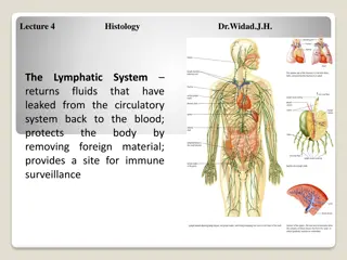
  4. Immune System The immune system is a system of barriers, cells, tissues and organs that work to fight invaders. Another important component of the immune system is the complement system. The major functions of the immune system are: 1. Differentiate self from non-self. 2. Eliminate foreign substances, cells and pathogens. Organization of the immune system Blood Primary (or central) lymphoid organs Secondary (or peripheral) lymphoid organs Cells vasculature and lymph systems connect the different organs

  5. Major organs Primary Lymphoid Organs Secondary (or peripheral) Lymph organs tonsils adenoids spleen Bone marrow Thymus Lymphatic vessels Lymph nodes 1. Immune cells mature to become immunocompetent in these organs. 2. Most cells mature to immune cells within the bone marrow and, after release, begin a life of patrol in the blood. The exception is the pre-T cell, which first undergoes maturation in the thymus before circulating in blood. peyer's patches appendix 1. These are the organs where mature immune cells participate in specific immune defense reactions.

  6. Major organs 2 2 1 2 2 2 2 2 1

  7. Types of Immunity Immune system Innate (non-specific; natural) immunity Adaptive (specific; acquired) immunity 1. Third line of defense. 2. Antigen specificity. It is activated by thousands of diverse antigens. 3. Responds with the proliferation of cells and the generation of antibodies. 4. Responds slowly, being fully activated about 4 days after the immunologic threat. 5. Exhibits immunologic memory, so that repeated exposure to the same infectious agent results in improved resistance against it. 1. Second line of defense 2. Is present at birth 3. Persists throughout life 4. Can be mobilized rapidly and act quickly 5. Attacks all antigens fairly equally

  8. First line or defence Epithelial Barriers

  9. The Complement System is Part of the 1stLine of Defense complement system: 1- The first part of the immune system that meets invaders such as bacteria 2-it is a group of proteins. 3- These proteins flow freely in the blood and can quickly reach the site of an invasion where they can react directly with antigens ( molecules that the body recognizes as foreign substances). functions of complement proteins (When activated ): 1. Trigger inflammation. 2. Attract eater cells such as macrophages to the area. 3. Coat intruders so that eater cells are more likely to devour (swallow and eat) them (a process called as opsonization). 4. Kill intruders.

  10. Cellular Elements of the Innate Immune System (2ndline of defense)

  11. Cells of the Immune System The cells of the immune system work together with different proteins to seek out and destroy anything foreign or dangerous that enters our body. Cells of the Immune System Inflammatory cells Antigen Specific cells Phagocytes Dendritic cells B cells or B lymphocyt es Basophils and Eosinophils Monocytes and macrophages T cells or T lymphocytes Neutrophils Mast cells

  12. Cells of the Immune System

  13. White Blood Cells (Leucocytes) Function Protection against infection by: Formation Secretion of antibodies bone marrow lymph tissue Phagocytosis

  14. Remember white blood cells 4000-11000/ml red blood cells (5-6-million/ml) 3

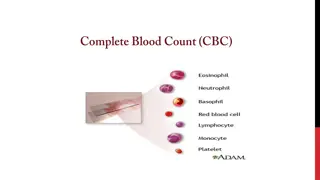
  15. Types of WBC Granular A granular granules (polymorphnuclear PMN) PMNs; because of multiple(lobes) nuclei Neutrophil - 62% - lobulated nucleus basic stain 2-5 - Purple cytoplasmic granules Basophil - 0.4% - rarely segmented nucleus - nucleus hidden by large round bluish granules Monocytes 5.3% kidney shape nucleus (make macrophage system) Eosinophil - 2.3% - 2 lobe nucleus eosin stain - coarse red granules Lymphocyte 30% round nucleus Small large 1-T lymphocyte 2-B lymphocyte 3-Natural killer cells (NKCs) Genesis of WBC: Myelocytic (granular+monocytes) Genesis of WBC: Lymphocytic sites of formation: bone marrow sites of formation: - Bone marrow - Thymus - Lymphoid tiss ( . ) ( )

  16. ( Types of WBC . ) 1- Granular (polymorphnuclear PMN): Neutrophil Neutrophil Eosinophil Eosinophil B Basophil asophil 62%. 2.3% 0.4%. 10-16um 12-18um 10-14um lobulated nucleus 2- 5. 2 lobe nucleus. rarely segmented nucleus. coarse red Purplecytoplasmic granules nucleus hidden by large round bluish granules. granules

  17. ( . Types of WBC cont. ) 2. A granular Monocytes Monocytes Lymphocyte Lymphocyte 5.3% 30% 15-20um small (5-8um) large (9-15um) kidney shape nucleus round nucleus

  18. Physiological Variations in WBCs Count Age: Infants: about 20,000 /mm3 Children:10,000 to 15,000/mm3 Adults: 4,000 and 11,000/mm3 Sex: males have more WBCs than females. Diurnal variation: Minimum in early morning and maximum in the afternoon. Exercise: Increases slightly. Sleep: Decreases. Emotional conditions like anxiety: Increases. Pregnancy: Increases. Menstruation: Increases. Parturition: Increases. Male Slide

  19. WBCs Concentrations (Normal Counts) and Life Span Percentage of Total WBCs Approximate Normal Range (/ L) Cells 4000 - 11000 Total WBCs 60 70% (62%) 3000 - 6000 Neutrophils 1 4% (2.3%) 150 - 300 Eosinophils 0.4% (0.4%) 0 - 100 Basophils 20 40% (30%) 1500 - 4000 Lymphocytes 2 8% (5.3%) 300 - 600 Monocytes (Macrophages) , .

  20. Life span of WBCs Granulocytes Monocytes Lymphocytes Neutrophil, Eosinophil, Basophil In blood: 10-20 hours Leave to the tissue: - transform into macrophage stay up to months weeks to months according to its type B > few weeks T > year (100-300day) in tissues 4-5 days During Infection: only few hours (they die after ingesting bacteria)

  21. Genesis of WBC (Leucopoiesis) Two major lineage of WBC are formed : Myelocytic: beginning with myelobast and giving rise to granular leucocytes and monocytes Lymphocytic: beginning with lymphoblast and giving rise to lymphocytes the WBC need to leave the blood to work unlike RBC. Sites of WBC Formation Granulocytes (neutrophil, basophil, eosinophil) Monocytes Lymphocytes bone marrow and lymphoid tissues especially the lymph glands, spleen, thymus, tonsils, and Peyer s patches bone marrow in bone marrow

  22. Genesis of WBC WBCs formed in the bone marrow are stored within the marrow until they are needed in the circulatory system. Various factors cause them to be released. Normally, about three times as many white blood cells are stored in the marrow as circulate in the entire blood. The lymphocytes are mostly stored in the various lymphoid tissues, except for a small number that are temporarily being transported in the blood.

  23. Genesis of WBC

  24. Genesis of WBC Leucopoiesis

  25. White Blood Cells NEUTROPHILLS Formation and Maturation of Neutrohils:in Bone Marrow ( Maturation of red blood cells ( ) Stem cells . ) Myeloblast Promyelocytes Neutrophil myelocytes Young neutrophil metamyelocytes Band neutrophil Polymorphnuclear neutrophil (MatureNeutrphils released to blood)

  26. White Blood Cells NEUTROPHILLS Polymorphonuclear. They contain small granules of both acidic and basic. They constitute the first line of defence against bacterial infection. Very important at clearing bacterial infections. Neutrophils Cells infected by bacteria usually get dealt with by

  27. Neutrophil Function Defense against infection: Neutrophil has the ability of engulfing bacteria or organism by a process of phyagocytosis Neutrophil Phagocytosis 1-neutrophil attaches itself to the particle and projects pseudopodia in all directions around the particle. 2-pseudopodia meet one another on the opposite side and fuse, creating an enclosed chamber that contains the phagocytized particle. 3-the chamber moves to the inside of the cytoplasmic cavity and breaks away from the outer cell membrane to form a free-floating phagocytic vesicle (also called a phagosome) inside the cytoplasm. 4-The phagocytized particle is then digested by intracellular enzymes Steps of Phygocytosis Engulfing and killing of a microbe Ameoboid movement Chemotaxis Like alarms Margination Diapedesis Females slide

  28. Chemotaxis The attraction of the neutrophils and monocytes to move to inflamed area following chemotactic substances release from infected site. The chemotactic agents include a component of the complement system (C5a); leukotrienes; and polypeptides from lymphocytes, mast cells, and basophils. Chemotactic substances: - Bacterial toxin - Degenerative products of inflamed tissue Chemotaxis is effective up to 100 micrometers away from an inflamed tissue. Therefore, because almost no tissue area is more than 50 micrometers away from a capillary, the chemo- tactic signal can easily move hordes of WBCs from the capillaries into the inflamed area. - Complement system - Reaction product of plasma clotting

  29. Margination & Diapedesis There are gabs inbetween the endothelial capillaries where the neutrophils squeeze neutrophils through 1- WBCs marginate (aggregate and stick) along the wall of blood capillaries. 2- WBC squeezes itself through endothelial holes leaving blood capillaries (diapedesis) 3- WBCs move by amoeboid motion towards inflammation area following chemotactic substance released from site of infection 4- Upon reaching the site of infection neutrophils start to engulf infecting organism

  30. Chemotaxis, margination & diapedesis

  31. Phagocytosis and Opsonization Phagocytosis: means cellular ingestion of the offending agent. opsonization is the coating of the invader Selective process: foreign substance recognize by: -Rough surface -No protective protein coat, which prevents phagocytosis. -Marked by certain substance (opsonization) name tag e.g Complement 3 or antibodies making them ready for killing. Neutrophils encircled the bacteria with pseudopodia and engulf it inside into a vacuole (phagosome), takes 3-20 bacteria Opsonization: Complement 3b or antibodies like IgG making them ready for killing a process known as opsonization. Some plasma factors act on the bacteria to make them tasty to the phagocytes (opsonization). The principal opsonins that coat the bacteria are immunoglobulins of a particular class (IgG) and complement proteins.

  32. Opsonization and Phagocytosis

  33. Microbial killing killing effect results from several powerful oxidizing agents formed by enzymes in the membrane of the phagosome or by a special organelle called the peroxisome. ese oxidizing agents include large quantities of superoxide (O2 ), hydrogen peroxide (H2O2), and hydroxyl ions (OH ), which are lethal to most bacteria, even in small quantities. Digestion of organism inside the phagosome: 1- Fusion of intracellular lysosomes with phagosome vacuole 2- Lysosomes discharge its proteolytic enzymes such as myeloperoxidase, catalase into the vacuole, killing and digesting the engulfed bacteria. And\or Release of bactericidal such as superoxide, hydrogen peroxide to kill the bacteria Some bacteria, notably the tuberculosis bacillus, have coats that are resistant to lysosomal digestion and also secrete substances that partially resist the killing effects of the neutrophils and macrophages.These bacteria are responsible for many of the chronic diseases, an example of which is tuberculosis.

  34. Microbial killing

  35. Formation and Maturation of Eosinophils: in Bone Marrow Stem cells ( Maturation of red blood cells ( ) Myeloblast . ) Promyelocytes Eosinophil myelocytes Eosinophil metamyelocytes polymorphnuclear eosinophil (Mature Eosinophil released to blood)

  36. Eosinophil Function 1- they are weak phagocytes. 1- they are weak phagocytes. 2- they are often produced in large numbers in people with parasitic infections, and they migrate in large numbers into tissues diseased by parasites., e.g., ascaris, hook worm, bilharzia. Eosinophils attach themselves to the parasites by way of special surface molecules and release substances that kill many of the parasites ex:(hydrolytic anzymes,superoxide) 3- They are increased in allergic conditions by the release of eosinophil chemotactic factor released from the mast cells and basophiles. 4- Eosinophils phagocytose the antigen-antibody complexes and release substances to neutralize the histamine. ex: (asthma, rhinitis, drug reaction) 4-They may produce profibrinolysin fibrinolysin which digest fibrin clot.

  37. Formation and Maturation of Basophils:in Bone Marrow Stem cells ) ( cells Myeloblast Maturation of red blood ( . ) Promyelocytes Basophil myelocytes Basophil metamyelocyt Polymorphnuclear Basophil (Mature Basophils released to blood) large round cytoplasmic polysaccharide granules which take base dye (methylene blue) and stain blue in color.

  38. Basophils Functions: 1-Both mast cells and basophils secrete: histamine (to prevent clotting), heparin, bradykinin, Serotonin (5HT) (contribute to inflammation response), slow-reacting substance of anaphylaxis (a mixture of three leukotrienes) and a number of lysosomalenzymes. The release of those substances cause local and vascular reactions characteristic of allergic manifestation 2-They are increased in allergic reaction: immediate-type hypersensitivity (allergic) reactions.

  39. Monocytes\ Macrophages Monocytes contain agranular cytoplasm, but when they convert into macrophages and enter tissue, they swell and their cytoplasm become filled with a large number of lysosomes. Monocytes leave the blood stream to tissues and turn into macrophages. Monocytes are young macrophages. Monocytes are big eaters. Monocytes are slower to respond to invaders than neutrophils, but they are larger, have greater capacities, and live longer. 15-20 m - Active cells 60- 80 m

  40. Monocytes\ Macrophages Anti-Inflammatory: - Directly: phagocytosis of bacteria, dead cells. - Indirectly: cooperating with lymphocytes by recognizing foreign body (take in foreign body process it and present it to lymphocytes). Functions of monocytes/macrophages: - First line of defense. - Phagocytosis and killing of microorganisms. They are more Efficient than Neutrophils (100 bacteria vs 3-20 by Neutrphil, larger particles like old RBCs & malarial parasites). - Activation of T cells and initiation of the immune response by presenting the antigen to these cells. - gets rid of waste and survives. Monocytes secrete: - Interleukin-1 (IL-1). - Colony stimulating factor (M-CSF). - Platelet-activating factor (PAF).

  41. Indirect anti-inflammatory Direct anti Inflammatory

  42. Formation and Maturation of Monocytesand Macrophages: in Bone Marrow ( Maturation of red blood cells ( ) Stem cells . ) Monoblast monoblast Promonocyte promonocyte mature monocytes released into blood Stay for 10-20 hours in circulation Then leave blood to tissues transforming into larger cells macrophage Macrophage life span is longer upto few months .

  43. Monocytes\ Macrophages There are two types of macrophages; motile and fixed. There are tissue-specific macrophages; fixed macrophages (monocyte-macrophage system; reticulo-endothelial system) - Alveolar macrophage - Peritoneal macrophage - Kuppfer cells in liver sinuses - Osteoclasts in bone - Microglial cells in brain - Histiocytes in skin and subcutaneous tissue - Mesengial cells in the kidneys - Few specialized endothelial cells in lymph nodes bone marrow, spleen and

  44. Reticuloendothelial System Consist of: Monocytes Macrophage Endothelial cells (bone marrow,spleen, lymph node) Located in: all tissues especially: skin (histocytes), liver (kupffer), spleen, bone marrow, lymph nodes, lung ( . )

  45. Functions of Reticuloendothelialsystem -Phagocytosis: Bacterial, dead cells, foreign particles. -Breakdown of Hb -Immune function: processing antigen and antibodies production (indirect) -Storage of iron in the liver and bone marrow in the form of fe3 (mostly),fe2

  46. Macrophage and Neutrophil Responses During Inflammation Neutrophils = microphages 1st line of defense Tissue macrophages, barriers and complement system. 2nd line of defense Neutrophil invasion of the inflamed area. 3rdline of defense Monocytes macrophage invasion of inflamed area. 4thline of defense Increased production of granulocytes and monocytes by the bone marrow.

  47. LYMPHOCYTES Function and types Types: 1-Thymus dependent (T-lymphocytes) 2 -Thymus independent (B-lymphocytes) LYMPHOCYTES ARE RESPONSIBLE FOR ADAPTED/ACQUIRED IMMUNITY Function:

  48. LymphocytesFormation and Maturation: T lymphocyte and B lymphocyte produced by the bone marrow however, T get maturated in the thymus and the B cells in the bone marrow then they go to the lymphoid organs like spleen Lymph nodes etc Formed in bone marrow, thymus, lymphoid tissues Stem cells (thymus, lymphoid tissue & bone marrow) ( Maturation of red blood cells ( ) . ) lymphoblast intermediate pyronophilic blast cell lymphocytes Life Span Of Lymphocytes range from weeks to months according to its type. Both types of lymphocytes are derived in the embryo from (pluripotent hematopoietic stem cells) that form common (lymphoid progenitor cells).

More Related Content

giItT1WQy@!-/#giItT1WQy@!-/#giItT1WQy@!-/#giItT1WQy@!-/#giItT1WQy@!-/#giItT1WQy@!-/#giItT1WQy@!-/#giItT1WQy@!-/#giItT1WQy@!-/#giItT1WQy@!-/#giItT1WQy@!-/#giItT1WQy@!-/#giItT1WQy@!-/#giItT1WQy@!-/#giItT1WQy@!-/#giItT1WQy@!-/#giItT1WQy@!-/#giItT1WQy@!-/#giItT1WQy@!-/#