Welcome to Pre-K at Prattville Primary School 2020-2021
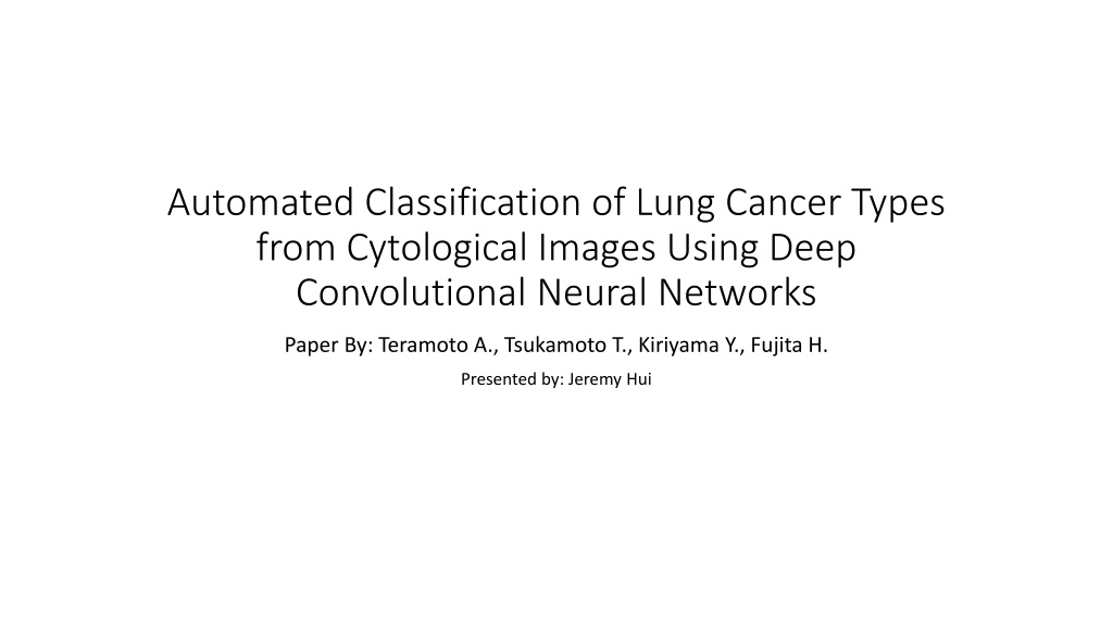
Automated Classification of Lung Cancer Types
from Cytological Images Using Deep
Convolutional Neural Networks
Paper By: Teramoto A., Tsukamoto T., Kiriyama Y., Fujita H.
Presented by: Jeremy Hui
Introduction
•
Lung Cancer is one of the leading causes of death worldwide
•
Accurate classification of cancer types is required for accurate and
stable diagnosis
•
Primary Lung Cancers
•
Small cell lung cancer
•
Non-small cell lung cancer
Introduction
•
Non-small cell lung cancer can be further classified into
•
Adenocarcinoma
•
Squamous cell carcinoma
•
Large cell carcinoma
•
Large cell carcinoma is easiest to detect
•
Focuses on adenocarcinoma, squamous cell carcinoma and small cell
carcinoma
Previous work
•
D. C. Ciresan, A. Giusti, L. M. Gambardella, and J. Schmid- huber,
“Mitosis detection in breast cancer histology images with deep neural
networks,” in
Medical Image Computing and Computer-Assisted
Intervention—MICCAI 2011
, vol. 8150 of
Lecture Notes in Computer
Science
, pp. 411–418, Springer, Berlin, Germany, 2013.
•
H. Wang, A. Cruz-Roa, A. Basavanhally et al., “Mitosis detec- tion in
breast cancer pathology images by combining hand- crafted and
convolutional neural network features,”
Journal of Medical Imaging
,
vol. 1, no. 3, p. 034003, 2014.
Approach
•
Automated classification for lung cancers presented in microscopic
images using a deep convolutional neural network (DCNN)
•
Architecture
•
3 convolutional layers
•
3 pooling layers
•
2 fully connect layers
Image Dataset
•
76 cases of cancer cells collected
•
40 cases of adenocarcinoma
•
20 cases of squamous cell carcinoma
•
16 cases of small cell carcinoma
•
Digital camera attached to microscope with x40 objective lens used to
take images
•
82 images of adenocarcinoma
•
125 images of squamous cell carcinoma
•
91 images of small cell carcinoma
Image Dataset
•
Images in JPEG format of matrix size 2040 x 1536 pixels
•
768 x 768 square images were cropped
•
Resized to 256 x 256 pixels
Data Augmentation
•
DCNN requires a sufficient amount of training data
•
Rotating, inverting and filtering applied to the original images
•
Images were flipped, resulting in twice the original number
•
Gaussian filter applied in filtering
•
S.D of Gaussian kernel = 3 pixels
•
Convolutional edge enhancement filter was applied
•
Center weight 5.4, 8-surrounding weight -0.55
Architecture
•
3 convolutional layers, 3 pooling layers, 2 fully connected layers
•
Rectified linear unit (ReLU) activation function after every convolution
layer
•
Probabilities of cancer types (adenocarcinoma, squamous cell
carcinoma, small cell carcinoma) obtained using softmax function
•
Dropout rate = 50% for full connection layers
•
Number of epochs = 60,000
Architecture
Results
•
3 fold cross-validation
Results
Results
Welcome to Pre-K at Prattville Primary School 2020-2021! Visit our school website for important updates and information. Access Pre-K information, meet the teachers, and stay informed about COVID-19 guidelines. Find details about the first day of school, arrival, and dismissal procedures. Stay connected with the school community through valuable resources. Get ready for an exciting educational journey at Prattville Primary School!

Download Presentation

Please find below an Image/Link to download the presentation.
The content on the website is provided AS IS for your information and personal use only. It may not be sold, licensed, or shared on other websites without obtaining consent from the author.If you encounter any issues during the download, it is possible that the publisher has removed the file from their server.
You are allowed to download the files provided on this website for personal or commercial use, subject to the condition that they are used lawfully. All files are the property of their respective owners.
The content on the website is provided AS IS for your information and personal use only. It may not be sold, licensed, or shared on other websites without obtaining consent from the author.
E N D
Presentation Transcript
Automated Classification of Lung Cancer Types from Cytological Images Using Deep Convolutional Neural Networks Paper By: Teramoto A., Tsukamoto T., Kiriyama Y., Fujita H. Presented by: Jeremy Hui
Introduction Lung Cancer is one of the leading causes of death worldwide Accurate classification of cancer types is required for accurate and stable diagnosis Primary Lung Cancers Small cell lung cancer Non-small cell lung cancer
Introduction Non-small cell lung cancer can be further classified into Adenocarcinoma Squamous cell carcinoma Large cell carcinoma Large cell carcinoma is easiest to detect Focuses on adenocarcinoma, squamous cell carcinoma and small cell carcinoma
Previous work D. C. Ciresan, A. Giusti, L. M. Gambardella, and J. Schmid- huber, Mitosis detection in breast cancer histology images with deep neural networks, in Medical Image Computing and Computer-Assisted Intervention MICCAI 2011, vol. 8150 of Lecture Notes in Computer Science, pp. 411 418, Springer, Berlin, Germany, 2013. H. Wang, A. Cruz-Roa, A. Basavanhally et al., Mitosis detec- tion in breast cancer pathology images by combining hand- crafted and convolutional neural network features, Journal of Medical Imaging, vol. 1, no. 3, p. 034003, 2014.
Approach Automated classification for lung cancers presented in microscopic images using a deep convolutional neural network (DCNN) Architecture 3 convolutional layers 3 pooling layers 2 fully connect layers
Image Dataset 76 cases of cancer cells collected 40 cases of adenocarcinoma 20 cases of squamous cell carcinoma 16 cases of small cell carcinoma Digital camera attached to microscope with x40 objective lens used to take images 82 images of adenocarcinoma 125 images of squamous cell carcinoma 91 images of small cell carcinoma
Image Dataset Images in JPEG format of matrix size 2040 x 1536 pixels 768 x 768 square images were cropped Resized to 256 x 256 pixels
Data Augmentation DCNN requires a sufficient amount of training data Rotating, inverting and filtering applied to the original images Images were flipped, resulting in twice the original number Gaussian filter applied in filtering S.D of Gaussian kernel = 3 pixels Convolutional edge enhancement filter was applied Center weight 5.4, 8-surrounding weight -0.55
Architecture 3 convolutional layers, 3 pooling layers, 2 fully connected layers Rectified linear unit (ReLU) activation function after every convolution layer Probabilities of cancer types (adenocarcinoma, squamous cell carcinoma, small cell carcinoma) obtained using softmax function Dropout rate = 50% for full connection layers Number of epochs = 60,000
Results 3 fold cross-validation