Update on Multiple Myeloma: Diagnosis, Risk Stratification, and Management in 2018
Multiple myeloma is a significant hematologic malignancy with varying stages of progression. From the asymptomatic MGUS to the more advanced SMM, early diagnosis and risk stratification are crucial. The presence of myeloma defining events, specific biomarkers, and established criteria guide the diagnosis and management of this condition. The updated criteria allow for timely initiation of therapy to prevent end organ damage, representing a paradigm shift in patient care.
Download Presentation

Please find below an Image/Link to download the presentation.
The content on the website is provided AS IS for your information and personal use only. It may not be sold, licensed, or shared on other websites without obtaining consent from the author.If you encounter any issues during the download, it is possible that the publisher has removed the file from their server.
You are allowed to download the files provided on this website for personal or commercial use, subject to the condition that they are used lawfully. All files are the property of their respective owners.
The content on the website is provided AS IS for your information and personal use only. It may not be sold, licensed, or shared on other websites without obtaining consent from the author.
E N D
Presentation Transcript
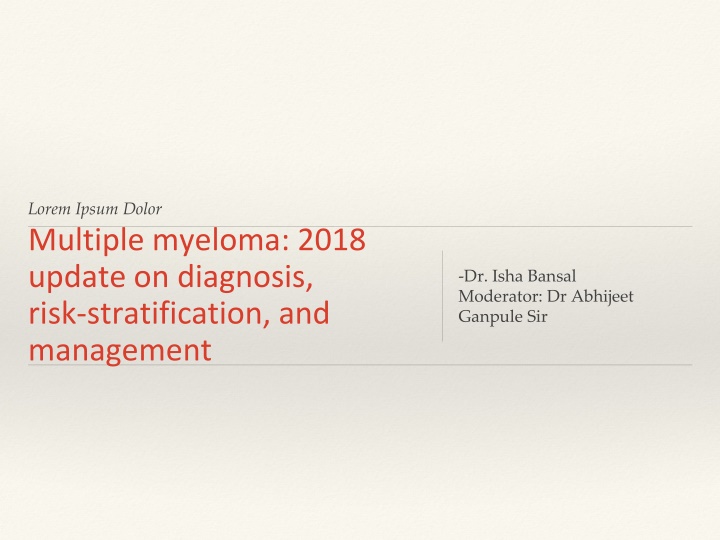
Lorem Ipsum Dolor Multiple myeloma: 2018 update on diagnosis, risk stratification, and management -Dr. Isha Bansal Moderator: Dr Abhijeet Ganpule Sir
This article has been taken from American Journal of Haematology, Volume 93, Issue 8 Published in the month of August, 2018
Introduction Multiple myeloma accounts for 1% of all cancers and approximately 10% of all hematologic malignancies. Men>Women, Median age- 65 years Common in African Americans compared with Caucasians
Almost all patients with multiple myeloma evolve from an asymptomatic premalignant stage termed monoclonal gammopathy of undetermined significance (MGUS) 1% every year More advanced premalignant stage referred to as smoldering multiple myeloma (SMM) can be recognized clinically 10% per year over the first 5 years following diagnosis, 3% per year over the next 5 years, and 1.5% per year thereafter t(4;14) translocation, del(17p), and gain(1q)-higher risk
Diagnosis The diagnosis of multiple myeloma requires the presence of one or more myeloma defining events (MDE) in addition to evidence of either 10% or more clonal plasma cells on bone marrow examination or a biopsy proven plasmacytoma. MDE consists of established CRAB (hypercalcemia, renal failure, anemia, or lytic bone lesions) features as well as 3 specific biomarkers: clonal bone marrow plasma cells 60%, serum free light chain (FLC) ratio 100 (provided involved FLC level is 100 mg/L), and more than one focal lesion on MRI.
Each of the new biomarkers is associated with an approximately 80% risk of progression to symptomatic end organ damage in two or more independent studies. The updated criteria represent a paradigm shift since they allow early diagnosis and initiation of therapy before end organ damage.
International Myeloma Working Group criteria Non IgM monoclonal gammopathy of undetermined significance (MGUS ) All 3 criteria must be met: Serum monoclonal protein (non IgM type) <3 gm/dL Clonal bone marrow plasma cells <10% Absence of end organ damage such as hypercalcemia, renal insufficiency, anemia, and bone lesions (CRAB) that can be attributed to the plasma cell proliferative disorder
Smoldering multiple myelomaBoth criteria must be met: Serum monoclonal protein (IgG or IgA) 3 g/dL, or urinary monoclonal protein 500 mg per 24 hours and/or clonal bone marrow plasma cells 10% 60% Absence of myeloma defining events or amyloidosis
Multiple myeloma Both criteria must be met: Clonal bone marrow plasma cells 10% or biopsy proven bony or extramedullary plasmacytoma Any one or more of the following myeloma defining events: Evidence of end organ damage that can be attributed to the underlying plasma cell proliferative disorder, specifically: Hypercalcemia: serum calcium >0 25 mmol/L (>1 mg/dL) higher than the upper limit of normal or >2 75 mmol/L (>11 mg/dL)
Renal insufficiency: creatinine clearance <40 mL per minute or serum creatinine >177 mol/L (>2 mg/dL) Anemia: hemoglobin value of >2 g/dL below the lower limit of normal, or a hemoglobin value <10 g/dL Bone lesions: one or more osteolytic lesions on skeletal radiography, computed tomography (CT), or positron emission tomography CT (PET CT) Clonal bone marrow plasma cell percentage 60% Involved: uninvolved serum free light chain (FLC) ratio 100 (involved free light chain level must be 100 mg/L) >1 focal lesions on magnetic resonance imaging (MRI) studies (at least 5 mm in size)
IgM Monoclonal gammopathy of undetermined significance (IgM MGUS) All 3 criteria must be met: Serum IgM monoclonal protein <3 g/dL Bone marrow lymphoplasmacytic infiltration <10% No evidence of anemia, constitutional symptoms, hyperviscosity, lymphadenopathy, or hepatosplenomegaly that can be attributed to the underlying lymphoproliferative disorder.
Light Chain MGUS All criteria must be met: Abnormal FLC ratio (<0.26 or >1.65) Increased level of the appropriate involved light chain (increased kappa FLC in patients with ratio > 1.65 and increased lambda FLC in patients with ratio < 0.26) No immunoglobulin heavy chain expression on immunofixation Absence of end organ damage that can be attributed to the plasma cell proliferative disorder Clonal bone marrow plasma cells <10% Urinary monoclonal protein <500 mg/24 hours
Solitary Plasmacytoma All 4 criteria must be met Biopsy proven solitary lesion of bone or soft tissue with evidence of clonal plasma cells Normal bone marrow with no evidence of clonal plasma cells Normal skeletal survey and MRI (or CT) of spine and pelvis (except for the primary solitary lesion) Absence of end organ damage such as hypercalcemia, renal insufficiency, anemia, or bone lesions (CRAB) that can be attributed to a lympho plasma cell proliferative disorder
Solitary Plasmacytoma with minimal marrow involvement All 4 criteria must be met Biopsy proven solitary lesion of bone or soft tissue with evidence of clonal plasma cells Clonal bone marrow plasma cells <10% Normal skeletal survey and MRI (or CT) of spine and pelvis (except for the primary solitary lesion) Absence of end organ damage such as hypercalcemia, renal insufficiency, anemia, or bone lesions (CRAB) that can be attributed to a lympho plasma cell proliferative disorder
serum protein electrophoresis (SPEP), serum immunofixation, and the serum FLC assay. FISH-t(11;14), t(4;14), t(14;16), t(6;14), t(14;20), trisomies, and del(17p) low dose WB CT or PET/CT imaging Serum CrossLaps to measure carboxy terminal collagen crosslinks (CTX) may be useful in assessing bone turnover and to determine adequacy of bisphosphonate therapy.
MOLECULAR CLASSIFICATION it is a collection of several different cytogenetically distinct plasma cell malignancies. 40%-trisomic multiple myeloma a translocation involving the immunoglobulin heavy chain (IgH) locus on chromosome 14q32 both trisomies and IgH translocations. gain(1q), del(1p), del(17p), del(13), RAS mutations
Recurrent trisomies involving odd numbered chromosomes with the exception of chromosomes 1, 13, and 21 Trisomic multiple myeloma t(11;14) (q13;q32) t(4;14) (p16;q32) t(14;16) (q32;q23) t(14;20) (q32;q11) IgH translocated multiple myeloma Few cases may represent 14q32 translocations involving unknown partner chromosomes Isolated Monosomy 14 Presence of trisomies and any one of the recurrent IgH translocations in the same patient Combined IgH translocated/trisomic multiple myeloma
PROGNOSIS AND RISK STRATIFICATION median survival in multiple myeloma is approximately 6 years. In the subset of patients eligible for ASCT, 4 year survival rates are more than 80%, OS 8 years. More precise estimation of prognosis requires an assessment of multiple factors. As in other cancers, OS in multiple myeloma is affected by host characteristics, tumor burden (stage), biology (cytogenetic abnormalities), and response to therapy Tumor burden in multiple myeloma has traditionally been assessed using the Durie Salmon Staging44 and the International staging system (ISS)
Two other markers that are associated with aggressive disease biology are elevated serum lactate dehydrogenase and evidence of circulating plasma cells on routine peripheral smear examination (plasma cell leukemia). The Revised International Staging System (RISS) combines elements of tumor burden (ISS) and disease biology (presence of high risk cytogenetic abnormalities or elevated lactate dehydrogenase level) to create a unified prognostic index that and helps in clinical care
Stage I Stage III Stage II Serum albumin 3.5 gm/dL Serum beta 2 microglobu lin >5.5 mg/L Not fitting Stage I or III Serum beta 2 microglobu lin <3.5 mg/L High risk cytogenetics [t(4;14), t(14;16), or del(17p)] or Elevated serum lactate dehydrogenase level No high risk cytogenetics Normal serum lactate dehydrogenase level
Mayo clinic risk stratification for multiple myeloma (mSMART) Standard Risk 75% Trisomies -t(11;14) t(6;14) Intermediate Risk 10% t(4;14) ,Gain(1q) High Risk 15% t(14:16) t(14;20) del(17p)
INDICATIONS FOR THERAPY To initiate therapy, patients must meet criteria for multiple myeloma. treatment of asymptomatic patients with SMM early therapy with lenalidomide and dexamethasone in patients with high risk SMM
TREATMENT OF NEWLY DIAGNOSED MYELOMA The initial impact came from the introduction of thalidomide, bortezomib, and lenalidomide. Thalidomide, lenalidomide, and pomalidomide are termed immunomodulatory agents (IMiDs).
VRd Bortezomib Lenalidomide Dexamethasone (VRd) Bortezomib 1.3 mg/m2 subcutaneous days 1, 8, 15 Lenalidomide 25 mg oral days 1 14 Dexamethasone 20 mg oral on day of and day after bortezomib (or 40 mg days 1, 8, 15, 22) Repeated every 3 weeks
KRd Carfilzomib 20 mg/m2 (days 1 and 2 of Cycle 1) and 27 mg/ m2 (subsequent doses) intravenously on days 1, 2, 8, 9, 15, 16 Lenalidomide 25 mg oral days 1 21 Dexamethasone 40 mg oral days 1, 8, 15, 22 Repeated every 4 weeks
Rd Lenalidomide 25 mg oral days 1 21 every 28 days Dexamethasone 40 mg oral days 1, 8, 15, 22 every 28 days Repeated every 4 weeks
DRd Daratumumab 16 mg/ kg intravenously weekly x 8 weeks, and then every 2 weeks for 4 months, and then once monthly Lenalidomide 25 mg oral days 1 21 Dexamethasone 40 mg intravenous days 1, 8, 15, 22 (given oral on days when no daratumumab is being administered) Lenalidomide Dexamethasone repeated in usual schedule every 4 weeks
ERd Elotuzumab 10 mg/ kg intravenously weekly x 8 weeks, and then every 2 weeks Lenalidomide 25 mg oral days 1 21 Dexamethasone per prescribing information Lenalidomide Dexamethasone repeated in usual schedule every 4 weeks
IRd Ixazomib 4 mg oral days 1, 8, 15 Lenalidomide 25 mg oral days 1 21 Dexamethasone 40 mg oral days 1, 8, 15, 22 Repeated every 4 weeks
DPd Daratumumab 16 mg/kg intravenously weekly 8 weeks, and then every 2 weeks for 4 months, and then once monthly Pomalidomide 4 mg oral on days 1 21 Dexamethasone 40 mg intravenous days 1, 8, 15, 22 (given oral on days when no daratumumab is being administered)
DVd Daratumumab 16 mg/ kg intravenously weekly x 8 weeks, and then every 2 weeks for 4 months, and then once monthly Bortezomib 1.3 mg/m2 subcutaneous on days 1, 8, 15, 22 Dexamethasone 40 mg intravenous days 1, 8, 15, 22 (given oral on days when no daratumumab is being administered) Bortezomib Dexamethasone repeated in usual schedule every 4 weeks
VCd Cyclophosphamide 300 mg/m2 orally on days 1, 8, 15, and 22 Bortezomib 1.3 mg/m2 subcutaneous on days 1, 8, 15, 22 Dexamethasone 40 mg oral on days on days 1, 8, 15, 22 Repeated every 4 weeksc
Pd Pomalidomide 4 mg days 1 21 Dexamethasone 40 mg oral on days on days 1, 8, 15, 22 Repeated every 4 weeks
Summary Multiple myeloma accounts for approximately 10% of hematologic malignancies. The diagnosis requires 10% clonal bone marrow plasma cells or a biopsy proven plasmacytoma plus evidence of one or more multiple myeloma defining events: CRAB(hypercalcemia, renal failure, anemia, or lytic bone lesions) features felt related to the plasma cell disorder, bone marrow clonal plasmacytosis 60%, serum involved/uninvolved free light chain (FLC) ratio 100(provided involved FLC is 100 mg/L), or >1 focal lesion on magnetic resonance imaging. Patients with del(17p), t(14;16), and t(14;20) have high risk multiple myeloma. Patients with t(4;14) translocation and gain(1q) have intermediate risk. All others are considered standard risk.
Initial treatment consists of bortezomib, lenalidomide, dexamethasone (VRd). In high risk patients, carfilzomib, lenalidomide, dexamethasone (KRd) is an alternative to VRd. In eligible patients, initial therapy is given for approximately 3 4 cycles followed by autologous stem cell transplantation (ASCT). Standard risk patients can opt for delayed ASCT at first relapse. Patients not candidates for transplant are treated with VRd for approximately 8 12 cycles followed by lenalidomide or lenalidomide plus dexamethasone. After ASCT, lenalidomide maintenance is recommended for standard risk patients, while maintenance with a bortezomib based regimen is needed for patients with intermediate or high risk disease. Most patients require a triplet regimen at relapse, with the choice of regimen varying with each successive relapse. Aggressive relapse with extramedullary plasmacytomas or plasma cell leukemia may require anthracycline containing combination chemotherapy regimens.
Thank You Johnny Appleseed
