Understanding the Human Urinary System
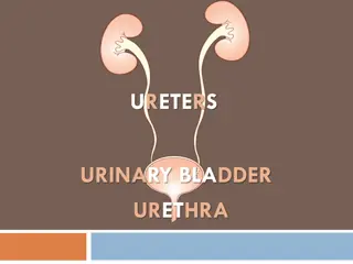
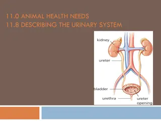
The human urinary system consists of the kidneys, ureters, bladder, and urethra. The kidneys filter blood to produce urine, which is carried to the bladder through the ureters for storage until excretion. Each kidney is structured with renal pyramids and nephrons for blood filtration and waste removal. The intricate system of uriniferous tubules, renal corpuscles, and podocytes within the nephrons plays a vital role in maintaining homeostasis through regulation of blood composition, volume, and pressure. Understanding the anatomy and functions of the urinary system is essential for overall health.
Download Presentation

Please find below an Image/Link to download the presentation.
The content on the website is provided AS IS for your information and personal use only. It may not be sold, licensed, or shared on other websites without obtaining consent from the author. Download presentation by click this link. If you encounter any issues during the download, it is possible that the publisher has removed the file from their server.
E N D
Presentation Transcript
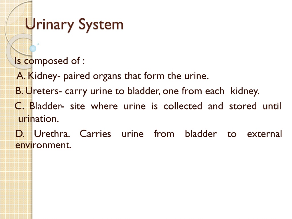
Urinary System Is composed of : A.Kidney- paired organs that form the urine. B.Ureters- carry urine to bladder,one from each kidney. C. Bladder- site where urine is collected and stored until urination. D. Urethra. Carries urine environment. from bladder to external
Kidney _Is bean shaped organ covered irregular by dense collage nous connective tissue capsule. _Has a convex border&a medial concave border have hilum,where the arteries enter,ureter& veins leave the kidney. _The upper part of ureter is expanded to form renal pelvis that branches to several large cup- shaped structure about (2-3) in number (major calyces), and smaller calyces (8-10) (minor calyces).
Each kidney is divided in to : _Outer cortex _Inner medulla :which is composed of 10-12 renal pyramids, the base of pyramid is toward the cortex as (medullar rays). _Functions of kidney : 1-Regulattion of blood ionic composition, regulate of blood ph, regulation of blood volume, regulation of blood pressure. 2-Maintenance of blood osmolarity. 3-Production of hormones erythropoietin. 4-Excretion of waste products.
Uriniferous tubules Is composed of : 1- Nephron 2-Collecting tubules and duct _Nephron Basic structural and functional unit of kidney, about (30-40 mm) long, (1-4) million morphologically distinct segments: _Glomerulus (Renal corpuscles). _Convoluted portion,straight portion of proximal tubules. _Convoluted portion,straight portion of distal tubules. _Thin &Thick segments (Henles loop). in each kidney, has six
Note-the proximal and distal convoluted tubules are located in the cortex, the straight portion of proximal and distal tubules &the thick, thin segments make a loop that extend in to the medulla called loop of henles. _Renal corpuscle :composed of: A-Glomerulus- a network of blood capillaries (fenestrated) about (10-20) capillaries loop surrounded by Bowman's capsule. B-Bowmans capsule-enclosed the glomerulus. It has two layers: *Parietal layer formed by simple squamous epithelium. *Visceral layer the podocytes.
_Podocytes *Are modified cells with multiple processes and pedicles. *Their processes and pedicles surround the glomerular capillaries. *The spaces between adjacent pedicles,called the filtration slits. *The pedicels interdigitate with each other and attach to the basal lamina endothelium. _Function of podocytes: -Mechanical support; they act to prevent the rupture of the glomerular capillaries due to blood pressure and at the same time allow filtration to proceed. -The secondary processes support the hydrostatic pressure exerted on the of capillary basement filtration. membrane
-The epithelium covering each capillary is the glomerula epithelium and that lining the opposing wall is known as capsular epithelium. -Bowman's space is the space between capillaries and capsular. -Vascular pole represent the region of the renal corpuscle where the efferent and afferent arterioles glomerulues. are located for composed of -Urinary pole opposite the vascular pole located where the capsular epithelium is continues with the cub dial epithelium of the proximal convoluted tubule. -The glomerular capillaries are situated between two arterioles (afferent &efferent) ,however,
blood through the capillaries it is under considerable pressure, and a clear fluid, the blood ultra filtrate, filter through the glomerular vessels walls and the glomerular epithelium in to capsular space, urine is formed from the blood ultra filtrate as flow through the various nephron segments. -Capsular epithelium is epithelium, it become cuboidal pole. typical simple at squamous urinary the
_Proximal tubules Has two segments : 1-Convoluted part, which makes several loops in the cortex near the renal corpuscles. 2-Straight portion, extending in to the outer zone of the medulla. _Histological features: -are lined by high cuboidal epithelium cells that have rounded nucleus located. -The apical surface there is microvilli (brush border) required for reabsorption. -more acidophilic than those other parts of nephron, the cytoplasm appears striated mitochondria. centrally due to elongated
-The lateral surface of cells contains folds 9 they are flattened processes, joined with processes of adjacent cells. these epithelial cells are adapted for fluid and exchange. ion _Distal convoluted tubules: -The last part of nephron, are found in cortex. -They are lined by low cuboidal epithelial cells. -There is no brush border, but occasionally one long cilium project in to the lumen. -Have large lumen because the epithelium is lower and the cells narrow there are more nuclei than in cross section of proximal segment. -Less acidophilic than p.c.t, the cells are adapted for fluid and ion exchange
_Loops of henle U shaped structure,composed of thin and thick segments: _Thin segment : consist of descending and ascending limb of henles loop, is lined by simple squamous epithelium about (3-50) cells are capillary except, their nuclei protrude more in to the lumen,the surface of cells has a few short microvilli. _Thick segment : consist of ascending limb of H. l, is lined by simple cuboidal epithelium, the cytoplasm is faintly stained, they similar to distal convoluted tubules, the loops maintain the osmotic parenchyma. seen, they resemble the gradient in renal
Kidney - Internal Gross Anatomy Know these terms for lecture and lab exams!
Histology 13
Kidney - Internal Micro Anatomy ~A million nephrons are located in the cortex The filtrate is carried by the collecting duct system through the medulla The urine is collected at the papillae into the minor and major Nephron Papilla Minor Calyx calyxes
Nephron 2 major parts to the nephron Renal Corpuscle Renal Tubule
