Understanding Shock and Blood Transfusion in Surgery
Shock is a state of low tissue perfusion leading to metabolic changes like anaerobic metabolism and acidosis. As cellular hypoxia progresses, immune and coagulation responses are activated, impacting cardiovascular, respiratory, renal, and endocrine systems. Shock can be classified into hypovolaemic shock due to reduced circulating volume. This information is crucial for medical professionals involved in managing shock and blood transfusion cases.
Download Presentation

Please find below an Image/Link to download the presentation.
The content on the website is provided AS IS for your information and personal use only. It may not be sold, licensed, or shared on other websites without obtaining consent from the author. Download presentation by click this link. If you encounter any issues during the download, it is possible that the publisher has removed the file from their server.
E N D
Presentation Transcript
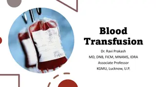
Shock and blood transfusion BY dr.Firas majeed katoof Assitant professor Consultant surgeon
Shock is a systemic state of low tissue perfusion that is inadequate for normal cellular respiration. With insuffcient delivery of oxygen and glucose, cells switch from aerobic to anaerobic metabolism. Pathophysiology Cellular As perfusion to the tissues is reduced, cells are deprived of oxygen and must switch from aerobic to anaerobic metabolism. The product of anaerobic respiration is not carbon dioxide but lactic acid, the accumulation of lactic acid in the blood produces a systemic metabolic acidosis. As glucose within cells is exhausted, anaerobic respiration ceases and there is failure of sodium/potassium pumps in the cell membrane . Intracellular lysosomes release autodigestive enzymes and cell lysis ensues. Intracellular contents, including potassium, are released into the blood stream
Microvascular As tissue ischaemia progresses, result in activation of the immune and coagulation systems. Hypoxia and acidosis activate complement and prime neutrophils, resulting in the generation of oxygen free radicals and cytokine release. These mechanisms lead to injury of the capillary endothelial cells.. Damaged endothelium loses its integrity and becomes leaky . allow fluid to leak out and tissue oedema ensues, exacerbating cellular hypoxia.
Systemic CARDIOVASCULAR As preload and afterload decrease, there is a compensatory increased sympathetic activity and release of catecholamines into the circulation. This results in tachycardia and systemic vasoconstriction (except in sepsis ). RESPIRATORY The metabolic acidosis and increased sympathetic response result in an increased respiratory rate and minute ventilation to increase the excretion of carbon dioxide (and so produce a compensatory respiratory alkalosis). RENAL Decreased perfusion pressure in the kidney leads to Reduced fltration at the glomerulus and a decreased urine output. The renin angiotensin aldosterone axis is stimulated, resulting in further vasoconstriction and increased sodium and water reabsorption by the kidney
ENDOCRINE activation of the adrenal and renin angiotensin systems, vasopressin (antidiuretic hormone) is released from the hypothalamus results in vasoconstriction and resorption of water in the renal collecting system. Cortisol is also released from the adrenal cortex, contributing to the sodium and water resorption and sensitising cells to catecholamines.
Classification of shock Hypovolaemic shock Hypovolemic shock is due to a reduced circulating volume. Hypovolemic may be due to haemorrhagic causes. Nonhaemorrhagic causes include poor fluid intake (dehydration), excessive fluid loss due to vomiting, diarrhoea, urinary loss (e.g. diabetes), evaporation, or thirdspacing where fluid is lost into the gastrointestinal tract and interstitial spaces, as for example in bowel obstruction or pancreatitis. Hypovolaemia is probably the most common form of shock, and to some degree is a component of all other forms of shock. Absolute or relative hypovolaemia must be excluded or treated in the management of the shocked state, regardless of cause.
Cardiogenic shock is due to primary failure of the heart to pump blood to the tissues. Causes of cardiogenic shock include myocardial infarction, cardiac dysrhythmias, valvular heart disease, blunt myocardial injury and cardiomyopathy myocardial depression caused by endogenous factors (e.g.bacterial and humoral agents released in sepsis) , exogenous factors, such as pharmaceutical agents or drug abuse. Evidence of venous hypertension with pulmonary or systemic edema may coexist with the classical signs of shock.
Obstructive shock there is a reduction in preload due to mechanical obstruction of cardiac flling. Common causes of obstructive shock include cardiac tamponade, Tension pneumothorax, massive pulmonary embolus or air embolus. there is reduced filling of the left and/or right sides of the heart leading to reduced preload and a fall in cardiac output.
Distributive shock Distributive shock describes the pattern of cardiovascular responses characterizing a variety of conditions, including septic shock, anaphylaxis spinal cord injury. Inadequate organ perfusion is accompanied by vascular dilatation with hypotension, low systemic vascular resistance, inadequate afterload and a resulting abnormally high cardiac output. In anaphylaxis, vasodilatation is due to histamine release, while in high spinal cord injury there is failure of sympathetic outflow and adequate vascular tone (neurogenic shock). The cause in sepsis is less clear but is related to the release of bacterial products (endotoxin) and the activation of cellular and humoral components of the immune system. There is maldistribution of blood flow at microvascular level with arteriovenous shunting and dysfunction of cellular utilization of oxygen. In the later phases of septic shock there is hypovolaemia from fluid loss into interstitial spaces and there may be concomitant myocardial depression, complicating the clinical picture
Endocrine shock Endocrine shock may present as a combination of hypovolaemic, cardiogenic or distributive shock. Causes include hypo and hyperthyroidism and adrenal insuffciency. Hypothyroidism causes a shock state similar to that of neurogenic shock due to disordered vascular and cardiac responsiveness to circulating catecholamines. Cardiac output falls due to low inotropy and bradycardia. There may also be an associated cardiomyopathy. Thyrotoxicosis may cause a highoutput cardiac failure. Adrenal insuffciency leads to shock due to hypovolaemia and a poor response to circulating and exogenous catecholamines. due to preexisting Addison s disease or be a relative insuffciency due to a pathological disease state, such as systemic sepsis.
Consequences Unresuscitatable shock Multiple organ failure is defned as two or more failed organ systems. There is no specifc treatment for multiple organ failure. Management is supporting of organ systems, with ventilation, cardiovascular support and haemofltration/dialysis until there is recovery of organ function. Multiple organ failure currently carries a mortality of 60%; thus, prevention is vital by early aggressive identifcation and reversal of shock.
TACHYCARDIA Tachycardia may not always accompany shock. Patients who are on beta- blockers or who have implanted pacemakers are unable to mount a tachycardia. A pulse rate of 80 in a fit young adult who normally has a pulse rate of 50 is very abnormal. Furthermore, in some young patients with penetrating trauma, where there is haemorrhage but little tissue damage,there may be a paradoxical bradycardia rather than tachycardia , BLOOD PRESSURE It is important to recognize that hypotension is one of the last signs of shock. Children and fit young adults are able to maintain blood pressure until the final stages of shock by dramatic increases in stroke volume and peripheral vasoconstriction. These patients can be in profound shock with a normal blood pressure. Elderly patients who are normally hypertensive may present with a normal blood pressure for the general population but be hypovolaemic and hypotensive relative to their usual blood pressure. Betablockers or other medications may prevent a tachycardic response.
RESUSCITATION ensure a patent airway and adequate oxygenation and ventilation. Once airway and breathing are assessed and controlled, attention is directed to cardiovascular resuscitation. If there is initial doubt about the cause of shock, it is safer to assume the cause is hypovolaemia and begin with fluid resuscitation, and then assess the response. In patients who are actively bleeding (major trauma, aortic aneurysm rupture, gastrointestinal haemorrhage), operative haemorrhage control should not be delayed and resuscitation should proceed in parallel with surgery Conversely, a patient with bowel obstruction and hypovolaemic shock must be adequately resuscitated before undergoing surgery otherwise the additional surgical injury and hypovolaemia induced
Fluid therapy In all cases of shock, regardless of classifcation, hypovolaemia and inadequate preload must be addressed before other therapy is instituted First line therapy, therefore, is intravenous access and administration of intravenous fluids. Access should be through short, wide bore catheters that allow rapid infusion of fluids as necessary. Long, narrow lines, such as central venous catheters, have too high a resistance to allow rapid infusion and are more appropriate for monitoring than fluid replacement therapy.
Type of fluids there is no overt difference in response or outcome between crystalloid solutions (normal saline, Hartmann s solution, Ringer s lactate) or colloids (albumin or commercially available products). Furthermore, administration of colloids had less volume benefit more expensive worse side effect profiles. Most importantly, the oxygen carrying capacity of crystalloids and colloids is zero. If blood is being lost, the ideal replacement fluid is blood, although crystalloid therapy may be required while awaiting blood products. Hypotonic solutions (dextrose etc.) are poor volume expanders and should not be used in the treatment of shock unless the defcit is free water loss (e.g. diabetes insipidus) or patients are sodium overloaded (e.g. cirrhosis)
Dynamic fluid response The shock status can be determined dynamically by the cardiovascular response to the rapid administration of a fluid bolus. In total, 250 500 mL of fluid is rapidly given (over 5 10minutes) and the cardiovascular responses in terms of heart rate, blood pressure and central venous pressure are observed. Patients can be divided into responders , transient responders and non- responders . Responders have an improvement in their cardiovascularstatus that is sustained. These patients are not actively losing fluid but require flling to a normal volume status. Transient responders have an improvement, but this then reverts to the previous state over the next 10 20 minutes. These patients have moderate ongoing fluid losses (either overt haemorrhage or further fluid shifts reducing intravascular volume). Non-responders are severely volume depleted and are likely to have major ongoing loss of intravascular volume, usually through persistent uncontrolled haemorrhage.
Vasopressor and inotropic support Vasopressor or inotropic therapy is not indicated as first line therapy in hypovolaemia. As ,administration of these agents in the absence of adequate preload rapidly leads to decreased coronary perfusion and depletion of myocardial oxygen reserves. Vasopressor agents (phenylephrine, noradrenaline) Where the vasodilatation is resistant to catecholamines (e.g. absolute or relative steroid deficiency) vasopressin may be used as an alternative vasopressor. And these are indicated in distributive shock states (sepsis, neurogenic shock) where there is peripheral vasodilatation, and a low systemic vascular resistance, leading to hypotension despite a high cardiac output. cardiogenic shock, or where myocardial depression has complicated a shock state (e.g. severe septic shock with low cardiac output), inotropic therapy may be required to increase cardiac output and therefore oxygen delivery. The inodilator dobutamine is the agent of choice.
CENTRAL VENOUS PRESSURE There is no normal central venous pressure (CVP) for a shocked patient, and reliance cannot be placed on an individual pressure measurement to assess volume status. Some patients may require a CVP of 5 cmH2O, whereas some may require a CVP of 15 cmH2O or higher. Further, ventricular compliance can change from minute to minute in the shocked state, and CVP is a poor reflection of end diastolic volume (preload). CVP measurements should be assessed dynamically as response to a fluid challenge. A fluid bolus (250 500 mL) is infused rapidly over 5 10 minutes. The normal CVP response is a rise of 2 5 cmH2O which gradually drifts back to the original level over 10 20 minutes. Patients with no change in their CVP are empty and require further fluid resuscitation. Patients with a large, sustained rise in CVP have high preload and an element of cardiac insuffciency or volume overload.
HAEMORRHAGE Haemorrhage must be recognised and managed aggressively to reduce the severity and duration of shock and avoid death and/ or multiple organ failure. Haemorrhage is treated by arresting the bleeding not by fluid resuscitation or blood transfusion. Although necessary as supportive measures to maintain organ perfusion, attempting to resuscitate patients who have ongoing haemorrhage will lead to physiological exhaustion (coagulopathy, acidosis and hypothermia) and subsequently death.
Defnitions Revealed and concealed haemorrhage Haemorrhage may be revealed or concealed. Revealed haemorrhage is obvious external haemorrhage, such as exsanguination from an open arterial wound or from massive haematemesis from a duodenal ulcer. Concealed haemorrhage is contained within the body cavity and must be suspected, actively investigated and controlled. trauma, haemorrhage may be concealed within the chest, abdomen, pelvis, retroperitoneum or in the limbs with contained vascular injury or associated with longbone fractures. Examples of non- traumatic concealed haemorrhage include occult gastrointestinal bleeding or ruptured aortic aneurysm.
Primary, reactionary and secondary haemorrhage Primary haemorrhage is haemorrhage occurring immediately due to an injury (or surgery). Reactionary haemorrhage is delayed haemorrhage (within 24 hours) and is usually due to dislodgement of a clot by resuscitation, normalisation of blood pressure and vasodilatation. Reactionary haemorrhage may also be due to technical failure, such as slippage of a ligature. Secondary haemorrhage is due to sloughing of the wall of a vessel. It usually occurs 7 14 days after injury and is precipitated by factors such as infection, pressure necrosis (such as from a drain) or malignancy
Surgical and non-surgical haemorrhage Surgical hemorrhage is due to a direct injury and is amenable to surgical control (or other techniques such as angioembolisation). Nonsurgical hemorrhage is the general ooze from all raw surfaces due to coagulopathy and cannot be stopped by surgical means (except packing). Treatment requires correction of the coagulation abnormalities.
Degree and classifcation The adult human has approximately 5 litres of blood (70 mL/ kg children and adults, 80 mL/kg neonates). Estimation of the amount of blood that has been lost is difficult, inaccurate and usually underestimates the actual value. External hemorrhage is obvious, but it may be difficult to estimate the actual volume lost. In the operating room, blood collected in suction apparatus can be measured and swabs soaked in blood weighed. The hemoglobin level is a poor indicator of the degree of hemorrhage because it represents a concentration and not an absolute amount. In the early stages of rapid hemorrhage, the hemoglobin concentration is unchanged (as whole blood is lost). Later, as fluid shifts from the intracellular and interstitial spaces into the vascular compartment, the hemoglobin and hematocrit levels will fall
Management Identify haemorrhage External haemorrhage may be obvious, but the diagnosis of concealed haemorrhage may be more diffcult. Any shock should be assumed to be hypovolaemic until proven otherwise and, similarly, hypovolaemia should be assumed to be due to haemorrhage until this has been excluded. Immediate resuscitative manoeuvres Direct pressure should be placed over the site of external haemorrhage. Airway and breathing should be assessed and controlled as necessary. Largebore intravenous access should be instituted and blood drawn for crossmatching (see Cross-matching below). Emergency blood should be requested if the degree of shock and ongoing haemorrhage warrants this.
Identify the site of haemorrhage Once haemorrhage has been considered, the site of haemorrhage must be rapidly identifed. to defne the next step in haemorrhage control (operation, angioembolisation, endoscopic control). history (previous episodes, known aneurysm, nonsteroidal therapy for gastrointestinal [GI] bleeding) examination (nature of blood fresh, melaena; abdominal tenderness, etc.). For shocked trauma patients, the external signs of injury may suggest internal haemorrhage, Investigation haemorrhage into a body cavity (thorax, abdomen) must be excluded with rapid s (chest and pelvis xray, abdominal ultrasound or diagnostic peritoneal aspiration). Rapid bedside tests are more appropriate for profound shock and exsanguinating haemorrhage than investigations such as computed tomography (CT) which take time..
Hemorrhage control The bleeding, shocked patient must be moved rapidly to a place of haemorrhage control. This will usually be in the operating room but may be the angiography or endoscopy suite. These patients require surgical and anaesthetic support and full monitoring and equipment must be available.
Blood and blood products whole blood transfusion has significant advantages over packed cells as it is coagulation factor rich and, if fresh, more metabolically active than stored blood. Packed red blood cells are spun down and concentrated packs of red blood cells. Each unit is approximately 330 mL and has a haematocrit of 50 70%. Packed cells are stored in a SAGM solution (saline adenine glucose mannitol) to increase shelf life to 5 weeks at 2 6 C. (Older storage regimes included storage in CPD: citrate phosphate dextrose solutions, which have a shelf life of 2 3 weeks.) Fresh frozen plasma (FFP) is rich in coagulation factors and is removed from fresh blood and stored at 40 to 50 C with a 2year shelf life. It is the first line therapy in the treatment of coagulopathic haemorrhage .Rhesus D- positive FFP may be given to a rhesus D negative woman although it is possible for seroconversion to occur with large volumes owing to the presence of red cell fragments, and RhD immunisation should be considered.
Cryoprecipitate is a supernatant precipitate of FFP and is rich in factor VIII and fbrinogen. It is stored at 30 C with a 2year shelf life. It is given in low fbrinogen states or factor VIII defciency Platelets Platelets are supplied as a pooled platelet concentrate and contain about 250 109/L. Platelets are stored on a special agitator at 20 24 C and have a shelf life of only 5 days. Platelet transfusions are given to patients with thrombocytopenia or with platelet dysfunction who are bleeding or undergoing surgery
Indications for blood transfusion Acute blood loss, to replace circulating volume and maintain oxygen delivery; Perioperative anaemia, to ensure adequate oxygen delivery during the perioperative phase; Symptomatic chronic anaemia, without haemorrhage or impending surgery. Blood groups and cross-matching Human red cells have on their cell surface many different antigens. Two groups of antigens are of major importance in surgical practice the ABO and rhesus systems. Blood group O is the universal donor type as it contains no antigens to provoke a reaction. Conversely, group AB individuals are universal recipients and can receive any ABO blood type because they have no circulating antibodies.
Rhesus system The rhesus D (Rh(D)) antigen is strongly antigenic and is present in approximately 85% of the population in the UK. Antibodies to the D antigen are not naturally present in the serum of the remaining 15% of individuals, but their formation may be stimulated by the transfusion of Rh positive red cells, or acquired during delivery of a Rh(D)positive baby. Acquired antibodies are capable, during pregnancy, of crossing the placenta and, if present in a Rh(D)negative mother, may cause severe haemolytic anaemia and even death (hydrops fetalis) in a Rh(D)positive fetus in utero.
Transfusion reactions If antibodies present in the recipient s serum are incompatible with the donor s cells, a transfusion reaction will result. This usually takes the form of an acute haemolytic reaction. Severe immunerelated transfusion reactions due to ABO incompatibility result in potentially fatal complement mediated intravascular haemolysis and multiple organ failure. Transfusion reactions from other antigen systems are usually milder and selflimiting. Febrile transfusion reactions are nonhaemolytic and are usually caused by a graft versus host response from leukocytes in transfused components. Such reactions are associated with fever, chills or rigors. The blood transfusion should be stopped immediately. This form of transfusion reaction is rare with leukodepleted blood
Cross-matching To prevent transfusion reactions, all transfusions are preceded by ABO and rhesus typing of both donor and recipient blood to ensure compatibility. The recipient s serum is then mixed with the donor s cells to confrmABO compatibility and to test for rhesus and any other blood group antigen antibody reaction. Full cross matching of blood may take up to 45 minutes in most laboratories. In more urgent situations, type specifc blood is provided which is only ABO/rhesus matched and can be issued within 10 15 minutes. Where blood must be given emergently, group O (universal donor) blood is given (O to females, O+ to males)
Complications from a single transfusion Complications from a single transfusion include: incompatibility haemolytic transfusion reaction; febrile transfusion reaction; allergic reaction; infection: bacterial infection (usually due to faulty storage); hepatitis; HIV; malaria; air embolism; thrombophlebitis; transfusion related acute lung injury (usually from FFP).
Complications from massive transfusion include: coagulopathy; hypocalcaemia; hyperkalaemia; hypokalaemia; hypothermia. In addition, patients who receive repeated transfusions over long periods of time (e.g. patients with thalassaemia) may develop iron overload. (Each transfused unit of red blood cells contains approximately 250 mg of elemental iron.)
Management of coagulopathy Correction of coagulopathy is not necessary if there is no active bleeding and haemorrhage is not anticipated (not due for surgery). However, coagulopathy following or during massive transfusion should be anticipated and managed aggressively. Prevention of dilutional coagulopathy is central to the damage control resuscitation of patients who are actively bleeding. This is the prime reason for delivering balanced transfusion regimes matching red blood cell packs with plasma and platelets. Based on moderate evidence, when red cells are transfused for active haemorrhage, it is best to match each red cell unit with one unit of FFP and one of platelets (1:1:1). This will reduce the incidence and severity of subsequent dilutional coagulopathy. Crystalloids and colloids should be avoided for the same reason. The balanced transfusion approach cannot, however, correct coagulopathy. Therefore, coagulation should be monitored routinely, either with point o f care testing (thromboelastometry) or with laboratory tests (fbrinogen, clotting times). Underlying coagulopathies should be treated in addition to the administration of 1:1:1 balanced transfusions. There are pharmacological adjuncts to blood component therapy. The antifbrinolytic tranexamic acid is the most commonly administered. It is usually administered empirically to bleeding patients because effective point of care tests of fbrinolysis are not yet routinely available. There is little evidence to support the use of other coagulation factor concentrates at this time


