Understanding Blood Transfusion: Components, Preparation, and Safety Measures
Blood transfusion is a crucial medical procedure involving the transfusion of different blood components to patients in need. This practice has evolved to include various components such as packed red blood cells, plasma, platelets, and more. The preparation of these components involves specific techniques like differential centrifugation. Safety measures, including reducing leukocytes and irradiating PRBCs, are taken to prevent adverse reactions. Blood products are transfused using intravenous tubing with filters to ensure patient safety. Proper equipment usage and monitoring are essential for a successful transfusion process.
Download Presentation

Please find below an Image/Link to download the presentation.
The content on the website is provided AS IS for your information and personal use only. It may not be sold, licensed, or shared on other websites without obtaining consent from the author. Download presentation by click this link. If you encounter any issues during the download, it is possible that the publisher has removed the file from their server.
E N D
Presentation Transcript
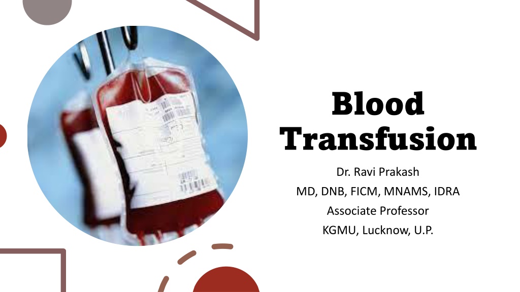
Blood Transfusion Dr. Ravi Prakash MD, DNB, FICM, MNAMS, IDRA Associate Professor KGMU, Lucknow, U.P.
Introduction About 85 million units are transfused worldwide annually. Blood is typically stored in components. Earlier Fresh whole blood has been thought was standard for transfusion. However, medical advancement has allowed the efficient use of the different components, such as packed red blood cells (PRBCs), individual factor concentrates, fresh frozen plasma (FFP), platelet concentrates, and cryoprecipitate.
Blood components Cryoprecipitate FFP Cryo deficient plasma PRBC Fibrinogen concentrate Whole blood Platelets
Component preparation Principle - Differential centrifugation Red cells Packed cells Red cells + additive Plasma Bank plasma Fresh frozen Cryo supernate Platelets Platelet rich concentrate Platelet rich plasma Cryoprecipitate Plasma + Platelets Whole blood Buffy RBC
Blood components 1. Single donor platelet 2. Single donor plasma 3. Therapeutic plasmapheresis Apheresis:
Infections: HIV, HBV, HCV, Syphilis, Malaria Blood grouping Antibody screening Pre- transfusion testing Cross matching Leukoreduction Irradiation Washed RBC NAT testing
Leucocyte depleted red blood cells concentrate should be prepared by a method known to reduce leucocytes in the final component to less than 5x10⁸ when intended to prevent febrile reactions and to less than 5x10⁶ when it is required to prevent alloimmunization or CMV infection. Pre- transfusion preparation Irradiated PRBC is 100% leucoreduced and used to prevent Graft Vs Host Disease (GVHD)
Blood products are transfused through intravenous tubing with filters. The filters, which typically have pore diameters of 170 to 260 microns, are also used to prevent particulate debris from being administered. Equipment However, the trapped particulate leads to bacterial growth, and the American Association of Blood Banks (AABB) advises against using a filter for more than four hours. Ideally, each unit should be transfused with separate blood transfusion set.
There is no evidence that warming blood is beneficial to the patient when infusion is slow. At infusion rates greater than 100 ml/minute, cold blood may be a contributing factor in cardiac arrest. Keeping the patient warm is probably more important than warming the infused blood. Warmed blood is most commonly required in: Warming of blood 1. large volume rapid transfusions (In adults: infusion greater than 50 ml/kg/hour, In children: infusion greater than 15 ml/kg/hour) 2. Exchange transfusion in infants 3. Patients with clinically significant cold agglutinins. It is important to note that Blood should only be warmed in a blood warmer. Blood should never be warmed in a bowl of hot water as this could lead to haemolysis of the red cells which could be life-threatening.
Time Limits for Infusion Blood/ blood product Start infusion Complete infusion Whole blood/ within 30 min. of within 4 hour red cells removing pack (less in high from ambient temperature) refrigerator Platelet concentrates immediately within 20 min FFP within 30 min within 20 min 10
Whole blood and PRBC: ABO and Rh compatible, cross matched Compatibility testing In the absence of ABO type specific blood, group O packed red cells should be transfused. Rh(D) negative recipient should receive Rh(D)negative whole blood or red blood cell components Rh(D) positive recipient can receive either Rh(D) positive or negative whole blood or red blood cell components.
Single donor plasma or fresh frozen plasma should be ABO compatible . Rh compatibility is not compulsory. Compatibility testing Cryoprecipitate should not require ABO/ Rh grouping. Platelet concentrate should be ABO and Rh(D) type specific with the recipient blood as far as possible. In case of shortage random donor platelets of any ABO/Rh group should be used provided there is no visual red cell contamination of the platelet concentrate. In case of single donor platelets prepared by apheresis, plasma should be reduced when plasma incompatible concentrate is in use (e.g. use of 'O' group SDP to B patient).
Whole Blood should be stored at 40 C 20 C in plastic blood bags. Expiry of Whole blood/PRBC collected in: A. Citrate-phosphate-dextrose solution (CPD) : 21 days Whole Blood B. Citrate-phosphatedextrose with adenine (CPDA- 1) :35 days C. Heparin solution: 24 hours D. Red cells containing additive solutions (SAGM, ADSOL, NUTRICEL): 42 days
Whole blood Indications: Massive hemorrhage Neonatal exchange transfusion Blood preferably within 72 hours of collection, but not exceeding 5 days should be used for exchange transfusion. Whole blood does not contain any viable platelet or labile coagulation factors.
The guidelines and clinical trials on transfusion requirements in critical care (TRICC) also recommend a value of 7 g/dl as the threshold for critically ill patients. The guidelines recommend a value of 8 g/dL as the threshold in patients with coronary artery disease Packed RBC Transfusion may also be indicated in patients with active or acute bleeding and patients with symptoms related to anemia (for example, tachycardia, weakness, dyspnea on exertion) and hemoglobin less than 8 g/dL. The ASA Task Force recommends a perioperative transfusion trigger of 6.0 10.0 g/dL, based on potential or actual ongoing bleeding, intravascular volume status, signs of organ ischemia and adequacy of cardiopulmonary reserve
One unit PRBC increases haematocrit by 3% and Hb by 1 g/dL. Unless the patient is actively bleeding, it is recommended to transfuse 1 unit of packed red cells at a time, follow up by checking post-transfusion hemoglobin. Storage changes like 2,3 diphosphoglycerate (DPG) depletion, pro-inflammation, poor deformability and change in nitric oxide physiology may alter effectiveness of transfusion. PRBC
Platelet concentrate 1. Random Donor Platelets (RDP) 2. Single Donor Platelet (SDP) RDP is prepared by centrifugation of a single unit of whole blood collected while SDP is collected by apheresis of donor blood. Platelet concentrate should be separated from whole blood within 8 hours of collection by centrifugation at 220 C using either platelet rich plasma (PRP) or buffy coat (BC) method Platelets is suspended in approximately 50 ml of plasma and stored at 220 C 20 C .
Platelet concentrate To prevent spontaneous bleeding - transfuse at less than 10,000 cells/microliter; some recommend less than 5,000 cells/microliter It is generally accepted that a count of 50,000/uL is sufficient for most invasive procedures including most surgeries. Platelet counts of >100,000/uL are recommended for ophthalmic and neurosurgery. Platelet transfusion Threshold in Bleeding Patients <50,000 cells/microliter in severe bleeding, including disseminated intravascular coagulation (DIC) <30,000 cells/microliter when bleeding, not life-threatening, or considered not severe <100,000 cells/microliter for bleeding in multiple trauma patients or patients with intracranial bleeding
Platelet concentrate Group specific transfusion is recommended Indication: 1. Thrombocytopenia 2. Massive transfusion protocol TTP and HIT are considered relative contraindication to platelet transfusion. 10 ml/kg of RDP increases platelet count by approx. 5000/ L 10 ml/kg of SDP increases platelet count by approx. 25000-30000/ L Dose is 10-15 ml/kg
Platelet concentrate Shelf life is 5 days In-line filter of 170-260 m is used for transfusion. It should be stored at room temperature Chance of bacterial contamination is high due to room temperature storage and they are WBC depleted.
Fresh Frozen Plasma Fresh Frozen Plasma (FFP) One unit=250 mL One apheresis unit=400-600 mL Usual PRBCs/FFP transfusion ratio is 3:1 to 1:1. One 250 mL unit raises the fibrinogen level by 10-15 mg/dL. Contains fibrinogen 400-900 mg/unit, albumin, protein C, protein S, anti-thrombin and tissue factor pathway inhibitor. Factor VII has shortest half life
Fresh Frozen Plasma FFP should be separated from the whole blood not later than 6 - 8 hours of collection and frozen to -300 C as early as possible. Prior to infusion the frozen plasma should be thawed rapidly at 30-370 C in a water bath with shaker. Once thawed it should be used within 6 hours. It should be stored at -300 C and once thawed should be used within 6 hours. Cryo poor plasma or Factor VIII Deficient Plasma: This is plasma from which cryoprecipitate has been removed. FFP can be kept up to 1 year at -300 C or below and up to 5 years at -600 C or below However, after 1 year its labelled as unused plasma.
Fresh Frozen Plasma Indications: 1. Coagulopathy: Inherited or acquired 2. Plasmapheresis 3. Antithrombin III deficiency 4. Pseudocholinesterase deficiency 5. Urgent reversal of Warfarin FFP should not be used as plasma expander or protein substitute or wound healing
Cryoprecipitate Prepared from the cryoglobulin fraction obtained by thawing FFP. After removal of the supernatant, the precipitate is refrozen in approximately 30 mL of plasma, which can be stored at 25 C or below for up to 36 months. Contains factor VIIIC, von Willi brand factor (vWF) s, fibrinogen, fibronectin and factor XIII. This is usually packed in 15 mL of plasma Each unit of cryoprecipitate contains 150-250 mg of fibrinogen (one unit of FFP contains 2-4 mg/mL of fibrinogen, which is equivalent to fibrinogen content of 2 units of cryoprecipitate). 10 units of cryoprecipitate should restore the fibrinogen level to 200 mg/dL. Cryoprecipitate administration is being replaced by the use of lyophilized human fibrinogen concentrates.
Fibrinogen concentrate Normal plasma concentration of fibrinogen is 200-400 mg/dL (maximum level among all coagulation factors). Fibrinogen is the major structural component of the clot. Minimum level required to maintain haemostasis is 100 mg/dL. Fibrinogen concentrates are superior to FFP or cryoprecipitate in management of fibrinogen deficiency. It is expensive.
Definition?? Massive transfusion is defined as replacement of >1 blood volume in 24 hours, or >50% of blood volume in 4 hours (adult blood volume is approximately 70 mL/kg), or > 10 units PRBC in 24 hours or > 150 ml/min in children: transfusion of >40 mL/kg (blood volume in children over 1 month old is approximately 80 mL/kg)
Massive blood transfusion When an amount of blood approaching or exceeding recipient's total blood volume is transfused within 24 hours, a fresh blood sample should be used for cross match at the time of subsequent transfusion of blood. Component therapy should be actively considered in these cases In case of a need for transfusion after 48 hours of earlier transfusion, a fresh sample should be asked for to perform a cross match.
Parameters Values to aim for Temperature >35 C pH >7.2, base excess < 6, lactate <4 mmol/L Acid-base status Ionised calcium (Ca) >1.1 mmol/L This should not be used alone as transfusion trigger; and, should be interpreted in context with haemodynamic status, organ & tissue perfusion. Haemoglobin (Hb) 50 x 10^9 /L (>100 x 10^9 if head injury/ intracranial haemorrhage) Platelet (Plt) PT/APTT 1.5x of normal Fibrinogen 1.0 g/L
Complications of blood transfusion
Adverse Event and Approximate Risk Per Unit Transfusion of RBC Febrile reaction: 1:60 Transfusion-associated circulatory overload: 1:100 Allergic reaction: 1:250 TRALI: 1:12,000 Hepatitis C infection: 1:1,149,000 Human immunodeficiency virus infection: 1:1,467,000 Fatal hemolysis: 1:1,972,000 Adverse effects
Adverse transfusion reactions Acute immune-mediated (<24 h after transfusion) Delayed immune-mediated (>24 h after transfusion) Delayed non- immune- mediated Acute non- immune-mediated Acute haemolytic transfusion reaction Delayed haemolytic transfusion reaction Iron overload Transfusion related sepsis Alloimmunisation to red cell antigens, platelets and leukocytes (Human Leukocyte Antigens) Febrile non-haemolytic transfusion reaction Non-immune haemolysis Transfusion related sepsis Transfusion related circulatory overload Transfusion-associated immunomodulation Urticarial Transfusion-associated graft versus host disease Anaphylactic Air embolism Transfusion-related acute lung injury Post-transfusion purpura
Febrile Non-Hemolytic Transfusion Reaction (FNHTR) Relatively common complication with a frequency of 4% to 30%. It is characterized by a temperature rise of 1 C or more within the first 4 hours of transfusion, which resolves within 48 hours, there can be associated with nausea, vomiting, dyspnoea, and hypotension. Antibodies against HLA or leukocyte antigen in donor plasma are most commonly implicated. Antigen present on donor white cells binds to the antibody in the recipient, which leads to pyrogen and cytokine release, such as TNF-alpha, IL1, and IL6. FNHTR can also occur due to biologic response modifiers (BRM) released during platelet storage, which is increased with storage.
Febrile Non-Hemolytic Transfusion Reaction (FNHTR) Platelet has maximum storage allowed for five days, during which the release of substances such as CD40L, IL6, and IL8 can occur. FNHTR is diagnosed with the above- mentioned features, and the possibility of chills and hemolytic causes are ruled out by further immunohematology workup and lab findings. FNHTR can be minimized by leukoreduction and by platelet additive solution (PAS), which replaces maximum plasma in stored platelets
Transfusion-associated Circulatory Overload It is characterized by respiratory distress secondary to cardiogenic pulmonary edema. This reaction is most common in patients already in a fluid-overloaded state, such as congestive heart failure or acute renal failure. Diagnosis is based on symptom onset within 6 to 12 hours of receiving a transfusion, clinical evidence of fluid overload, pulmonary edema, elevated brain natriuretic peptide, and response to diuretics. Preventive efforts and treatment include limiting the number of transfusions to the lowest amount necessary, transfusing over the slowest possible time, and administering diuretics before or between transfusions.
Allergic and Anaphylaxis An allergic reaction is similar to FNHTR, and its frequency is between 0.09% and 21%. It manifests as pruritus, urticaria, or systematically as bronchoconstriction and is seldom associated with fever. Pathogenesis is heterogeneous in origin ranging from proteins, antibodies and cytokines released during storage. Anaphylaxis can occur when the patient is IgA deficient and has antibodies against it, and receives IgA-containing platelets, and this occurs 1 in 50,000 transfusions. Diagnosis is made clinically, and it is an emergency. It can be prevented by giving washed platelets or platelets obtained from IgA-deficient donors. Allergic reactions can be minimized by depleting plasma in platelets
Transfusion-associated Graft vs. Host Disease This a rare but fatal complication characterized by fever and multiple systemic manifestations such as skin, gastrointestinal tract, liver, and others. It is caused by the transfusion of viable lymphocytes that get a favorable environment in the recipient, where they engraft and proliferate, subsequently attacking the host tissues. It can occur between 1 to 6 weeks from transfusion and occurs in an immunocompromised host, those with congenital T-cell defects, a population with low genetic diversity (homozygous HLA), or first-degree patient relatives. Symptoms involve multiple organs such as skin, the intestine, liver, and suppression of bone marrow. Diagnosis is that of exclusion and requires molecular methods of testing apart from microscopic features seen. This can be prevented by the irradiation of platelets.
This is uncommon, occurring in about 1:12,000 transfusions. Mainly caused by the transfusion of antibodies in the plasma from the donor against HLA or human neutrophil antigens. Transfusion- related Lung Patients will develop symptoms within 2 to 4 hours after receiving a transfusion. Injury (TRALI) Patients will develop acute hypoxemic respiratory distress, similar to acute respiratory distress syndrome (ARDS). Patients will have pulmonary edema, normal CVP, without evidence of left heart failure CVP. Diagnosis is made based on a history of recent transfusion, chest x-ray with diffuse patchy infiltrates, and the exclusion of other etiologies. While there is a 10% mortality, the remaining 90% will resolve within 96 hours with supportive care only.
Platelet concentrates are stored for up to 5 days at temperatures 22 to 24 degrees C. Sepsis and Bacterial Infection This temperature is favorable for the growth of bacteria like Staphylococcus aureus and gram-negative bacteria. Possible sources of infections can be improper cleansing of the donor phlebotomy site or if a donor has asymptomatic bacteremia. The risk of infection to the patient ranges from 0.14% to 1.41% with platelet transfusion. Pathogen reduction technology has been used to minimize infections in plasma and platelets.
Fatal Hemolysis This is extremely rare, occurring only in 1 out of nearly 2 million transfusions. It results from ABO incompatibility, and the recipient s antibodies recognize and induce hemolysis in the donor s transfused cells. Patients will develop an acute onset of fevers and chills, low back pain, flushing, dyspnea as well as becoming tachycardic and going into shock. Treatment is to stop the transfusion, leave the IV in place, intravenous fluids with normal saline, and keep urine output greater than 100 mL/hour, diuretics may also be needed. Cardiorespiratory support may be provided as appropriate.
Fatal Hemolysis A hemolytic workup should also be performed, including sending the donor blood and tubing and post-transfusion labs (see below for list) from the recipient to the blood bank. Retype and crossmatch Direct and indirect Coombs tests Complete blood count (CBC), creatinine, PT, and PTT (draw from the other arm) Peripheral smear Haptoglobin, indirect bilirubin, LDH, plasma-free hemoglobin Urinalysis for hemoglobin
Electrolyte Abnormalities Electrolyte Abnormalities They can also occur, although these are rare and more likely associated with large volume transfusion. Hypocalcemia can result as citrate, an anticoagulant in blood products, binds with calcium. Hyperkalemia can occur from the release of potassium from cells during storage. Higher risk in neonates and patients with renal insufficiency. Hypokalemia can result as a result of alkalinization of the blood as citrate is converted to bicarbonate by the liver in patients with normal hepatic function.
Differential diagnosis of cause of blood transfusion reaction
