Understanding Lenses and Vision Defects in Medical Physics
Explore the fascinating world of lenses and vision defects in this detailed lecture from Sabah Ibrahim Abbas at Al Karkh University. Discover the properties of convex and concave lenses, how light refraction forms images, and the workings of the human eye with its cornea, iris, lens, and retina. Gain insights into how images are processed in the brain and why they appear inverted on the retina.
Download Presentation

Please find below an Image/Link to download the presentation.
The content on the website is provided AS IS for your information and personal use only. It may not be sold, licensed, or shared on other websites without obtaining consent from the author. Download presentation by click this link. If you encounter any issues during the download, it is possible that the publisher has removed the file from their server.
E N D
Presentation Transcript
LECTURE TEN LENSES AND DEFECTS OF VISION Sabah Ibrahim Abbas Al Karkh university of sciences College of Sciences Medical physics department
LENSES The fact that most lenses have surfaces that is spherical in form. Some surfaces are convex, others are concave. When a light passes through any lens, refraction at each of its surfaces contributes to its image-forming properties. to calculate the image distance used the lens formula 1 = 1 +1 ? ; m=-? ? ? ?
LENSES Convex Lens Concave Lens
LENSES To understand how theses lenses work, in the case of the convex lens when arrange two prisms so that the rectangular surfaces corresponding to each other and the parallel rays incident on them after refracted these rays are intersect at a specific point. When using a number of parallel rectangular prisms between two prisms as shown in figure,
LENSES Concave lens when two prisms are placed in opposite direction and connected on the head with the addition of rectangular prisms at the sides of eachthem.
HUMAN EYE The front part of the eye called cornea, is made of transparent substance and its outer surface is convex in shape. It is through cornea that the light coming from objects enters the eyes. Just behind the cornea is iris which is also called colored diaphragm. A hole in the middle of the iris is called pupil. Then behind it is the eye lens which is a convex lens. It is due to the support of ciliary muscles that the eye lens is held in position. Eye lens is flexible and thus can change its focal length and shape with the help of ciliary muscles. Behind the eye lens is retina on which the image is formed in the eye
HUMAN EYE The light rays coming from the object enter the eyes through pupil and falls on eye lens. The eye lens then converge the light rays and produce an image of the object on retina which is real and inverted. Retina has large number of light sensitive cells which can generate electrical signals. After the image is formed on the retina it sends electrical signals to the brain and we have a sensation of image. Also, even though the image formed on retina is inverted out mind interprets it as erect
HUMAN EYE Function of iris andpupil The function of iris is to adjust the size of the pupil. If the amount of light entering the eye is less then pupil expands so that more light can enter the eye and in case the amount of light entering the eye is large then pupil contracts. The adjustment of the size of pupil takes some time and this is the reason when we go outside in the sunlight from a dark room we feel glare in our eyes or if we enter a dark room after coming from outside we see things clearly after some time
HUMAN EYE The light sensitive cells in the retina of our eye are of two shapes; rod shape and cone shape. The function of rod shaped cells is to respond to the brightness of light. And thefunction of cone shaped cells is to make us see colors and distinguish between them
HUMAN EYE Distant objects: When the rays of light are coming from distant object they are diverging at the beginning but become parallel when they reach our eye. Therefore to see a distant object, we need to have a convex eye-lens of low converging power to focus them to form an image on the retina of the eye. The convex eye-lens of low converging power has large focal length and is quite thin. Power of accommodation of the eye
HUMAN EYE When our eyes see distant objects then the ciliary muscles are relaxes and the focal length is maximum in this position. The eye-lens then converge the parallel rays of light to form an image of the distant object on retina. When the eye see distant object they are said to be accommodated.
HUMAN EYE Nearby objects: When the rays of light are coming from nearby object they diverge when they reach our eyes. Therefore, to see a nearby object we need to have a convex eye-lens of high converging power so as to focus and form an image on the retina. Convex eye-lens with high converging power has short focal length and is thick.
HUMAN EYE And when our eyes see nearby objects then the ciliary muscles get stretched and its focal length decreases. Due to this, the converging power of the eye lens increases and the diverging rays of light coming from object converge to form an image on retina. When the eyes see nearby object they are said to be accommodated
DEFECT OF VISION Myopia: The human eye is capable of about 40 diopters to start with... young people have about 50% added capacity "accomodation" until about the age of 25, which then decreases as we age. By 50, most of us will need glasses- this is called Presbyopia, or "old vision".
DEFECT OF VISION Myopia The defect of an eye in which it cannot see the distant objects clearly is called myopia. A person with myopia can see nearby objects clearly. Myopia is caused due to: High converging power of lens Eye-ball being too long Due to high converging of the eye-lens the image is formed in front of the retina and a person cannot see clearly the distant objects. In another case, if the eye- ball is too long than the retina is at larger distance from the eye-lens. In this case also the image is formed in front of the retina even though the eye-lens has correct convergingpower.
DEFECT OF VISION Hypermetropia or long-sightedness is a defect of an eye where a person cannot see nearby objects clearly. The near-point of hypermetropic eye is more than 25 cm away.This defect of eye is caused dueto: Low converging power of eye-lens Eye-ball being too short In case of hypermetropia the image of an object is formed behind the retina and therefore, a person cannot see clearly nearby objects.
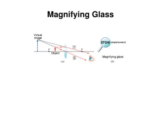
DEFECT OF VISION The near-point of an eye having hypermetropia is more than 25 cm. The condition of hypermetropia can be corrected by putting a convex lens in front of the eye. This is because when a convex lens of suitable power is placed in front of the hypermetropic eyes, then the convex lens first converge the diverging rays of light coming from a nearby object at the near point of the eye at which the virtual image of the nearby object is formed. Since the light rays now appear to be coming from the eye s near point, the eye-lens can easily focus and form the image on retina. Convex lens is used for hypermetropia so as to increase the converging power of the eye-lens.
DEFECTS OF VISION the cornea is less round than oval leading to an incomplete image striking the retina as some of the light is notfocused Astigmatism is a common eye focusing problem where light rays entering the eye become focused by the cornea and lens at different distances from the retina. This causes some parts of the image to be more out of focus than others, thereby affecting your visual acuity. Astigmatic errors are due to the shape of the eye being irregular, such that it is shaped more like an oblong football rather than a basketball.
DEFECTS OF VISION Astigmatism can occur due to an irregularity of the shape of the cornea or lens or both. This results in the light rays in the horizontal and vertical planes being focused at different locations. In an eye without astigmatism, the light rays in the horizontal and vertical planes become focused at one single point, giving a crisp and sharp image.
DEFECT OF VISION Spectacle glasses and contact lenses are the mainstays of treatment.
DEFECT OF VISION Your prescription will have 3 parts, and looks like this: +2.50 / -1.50 x 90 - Sphere number ( SPH): This indicates the overall amount of power (in diopters, D) to uniformly sharpen your focus to the most acceptable level. The plus sign indicates hyperopia. A minus sign indicates myopia. In the example above, the sphere has a value of +2.50D. - Cylinder number (CYL): This shows the amount of astigmatism (in diopters, D). The cylinder in the example above is -1.50 D. - Axis: This indicates the angle (in degrees)needed to correct the astigmatic error. The angle of astigmatism is 90 degrees in the exampleabove.
