Understanding Juvenile Hallux Valgus: Diagnosis and Management Insights
Delve into the world of juvenile hallux valgus, a condition affecting the alignment of the big toe in children. Learn about its different forms based on age groups, associated anatomical features, pathoanatomy, soft tissue, and bony deformities. Discover the hereditary and syndromic links, diagnostic considerations, and treatment approaches including prevention strategies and surgical indications.
- Juvenile Hallux Valgus
- Pediatric Orthopaedics
- Childrens Foot Deformity
- Management Strategies
- Anatomical Features

Download Presentation

Please find below an Image/Link to download the presentation.
The content on the website is provided AS IS for your information and personal use only. It may not be sold, licensed, or shared on other websites without obtaining consent from the author. Download presentation by click this link. If you encounter any issues during the download, it is possible that the publisher has removed the file from their server.
E N D
Presentation Transcript
JUVENILE HALLUX VALGUS Prasad Gourineni, MD Head of Pediatric Orthopaedics, Advocate Christ Hospital, Oak Lawn.
DISCLOSURE Own G2 Healthcare that sells calf stretching wedges.
HALLUX VALGUS Valgus of big toe by more than 15 degrees. Pediatric Hallux valgus is often bilateral Infantile Younger than 2. Juvenile 2-10 years. Adolescent More than 10 years
PEDIATRIC ANATOMY Physis at the proximal end of the first metatarsal and phalanges. Growth can worsen deformity.
INFANTILE HALLUX VALGUS Rare. May be congenital, hereditary, syndromic. Usually asymptomatic. Splinting may prevent worsening. Surgery is almost never indicated. High risk of recurrence.
JUVENILE HALLUX VALGUS Hereditary Autosomal dominant. Syndromic Down s, Marfan s. Neurological CP, CMT. Associated with Flat feet, Calf tightness, Metatarsus adductus, Splayfoot, Skewfoot, Generalized laxity. JRA. Progresses to adolescent and adult deformity.
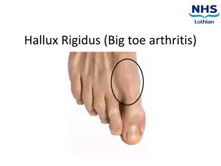
PATHOANATOMY Medial prominence of the first metatarsal head bunion. Redness, shoe fit issues. Flat foot, pronation of first ray, Lateral subluxation of the sesamoids. Plantarly displaced abductor erodes the cartilage.
SOFT TISSUE DEFORMITY Abductor hallucis, FHL, EHL can subluxate and further deform the bunion. Medial laxity, Lateral tightness incongruity. Achilles contracture.
BONY DEFORMITY First TMT deformity & Laxity. Metatarsus primus varus. IMA < 9 degrees. Distal Metatarsal articular angle. < 10 degrees. Hallux valgus interphalangeus. > 10 degrees. Long/short first metatarsal.
CLINICAL EVALUATION Foot, ankle, and leg mechanics Laxity of TMT and MTP Beighton score Shoe wear pattern
DEFORMITY LOCALIZATION Almost all primus varus seems to occur in the medial cuneiform.
NONOP TREATMENT Orthotics to decrease heel valgus and support arch height. Wide, flat shoes. Avoid high heels and tight toe box. Night splints to stretch the bunion. Stretching of Achilles, lateral heel, and big toe deformity.
SURGERY Delay surgery as much as possible. Simple bunionectomy, and medial capsulorraphy cause high recurrence. Correct bony deformity and realign soft tissues.
COMPLICATIONS Growth plate injury from proximal osteotomy. Recurrence & Over Correction / Hallux Varus. Other complications are less likely than in adults.
METATARSUS PRIMUS VARUS Deformity is usually from medial tilt of first TMT joint. Lateral laxity of TMT joint Medially tilted first metatarsal physis.
MEDIAL CUNEIFORM OSTEOTOMY Medial opening wedge osteotomy of the medial cuneiform corrects the TMT joint tilt and IMA, and pushes the first metatarsal distally.
HEMIEPIPHYSIODESIS Lateral half of first metatarsal. Medial half of the phalanges.
LAPIDUS Hypermobile 1stTMT joint
MTP FUSION Cerebral Palsy Down s Syndrome Ehler Danlos