Understanding Hallux Rigidus: Anatomy, Biomechanics, and Epidemiology
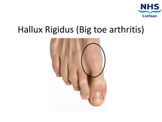
Hallux rigidus is a condition characterized by the loss of motion in the first metatarsophalangeal joint, primarily in adults, due to degenerative arthritis and osteophyte formation. This article delves into the anatomy, biomechanics, epidemiology, and etiologies of hallux rigidus, shedding light on its prevalence, impact on foot function, and potential causes such as trauma, degenerative conditions, and structural abnormalities. Understanding these aspects is crucial for effective management and treatment of hallux rigidus.
Download Presentation

Please find below an Image/Link to download the presentation.
The content on the website is provided AS IS for your information and personal use only. It may not be sold, licensed, or shared on other websites without obtaining consent from the author. Download presentation by click this link. If you encounter any issues during the download, it is possible that the publisher has removed the file from their server.
E N D
Presentation Transcript
Hallux Rigidus Maureen K Monda
Background Davis-Colley 1887: Hallux flexus Cotterill 1888: Hallux rigidus Hiss 1931: Hallux limitus Laird 1972: Functional Hallux limitus Stiff great toe A condition characterized by loss of motion of first MTP joint in adults due to degenerative arthritis osteophyte formation leads to dorsal impingement
Anatomy Tibial and fibula sesamoids FHL and FHB Plantar Plate Articular surface: -MTP -MT-Sesamoid
MTPJ ROM Nawoczenskiet al 1999 EM tracking to look at 3-D motion of 1st MTPJ 33 subjects 4 Clinical Tests for ROM of 1st MPJ compared to actual ROM of 1st MPJ during gait
Biomechanics 5,000 8,000 steps a day. Importance in push off phase of gait (MTP rocker) In sport: sprinting, jumping, ballet, dancing
Biomechanics Normal gait: up to 50% body weight transmitted through great toe complex Jogging, running: 2-3 times body weight Jumping: 8 times body weight.
Biomechanics Roll -first 20odorsiflexion -great toe fixed to ground -metatarsal rolls over PP -FHL, FHB, Abd H, Add H -Plantar fascia windlass
Biomechanics Slide ->20odorsiflexion -1stMT plantarflexion -peroneous longus Compression terminal dorsiflexion provides stability for transfer of forces and balance
Epidemiology 2ndmost common disorder after hallux valgus Coughlin and Shurnas 2003 -110 pts -80% bilateral -98% family history -68% female
Etiologies Multiple: -Traumatic (overuse/OCD injury sequelae, iatrogenic) -Degenerative (systemtic arthrosis, Inflammatory disorders) -Dorsal bunion (paralytic/neuro-muscular) -Metatarsus primus elevates (congenital) The prevailing thinking is that abnormality in dynamic foot function is the primary etiology of hallux rigidus. However, the true mechanism of dysfunction of the great toe joint during gait remains poorly understood.
Etiology Two distinct groups: Adolescents: -rigid swollen joint -painfull dorsiflexion -chondral lesion (traumatic) or OCD (atraumatic) Adults: -degenerative, overuse, traumatic etiology
Presentation Dorsal prominence Foot wear irritation Paraethesiae secondary to dorsal cutaneous nerve compression Painful ROM (DF and PF with pushoff) Pain less severe with disease progression
Physical examination Limited dorsiflexion -pain at extremes -at mid range poor prognosis Pain with grind test Tenderness on palpation -over sesamoids poor prognosis
Coughlin and Shurnas classification Grade Examination findings Xray findings 0 Stiffness normal 1 Mild pain at extremes of motion mild dorsal osteophyte, normal joint space 2 Moderate painwith range of motion increasingly more constant moderate dorsal osteophyte,<50% joint space narrowing 3 Significant stiffness, pain at extreme ROM,no pain at mid-range severe dorsal osteophyte,>50% joint space narrowing 4 severe dorsal osteophyte, >50% joint space narrowing Significant stiffness, pain at extreme ROM,pain at mid-range of motion
Treatment Lengthy discussion Is pain the issue Is Range of motion the issue Is push off strength important to them
Non-operative treatment Grade 0 and 1 disease Foot wear modification Activity modification Orthotics pressure on ostephytes MTPJ movement NSAIDs Steroid Inj selective Increased degeneration
Injections Injection Pons et al 2007 RCT Hyaluronatevs Triamcinolone 37 pts Improvement both groups No pain at rest Hyaluronate grp better -gait pain and passive ROM -AOFAS MTP score MUA+Injection Solan 2001 31pts MUA+ depomedrone/bupivacaine Blinded x-ray grading 1 yrfollow up Grade Survival surgery 1 6 months 4/12 2 3 months 12/18 3 2 months 5/5
Operative treatment Procedure Indication Notes Joint debridement and synovectomy acute osteochondral or chondral defects Dorsal cheilectomy Painwith dorsiflexion Shoe wear irritation Pain in mid ROM contraindicated Obtain 70-90% DF intraop. Dorsal closing wedge osteotomy of the proximal phalanx Runners with reduced dorsiflexion Failure of cheilectomy Decreases plantarflexion arc of motion Keller Procedure (resection arthroplasty) Elderly, low demand Significantjoint degeneration Risk of hyperextension Weakness with pushoff Transfer metatarsalgia MTP arthroplasty Controversial Poorlong term results with silicone implants Osteolysisand synovitis lead to joint destruction and pain Failed arthroplasty, transfer metatarsalgia MTP joint arthrodesis Significant joint arthritis (3 and 4) High fusion rate (70-100%) 15% IPJ degeneration
MTP joint arthrodesis dorsal plate with compression screw is biomechanically strongest construct preferred surgical alignment 10 to 15 degrees of valgusin relation to the metatarsal shaft 15 degrees of dorsiflexionin relation to the floor fusion in excessive dorsiflexion causes pain at tip of the toe, over the IP joint, and under the 1st metatarsal with excessive dorsiflexion fusion in excessive plantar flexion causes increased pressure at the tip of the toe fusion in excessive valgus increases the risk of IP joint degeneration


