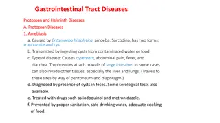
Understanding Entamoeba Histolytica: A Detailed Overview
Entamoeba Histolytica is a zoonotic protozoan parasite found in the digestive tract of various hosts. It mainly exists in two stages - Trophozoite and Cyst. Trophozoites are characterized by their size, clear ectoplasm, and movement via pseudopodia. Cysts are resistant structures with different contents based on maturity. Understanding its life cycle is crucial for diagnosis and treatment.

Download Presentation

Please find below an Image/Link to download the presentation.
The content on the website is provided AS IS for your information and personal use only. It may not be sold, licensed, or shared on other websites without obtaining consent from the author. Download presentation by click this link. If you encounter any issues during the download, it is possible that the publisher has removed the file from their server.
E N D
Presentation Transcript
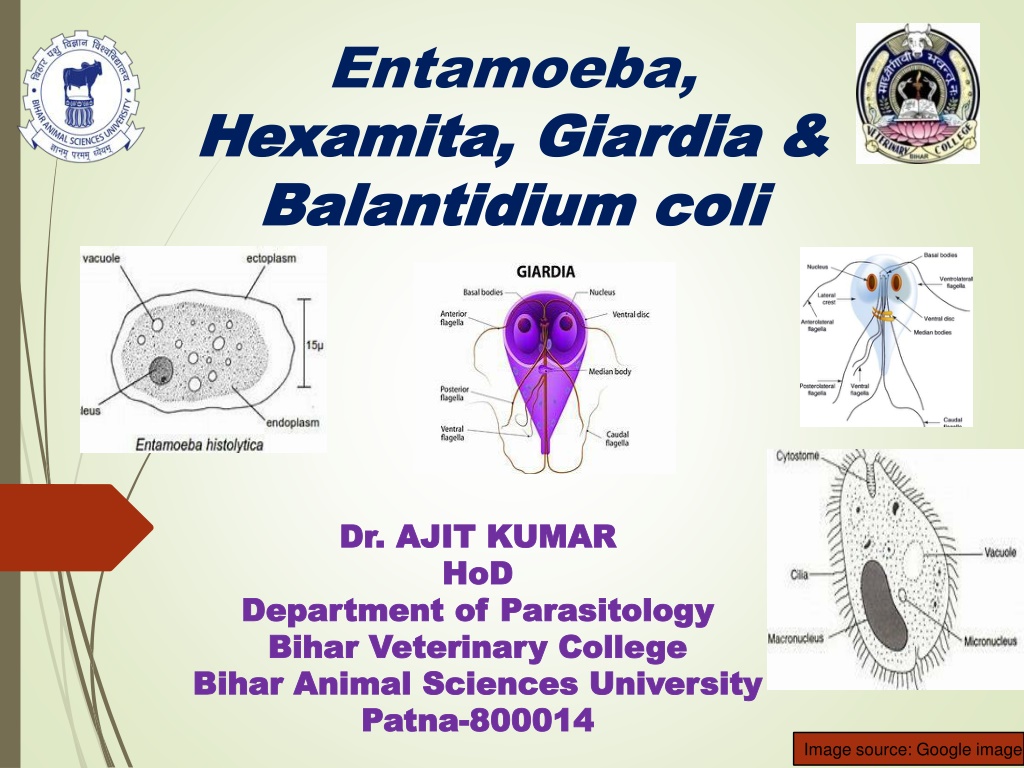
Entamoeba, Hexamita Hexamita, Giardia & , Giardia & B Balantidium alantidium coli Bihar Animal Sciences University | coli Dr. AJIT KUMAR Dr. AJIT KUMAR HoD HoD Department of Department of Parasitology Bihar Veterinary College Bihar Veterinary College Bihar Animal Sciences University Bihar Animal Sciences University Patna Patna- -800014 800014 Parasitology Image source: Google image
Sub Sub- -Phylum: Phylum: Sarcodina Family: Family: Endamoebidae Endamoebidae Sarcodina Family members are called parasitic amoebae which occur in the digestive tract of vertebrates and invertebrates. Movement by Pseudopodia. Holozoic type of nutrition. Binary fission type of asexual reproduction. Encystment is common feature.
Family: Family: Endamoebidae Endamoebidae Genus : Genus : Entamoeba Entamoeba Location: Digestive tract. Usually form cyst. Cysts have 1-8 nuclei depending upon the species.
Genus: Genus: Entamoeba Entamoeba Species Species Host Host No. of nucleus No. of nucleus present inside present inside cyst cyst Entamoeba histolytica Man, dog, cat, monkey, pig etc. Man, dog, monkey, pig etc. Quadrinucleated cyst Entamoeba coli 8-nucleated cyst Entamoeba bovis Cattle Uninucleated cyst
Entamoeba Entamoeba histolytica histolytica It is azoonotic protozoan parasite. Mainly two developmental stages are found :- Trophozoite and Cyst. Trophozoite :- Size about 10-60 m. It has clear ectoplasm and granular endoplasm. Endoplasm of trophozoite contain food vacuoles containing ingested erythrocytes and nucleus Trophozoites move by finger-like pseudopodia. Presence in freshly stool. a vesicular dysenteric Image source: Google image
Entamoeba Entamoeba histolytica histolytica Cyst : Trophozoite condenses into a spherical mass called cyst. Usually Round in shape and 5-15 m in size. Young cyst contains 1-4 chromatoid bars, glycogen vacuoles and single nucleus whereas Mature cyst contains four nuclei without chromatoid bars and glycogen vacuoles. Resistant to averse condition. Sensitive to desiccation, temperature above 400C and below -500C discharges undigested food and putrefaction and
Entamoeba Entamoeba histolytica histolytica Transmission: through cysts contaminated food or water , as trophozoites do not survive more than 30 minutes after passing with stools. Eating of raw and uncleaned vegetable usually in the form of salad. Eating of food contaminated by flies, use of night soil polluted water etc.
Entamoeba Entamoeba histolytica histolytica Life- cycle:- o Hosts get infection by the ingestion of cysts. o Excystment occurs in the intestine in the presence of trypsin enzyme and released metacystic form. o Meatcystic form undergo nuclear and cytoplasmic division and form eight trophozoites which migrate to the large intestine and grow into full size and invade the bowel mucosa. nucleated daughter
Life-cycle of Entamoeba histolytica Image source: Google image
Entamoeba Entamoeba histolytica histolytica Pathogenesis: o Pathogenesis depends on various factors like strain variation, presence of Escherichia coli or Aerobactor aerogenes , nutritional status of hosts, concurrent disease, immunity etc. o Parasites penetrates the intestinal wall by the lysis of the epithelium by the action of phosphohydrloae release by liposomes. o o Amoebae multiply and invade mucosa to produce inverted flask shaped ulcers. o Amoebae may reach other body parts like liver where it may produce inflammation and abscesses.
Entamoeba Entamoeba histolytica histolytica Symptoms: o Acute amoebic dysentery- faces consists of almost entirely blood and mucus with amoebae, abdominal pain, with straining. o Chronic occasional bowels, headache, flatulence etc. dysentery abdominal recurrent pain, dysentery, irregular nausea,
Entamoeba Entamoeba histolytica histolytica Diagnosis :- On the basis of symptoms like faeces with blood and mucus, abdominal pain etc. Microscopic examination of faeces reveals for trophozoites (in diarrhoeic faeces) or quadrinucleated (Entamoeba histolytica) or eight nucleated cysts (Entamoeba coli). the presence cysts 2 % iodine is also used for clearly Four nucleated cyst detection of cysts. Image source: Google image
Entamoeba Entamoeba histolytica histolytica Treatment : Metronidazole , Tinidazole etc. Control : Adoption of proper sanitation, personal hygiene, proper disposal of sewage avoidance of faecal contamination of food and water, supply of clean drinking water etc.. and night soil,
Family: Family: Hexamitidae Hexamitidae Genus: Hexamita and Giardia Both have two similar nuclei. Hexamita sp. cause catarrhal interitis in birs. Giardia :- o Two developmental stages - trophozoite and cyst. o Trophozoite : o Pyriform to elliptical in shape and bilaterally symmetrical. o Anterior end is posterior end is pointed. o A large adhesive disc is present on the ventral side whereas dorsal side convex. Inectious rounded and o It has two anterior nucleus, two median axostyle and eight flagella arranged in two pairs Image source: Google image
Genus: Genus: Giardia Giardia Cyst: Cysts are oval or elliptical in shape with 2 or 4 nuclei and a number of fibrillar remnants of the trophozoite organelles. Image source: Google image
Genus: Genus: Giardia Giardia Species Species Hosts Hosts Location Location Giardia intestinalis or Giardia lamblia Man, monkey and pig Upper digestive tract G. canis Dog G. bovis Ox G. cati Cat G. caprae Goat
Genus: Genus: Giardia Giardia Life-cycle : Direct Reproduction :- By binary fission Transmission: through contaminated food or water. the ingestion of cysts
Life-cycle of Giardia Image source: Google image
Giardia Giardia Pathogenesis: o Chronic diarrhoea in man especially children. o Interference deficiency in the fat soluble vitamins. in fat digestion which results Clinical signs : o Giardiosis dysentery and steatorrhoea (fats comes with stool) and deficiency of fat soluble vitamin. (beaver fever) results diarrhoea,
Giardia Giardia Diagnosis: o Microscopic faecal examination reveals oval or elliptical shaped cyst contain 2 or 4 nuclei. o Trophozoite stage may be found in diarrhoeic stool. Treatment: Trophozoites o Metroniodazole,Tinidazole, Fenbendazole, Chloroquine etc. Image source: Google image Control: o Good hygiene and proper sanitation. Cyst
Hexamita meleagridis Morphology Morphology oPyriform shaped, two similar nuceli, out of eight flagella, six flagella anteriorly directed in two groups of three in each and two caudal flagella, two axostyles but absent. Host and location o It occurs in the duodenum and small intestine of young turkey, peafowl, quail, partridge, chicken. Transmission Transmission o Organisms transmitted contaminated feed and drinking water. adhesive disc duck and also through
Hexamita meleagridis Diagnosis Diagnosis o Examination of intestinal contents or scraping of small intestine of sacrificed birds to microscopic detection of living Hexamita organisms. Treatment Treatment o Nithiazide @ 0.02 percent Furazolidone @ 0.01 percent in feed / Oxyteracycline drugs are used in treatment. Control Control o Adoption of hygienic measures in drinking water/
Hexamita meleagridis Pathogenesis Pathogenesis o Young turkey up to two months of age are most susceptible whereas old birds act as symptomless carrier. o It causes catarrhal enteritis in the small intestine and intestinal contents become thin, watery and foamy. Myriad of Hexamita are occur in the bulbous area of caeca. Clinical Clinical signs signs o Hexamitosis disease is known as Infectious catarrhal enteritis ion turkey poults. o Symptoms include foamy and watery diarrhoea, loss of body weight, nervousness and death of affected birds.
Phylum: Phylum: C Ciliophora iliophora Balantidium coli o Highly organized protozoan having two dissimilar nuclei. o A large macronucleus responsible for cytoplasmic activities of the organisms and a small vesicular micronucleus concerned process. with reproductive o Locomotion: by cilia. o Reproduction: Asexual reproduction by transverse binary fission and sexual reprocess by conjugation.
Balantidium Balantidium coli coli Host: Pig, dog, cattle, monkey and man. Location: large intestine ( caecum and colon). Two developmental stages: Trophozoite and cyst Trophozoite o Active motile stage and 50-60 m in length. o At the anterior end funnel shaped peristome which leads to cytopharynx or mouth. o Two dissimilar nuclei i.e. macronucleus kidney shaped and micronucleus lies in the notch of macronucleus. o Two contractile vacuoles one at each end. o An excretory organelle i.e. cytopyge present at the posterior end of the organism. Cytopyge Image source: Google image
Balantidium Balantidium coli coli Cyst: o Ovoid to spherical in shape, double walled, yellowish in colour and 40-60 m in diameter. o Cysts inside and outside the host body. are formed both Image source: Google image
Balantidium Balantidium coli coli Transmission: o Pig is considered as primary hot and in it B. coli exists as commensal in the large intestine of most pigs. o Pig is probable the persistent source of infection for man, cattle etc. o Hosts get infection through the ingestion of contaminated food and water with cyst of B. coli.
Balantidium Balantidium coli coli Pathogenesis and symptoms: o In pigs normally it is non-pathogenic but occasionally invade the mucosa and producing superficial to deep ulcer with enteritis. It produces diarrhoea and dysentery with hemorrhage. o In man, B. coli produce superficial to deep ulcer associated with enteritis. The initial lesions may resemble those produce by Entamoeba histolytica but do not show the similar tendency to enlarge or spread.
Balantidium Balantidium coli coli Diagnosis: o Microscopic faecal examination reveals cysts. o Trophozoite stage may be found in diarrhoeic stool. Cyst Treatment: Tetracycline, Metronidazole etc. Control: o Good hygiene and proper sanitation. o Proper disposal of faces of pig and man. Image source: Google image