Understanding Agarose Gel Electrophoresis: A Comprehensive Overview
Agarose gel electrophoresis is a vital technique used to separate DNA, RNA, or proteins based on their physical properties like size and charge. This seminar covers the definition, working process, and applications of agarose gel electrophoresis, shedding light on its importance in molecular biology research.
- Agarose Gel Electrophoresis
- DNA Separation
- RNA Analysis
- Molecular Biology Techniques
- Protein Size Sorting
Uploaded on Sep 27, 2024 | 0 Views
Download Presentation

Please find below an Image/Link to download the presentation.
The content on the website is provided AS IS for your information and personal use only. It may not be sold, licensed, or shared on other websites without obtaining consent from the author. Download presentation by click this link. If you encounter any issues during the download, it is possible that the publisher has removed the file from their server.
E N D
Presentation Transcript
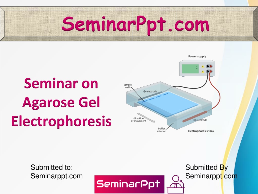
SeminarPpt.com Seminar on Agarose Gel Electrophoresis Submitted to: Seminarppt.com Submitted By Seminarppt.com
Table Contents Definition Introduction About Agarose Gel Electrophoresis? Working of Agarose Gel Electrophoresis Process of Agarose Gel Electrophoresis Conclusion 2
Definition Electrophoresis is a technique that uses electrical current to separate DNA, RNA or proteins based on their physical properties such as size and charge 3
Introduction Agarose gel electrophoresis is a form of electrophoresis used for the separation of nucleic acid (DNA or RNA) fragments based on their size. Negatively charged DNA/RNA migrates through the pores of an agarose gel towards the positively charged end of the gel when an electrical current is applied, with smaller fragments migrating faster. 4
About Agarose Gel Electrophoresis? RNA,1 however, tends to form secondary structures sometimes multiple different species for the same fragment that affect the way it migrates. Consequently, observed bands do not always represent their true sizes and images are blurry 6
About Agarose Gel Electrophoresis? Native agarose gels (where conditions do not disrupt the natural structures of analytes) therefore tend not to be used for the analysis of RNA sizes, although they can give an estimate of quantity and integrity. Alternatives include northern blotting and denaturing agarose gel electrophoresis2 that use conditions able to disrupt secondary structures. 7
About Agarose Gel Electrophoresis? Agarose gels may also be used to separate proteins3 based on their size and charge (unlike DNA/RNA, proteins vary in charge according to the amino acids incorporated). However, due to the large pore sizes in agarose gels, proteins are often separated on polyacrylamide gels that have smaller pores instead, offering greater resolution for small protein molecules. 8
Working of Agarose Gel Electrophoresis Agarose is a component of agar. It forms a 3D gel matrix of helical agarose molecules in supercoiled bundles held by hydrogen bonds, with channels and pores through which molecules are able to pass. When heated, these hydrogen bonds break, turning the agarose to liquid and allowing it to be poured into a mold before it resets. 9
Working of Agarose Gel Electrophoresis The percentage of agarose included in a gel impacts the pore sizes and thus the size of molecules that may pass through and speed at which they do so. The higher the percentage of agarose, the smaller the pore size, thus the smaller the molecules able to pass and the slower the migration. 10
Working of Agarose Gel Electrophoresis In the molecular biology lab, 0.7-1% agarose gel is typically used for day-to-day DNA separations, offering good, clear differentiation of fragments in the range of 0.2-10 kb. Larger fragments may be resolved using lower percentage gels, but they become very fragile and hard to handle, while higher percentage gels will give better resolution of small fragments but are brittle and may set unevenly. 11
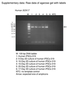
Working of Agarose Gel Electrophoresis As DNA is not visible to the naked eye, an intercalating dye such as ethidium bromide (EtBr) is incorporated in the gel during setting. This binds the DNA and fluoresces under UV light, allowing the DNA fragments to be visualized. The more DNA present, the brighter the band. 12
Working of Agarose Gel Electrophoresis When the samples have run far enough to obtain sufficient separation, the gel is removed from the tank and placed on a UV light box. The intercalating dye then allows the sample bands to be visualized and their size determined in comparison to a DNA ladder with known band sizes. 13
Process of Agarose Gel Electrophoresis Determine the required gel percentage 0.7 1% agarose gel is typically adequate for most applications, but it is important to choose a percentage appropriate for your samples and expected fragment sizes. Combine the agarose powder with the same buffer type to be used to run the gel and heat to melt the mixture, avoiding boiling. 14
Process of Agarose Gel Electrophoresis Pour a gel Choose a gel casting mold and comb of the desired size, giving a sufficient number of wells for all samples and ladders and well capacity to hold the quantity of each sample to be loaded. Secure the open ends of the mold with a casting frame or tape to contain the gel while it sets. 15
Process of Agarose Gel Electrophoresis Mix samples/ladders with loading dye Loading dyes perform multiple functions. They allow the user to see where their otherwise colorless sample is, making it easier to pipette the sample into the well accurately and thus reducing the likelihood of cross-contamination of samples between the wells. 16
Process of Agarose Gel Electrophoresis Load the gel Remove the casting frame/tape from the set gel and place it in the gel tank, ensuring that the wells are at the negative end (black electrodes). Fill the tank with running buffer (TAE or TBE) so that the gel is submerged. Carefully remove the comb and gently pipette the sample-dye (and water if used) mix into the wells. 17
Process of Agarose Gel Electrophoresis Run the gel Place the lid on the tank with the electrodes black to black and red to red and plug the electrodes into a power pack, also black to black and red to red. This, along with the gel tank, makes up the gel electrophoresis machine. 18
Process of Agarose Gel Electrophoresis Visualization Once the samples have run most of the way down the gel (the dye front will make this visible), turn off the power pack. Wearing gloves, gently remove the gel in the mold from the tank, draining off excess running buffer, and transfer to a UV box in an appropriate container for visualization. 19
Conclusion Agarose gels may be visualized on a UV light box in a dark room or using a self-contained light box linked to a camera. Whichever system is utilized, UV light is shone through the gel from below and bands of DNA fluoresce thanks to the intercalating dye bound to them. 20
References Wikipedia.org Google.com Seminarppt.com Studymafia.org
