Thyroid Gland Function and Hormones
The thyroid gland, essential for regulating metabolic rate, secretes thyroxine and triiodothyronine. Learn about their synthesis, secretion, and the importance of iodine in thyroid hormone formation. Explore the physiological anatomy of the thyroid gland and how it controls metabolism through hormone secretion.

Download Presentation

Please find below an Image/Link to download the presentation.
The content on the website is provided AS IS for your information and personal use only. It may not be sold, licensed, or shared on other websites without obtaining consent from the author.If you encounter any issues during the download, it is possible that the publisher has removed the file from their server.
You are allowed to download the files provided on this website for personal or commercial use, subject to the condition that they are used lawfully. All files are the property of their respective owners.
The content on the website is provided AS IS for your information and personal use only. It may not be sold, licensed, or shared on other websites without obtaining consent from the author.
E N D
Presentation Transcript
Thyroid Gland Thyroid Gland Dr. Dr. Noori Noori M M Luaibi Luaibi
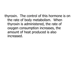
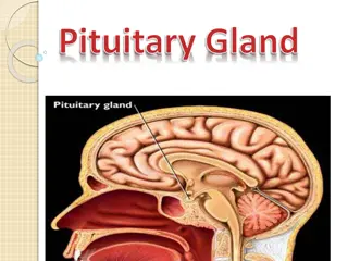
Thyroid Metabolic Hormones The thyroid gland, located immediately below the larynx on each side of and anterior to the trachea, is one of the largest of the endocrine glands, normally weighing 15 to 20 grams in adults. The thyroid secretes two major hormones, thyroxine and triiodothyronine, commonly called T4 and T3, respectively. Both of these hormones profoundly increase the metabolic rate of the body. Complete lack of thyroid secretion usually causes the basal metabolic rate to fall 40 to 50 per cent below normal, and extreme excesses of thyroid secretion can increase the basal metabolic rate to 60 to 100 per cent above normal. Thyroid secretion is controlled primarily by thyroid- stimulating hormone (TSH) secreted by the anterior pituitary gland. The thyroid gland also secretes calcitonin, an important hormone for calcium metabolism .
SYNTHESIS AND SECRETION OF THE THYROID METABOLIC HORMONES About 93 per cent of the metabolically active hormones secreted by the thyroid gland is thyroxine, and 7 per cent triiodothyronine. However, almost all the thyroxine is eventually converted to triiodothyronine in the tissues, so that both are functionally important. The functions of these two hormones are qualitatively the same, but they differ in rapidity and intensity of action. Triiodothyronine is about four times as potent as thyroxine, but it is present in the blood in much smaller quantities and persists for a much shorter time than does thyroxine.
Physiologic Anatomy of the Thyroid Gland. The thyroid gland is composed, Figure 76 1, of large numbers of closed follicles (100 to 300 micrometers in diameter) filled with a secretory substance called colloid and lined with cuboidal epithelial cells that secrete into the interior of the follicles.The major constituent of colloid is the large glycoprotein thyroglobulin, which contains the thyroid hormones within its molecule. Once the secretion has entered the follicles, it must be absorbed back through the follicular epithelium into the blood before it can function in the body.The thyroid gland has a blood flow about five times the weight of the gland each minute, which is a blood supply as great as that of any other area of the body, with the possible exception of the adrenal cortex.
IODINE IS REQUIRED FOR FORMATION OF THYROXINE To form normal quantities of thyroxine, about 50 milligrams of ingested iodine in the form of iodides are required each year, or about 1 mg/week. To prevent iodine deficiency, common table salt is iodized with about 1 part sodium iodide to every 100,000 parts sodium chloride. Fate of Ingested Iodides. Fate of Ingested Iodides. Iodides ingested orally are absorbed from the gastrointestinal tract into the blood in about the same manner as chlorides. Normally, most of the iodides are rapidly excreted by the kidneys, but only after about one fifth are selectively removed from the circulating blood by the cells of the thyroid gland and used for synthesis of the thyroid hormones.
IODIDE PUMP (IODIDE TRAPPING) The first stage in the formation of thyroid hormones, Figure 76 2, is transport of iodides from the blood into the thyroid glandular cells and follicles.The basal membrane of the thyroid cell has the specific ability to pump the iodide actively to the interior of the cell. This is achieved by the action of a sodium-iodide sympoter (NIS),which co-transport one iodide ion along with two sodium ions across the basolateral (plasma) membrane into the cell. The energy for transporting iodide against a concentration grained comes from the sodium-potassium ATPase pump, which pumps sodium out of the cell, thereby establishing low intracellular sodium concentration and a gradient for facilitated diffusion of sodium into the cell. This process of concentration the iodide in the cell is called iodide trapping. In a normal gland, the iodide pump concentrates the iodide to about 30 times its concentration in the blood. When the thyroid gland becomes maximally active, this concentration ratio can rise to as high as 250 times. The rate of iodide trapping by the thyroid is influenced by several factors, the most important being the concentration of TSH; TSH stimulates and hypophysectomygreatly diminishes the activity of the iodide pump in thyroid cells. Iodide is transport out of the thyroid cells across the apical membrane into the follicle by a chloride-iodide ion counter-transporter molecule called pendrin. The thyroid epithelial cells also secrete into the follicle thyroglobulin that contaims tyrosine amino acids to which the iodide ions will bind .
THYROGLOBULIN, AND CHEMISTRY OF THYROXINE AND TRIIODOTHYRONINE Formation Formationand Secretion of Thyroglobulin by the Thyroid Cells. The thyroid cells are typical protein-secreting glandular cells. The endoplasmic reticulum and Golgi apparatus synthesize and secrete into the follicles a large glycoprotein molecule called thyroglobulin, with a molecular weight of about 335,000. Each molecule of thyroglobulin contains about 70 tyrosine amino acids, and they are the major substrates that combine with iodine to form the thyroid hormones. Thus, the thyroid hormones form within the thyroglobulin molecule. That is, the thyroxine and triiodothyronine hormones formed from the tyrosine amino acids remain part of the thyroglobulin molecule during synthesis of the thyroid hormones and even afterward as stored hormones in the follicular colloid.
OXIDATION OF THE IODIDE ION. The first essential step in the formation of the thyroid hormones is conversion of the iodide ions to an oxidized form of iodine, either nascent iodine (I0) or I_3, that is then capable of combining directly with the amino acid tyrosine. This oxidation of iodine is promoted by the enzyme peroxidase and its accompanying hydrogen peroxide, which provide a potent system capable of oxidizing iodides. The peroxidase is either located in the apical membrane of the cell or attached to it, thus providing the oxidized iodine at exactly the point in the cell where the thyroglobulin molecule issues forth from the Golgi apparatus and through the cell membrane into the stored thyroid gland colloid. When the peroxidase system is blocked or when it is hereditarily absent from the cells, the rate of formation of thyroid hormones falls to zero.
IODINATION OF TYROSINE AND FORMATION OF THE THYROID HORMONES ORGANIFICATION OF THYROGLOBULIN. The binding of iodine with the thyroglobulin molecule is called organification of the thyroglobulin. Oxidized iodine even in the molecular form will bind directly but very slowly with the amino acid tyrosine. In the thyroid cells, however, the oxidized iodine is associated with an iodinase enzyme (Figure 76 2) that causes the process to occur within seconds or minutes.Therefore, almost as rapidly as the thyroglobulin molecule is released from the Golgi apparatus or as it is secreted through the apical cell membrane into the follicle, iodine binds with about one sixth of the tyrosine amino acids within the thyroglobulin molecule. (Figure 76 3 (shows the successive stages of iodination of tyrosine and final formation of the two important thyroid hormones, thyroxine and triiodothyronine. Tyrosine is first iodized to monoiodotyrosine and then to diiodotyrosine. Then, during the next few minutes, hours, and even days, more and more of the iodotyrosine residues become coupled with one another. The major hormonal product of the coupling reaction is the molecule thyroxine (T4), which is formed when two molecules of diiodotyrosine are joined together; the thyroxine then remains part of the thyroglobulin molecule . Or one molecule of monoiodotyrosine couples with one molecule of diiodotyrosine to form triiiodothyronine(T3), which represents about one fifteenth of the final hormones , small amounts of reverse T3 (RT3) are formed by coupling of diiodotyrosine with monoiodotyrosine , but RT3 dose not appear to be of functional significance in humans.
STORAGE OF THYROGLOBULIN. The thyroid gland is unusual among the endocrine glands in its ability to store large amounts of hormone. After synthesis of the thyroid hormones has run its course, each thyroglobulin molecule contains up to 30 thyroxine molecules and a few triiodothyronine molecules. In this form, the thyroid hormones are stored in the follicles in an amount sufficient to supply the body with its normal requirements of thyroid hormones for 2 to 3 months. Therefore, when synthesis of thyroid hormone ceases, the physiologic effects of deficiency are not observed for several months.
RELEASE OF THYROXINE AND TRIIODOTHYRONINE FROM THE THYROID GLAND Thyroglobulin itself is not released into the circulating blood in measurable amounts; instead, thyroxine and triiodothyronine must first be cleaved from the thyroglobulin molecule, and then these free hormones arereleased. This process occurs as follows: The apical surface of the thyroid cells sends out pseudopod extensions that close around small portions of the colloid to form pinocytic vesicles that enter the apex of the thyroid cell. Then lysosomes in the cell cytoplasm immediately fuse with these vesicles to form digestive vesicles containing digestive enzymes from the lysosomes mixed with the colloid. Multiple proteases among the enzymes digest the thyroglobulin molecules and release thyroxine and triiodothyronine in free form. These then diffuse through the base of the thyroid cell into the surrounding capillaries. Thus, the thyroid hormones are released into the blood. About three quarters of the iodinated tyrosine in the thyroglobulin never becomes thyroid hormones but remains monoiodotyrosine and diiodotyrosine. During the digestion of the thyroglobulin molecule to cause release of thyroxine and triiodothyronine, these iodinated tyrosinesalso are freed from the thyroglobulin molecules. However, they are not secreted into the blood. Instead, their iodine is cleaved from them by a deiodinase enzyme that makes virtually all this iodine available again for recycling within the gland for forming additional thyroid hormones. In the congenital absence of this deiodinase enzyme, many persons become iodine-deficient because of failure of this recycling process.
DAILY RATE OF SECRETION OF THYROXINE AND TRIIODOTHYRONINE. About 93 per cent of the thyroid hormone released from the thyroid gland is normally thyroxine and only 7 per cent is triiodothyronine. However, during the ensuing few days, about one half of the thyroxine is slowly deiodinated to form additional triiodothyronine. Therefore, the hormone finally delivered to and used by the tissues is mainly triiodothyronine, a total of about 35 micrograms of triiodothyronine per day.
Transport of Thyroxine and Triiodothyronine to Tissues Thyroxineand TriiodothyronineAre Bound to Plasma Proteins. On entering the blood, over 99 per cent of the thyroxine and triiodothyronine combines immediately with several of the plasma proteins, all of which are synthesized by the liver. They combine mainly with thyroxine-binding globulin and much less so with thyroxine- binding prealbumin and albumin. Thyroxine and Triiodothyronine Are Released Slowly to Tissue Cells. Because of high affinity of the plasma-binding proteins for the thyroid hormones, these substances in particular, thyroxine are released to the tissue cells slowly. Half the thyroxine in the blood is released to the tissue cells about every 6 days, whereas half the triiodothyronine because of its lower affinity is released to the cells in about 1 day. On entering the tissue cells, both thyroxine and triiodothyronine again bind with intracellular proteins, the thyroxine binding more strongly than the triiodothyronine. Therefore, they are again stored, but this time in the target cells themselves, and they are used slowly over a period of days or weeks.
THYROID HORMONES HAVE SLOW ONSET AND LONG DURATION OF ACTION. After injection of a large quantity of thyroxine into a human being, essentially no effect on the metabolic rate can be discerned for 2 to 3 days, thereby demonstrating that there is a long latent period before thyroxine activity begins. Once activity does begin, it increases progressively and reaches a maximum in 10 to 12 days Figure 76 4. Thereafter, it decreases with a half-life of about 15 days. Some of the activity persists for as long as 6 weeks to 2 months. The actions of triiodothyronine occur about four times as rapidly as those of thyroxine, with a latent period as short as 6 to 12 hours and maximal cellular activity occurring within 2 to 3 days. Most of the latency and prolonged period of action of these hormones are probably caused by their binding with proteins both in the plasma and in the tissue cells, followed by their slow release.
PHYSIOLOGIC FUNCTIONS OF THE THYROID HORMONES 1. Thyroid Hormones Increase the Transcription of Large Numbers of Genes 2. Thyroid Hormones Activate Nuclear Receptors. 3. Thyroid Hormones Increase Cellular Metabolic Activity 4. Thyroid Hormones Increase the Number and Activity of Mitochondria. 5. Thyroid Hormones Increase Active Transport of Ions Through Cell Membranes. 6. Effect of Thyroid Hormone on Growth.
7.Effects of Thyroid Hormone on Specific Bodily Mechanisms include: A) Stimulation of Carbohydrate Metabolism. Thyroid hormone stimulates almost all aspects of carbohydrate metabolism, including rapid uptake of glucose by the cells, enhanced glycolysis, enhanced gluconeogenesis, increased rate of absorption from the gastrointestinal tract, and even increased insulin secretion with its resultant secondary effects on carbohydrate metabolism. All these effects probably result from the overall increase in cellular metabolic enzymes caused by thyroid hormone. B) Stimulation of Fat Metabolism. Effect on Plasma and Liver Fats. Increased thyroid hormone decreases the concentrations of cholesterol, phospholipids, and triglycerides in the plasma, even though it increases the free fatty acids. Conversely, decreased thyroid secretion greatly increases the plasma concentrations of cholesterol, phospholipids, and triglycerides and almost always causes excessive deposition of fat in the liver as well.The large increase in circulating plasma cholesterol in prolonged hypothyroidism is often associated with severe atherosclerosis. C) Increased Requirement for Vitamins. D) Increased Basal Metabolic Rate. E) Decreased Body Weight.
8. Effect of Thyroid Hormones on the Cardiovascular System include: . Increased Heart Rate. . Increased Heart Strength. . Normal Arterial Pressure. . Increased Respiration. . Increased Gastrointestinal Motility 9. Excitatory Effects on the Central Nervous System. 10. Effect on the Function of the Muscles. Include Muscle Tremor. 11. Effect on Sleep.
12. Effect on Other Endocrine Glands. Increased thyroid hormone increases the rates of secretion of most other endocrine glands, but it also increases the need of the tissues for the hormones. For instance, increased thyroxine secretion increases the rate of glucose metabolism everywhere in the body and therefore causes a corresponding need for increased insulin secretion by the pancreas. Also, thyroid hormone increases many metabolic activities related to bone formation and, as a consequence, increases the need for parathyroid hormone. Thyroid hormone also increases the rate at which adrenal glucocorticoids are inactivated by the liver. This leads to feedback increase in adrenocorticotropic hormone production by the anterior pituitary and, therefore, increased rate of glucocorticoid secretion by the adrenal glands.
13. Effect of Thyroid Hormone on Sexual Function. For normal sexual function, thyroid secretion needs to be approximately normal In men, lack of thyroid hormone is likely to cause loss of libido; great excesses of the hormone, however, sometimes cause impotence. In women, lack of thyroid hormone often causes menorrhagia and polymenorrhea that is, respectively, excessive and frequent menstrual bleeding. Yet, strangely enough, in other women thyroid lack may cause irregular periods and occasionally even amenorrhea. A hypothyroid woman, like a man, is likely to have greatly decreased libido.To make the picture still more confusing. In the hyperthyroid woman, oligomenorrhea, which means greatly reduced bleeding, is common, and occasionally amenorrhea results.
REGULATION OF THYROID HORMONE SECRETION To maintain normal levels of metabolic activity in the body, precisely the right amount of thyroid hormone must be secreted at all times; to achieve this, specific feedback mechanisms operate through the hypothalamus and anterior pituitary gland to control the rate of thyroid secretion. These mechanisms are as follows. TSH (FROM THE ANTERIOR PITUITARY GLAND) INCREASES THYROID SECRETION. TSH, also known as thyrotropin, is an anterior pituitary hormone, a glycoprotein with a molecular weight of about 28,000., increases the secretion of thyroxine and triiodothyronine by the thyroid gland. Its specific effects on the thyroid gland are as follows:
1. Increased proteolysis of the thyroglobulin that has already been stored in the follicles, with resultant release of the thyroid hormones into the circulating blood and diminishment of the follicular substance itself. 2. Increased activity of the iodide pump, which increases the rate of iodide trapping in the glandular cells, sometimes increasing the ratio of intracellular to extracellular iodide concentration in the glandular substance to as much as eight times normal. 3. Increased iodination of tyrosine to form the thyroid hormones. 4. Increased size and increased secretory activity of the thyroid cells. 5. Increased number of thyroid cells plus a change from cuboidal to columnar cells and much in folding of the thyroid epithelium into the follicles. IN SUMMARY, TSH increases all the known secretory activities of the thyroid glandular cells. The most important early effect after administration of TSH is to initiate proteolysis of the thyroglobulin, which causes release of thyroxine and triiodothyronine into the blood within 30 minutes. The other effects require hours or even days and weeks to develop fully.
Cyclic Adenosine Monophosphate Mediates the Stimulatory Effect of TSH. In the past, it was difficult to explain the many and varied effects of TSH on the thyroid cell. It is now clear that most, if not all, of these effects result from activation of the second messenger cyclic adenosine monophosphate (cAMP) system of the cell. The first event in this activation is binding of TSH with specific TSH receptors on the basal membrane surfaces of the thyroid cell. This then activates adenylyl cyclase in the membrane, which increases the formation of cAMP inside the cell. Finally, the cAMP acts as a second messenger to activate protein kinase,which causes multiple phosphorylations throughout the cell. The result is both an immediate increase in secretion of thyroid hormones and prolonged growth of the thyroid glandular tissue itself. This method for control of thyroid cell activity is similar to the function of cAMP as a second messenger in many other target tissues of the body.
FEEDBACK EFFECT OF THYROID HORMONE TO DECREASE ANTERIOR PITUITARY SECRETION OF TSH Increased thyroid hormone in the body fluids decreases secretion of TSH by the anterior pituitary. When the rate of thyroid hormone secretion rises to about 1.75 times normal, the rate of TSH secretion falls essentially to zero. Almost all this feedback depressant effect occurs even when the anterior pituitary has been separated from the hypothalamus. Therefore, as show in Figure 76-7, it is probable that increased thyroid hormone inhibits anterior pituitary secretion of TSH mainly by a direct effect on the anterior pituitary gland itself. Regardless of the mechanism of the feedback, its effect is to maintain an almost constant concentration of free thyroid hormones in the circulating body fluids.
Antithyroid Substances Suppres Thyroid secretion Drugs that suppress thyroid secretion are called antithyroid substances.The best known of these substances are thiocyanate, propylthiouracil, and high concentrations of inorganic iodides. The mechanism by which each of these blocks thyroid secretion is different from the others, and they can be explained as follows. 1. Thiocyanate Ions Decrease Iodide Trapping. 2. Propylthiouracil Decreases Thyroid Hormone Formation. Propylthiouracil (and other, similar compounds, such as methimazoleand carbimazole) prevents formation of thyroid hormone from iodides and tyrosine. 3. Iodides in High Concentrations Decrease Thyroid Activity and Thyroid Gland Size.
Diseases of the Thyroid Hyperthyroidism Most effects of hyperthyroidism are obvious from the preceding discussion of the various physiologic effects of thyroid hormone. However, some specific effects should be mentioned in connection especially with the development, diagnosis, and treatment of hyperthyroidism. Causes of Hyperthyroidism (Toxic Goiter, Thyrotoxicosis, Graves Disease). In most patients with hyperthyroidism, the thyroid gland is increased to two to three times normal size, with tremendous hyperplasia and in folding of the follicular cell lining into the follicles, so that the number of cells is increased greatly. Also, each cell increases its rate of secretion several fold; adioactive iodine uptake studies indicate that some of these hyperplastic glands secrete thyroid hormone at rates 5 to 15 times normal.
GRAVES DISEASE , the most common form of hypothyroidism is an autoimmune disease in which antibodies called thyroid-stimulating immunoglobulin (TSIs) from against the TSH receptor in the Thyroid gland These antibody that bind with the same membrane receptors that bind TSH. They induce continual activation of the cAMP system of the cells,with resultant development of hyperthyroidism. These antibodies TSI have a prolonged stimulating effect on the thyroid gland, lasting for as long as 12 hours, in contrast to a little over 1 hour for TSH. The high level of thyroid hormone secretion caused by TSI in turn suppresses anterior pituitary formation of TSH. Therefore, TSH concentration are less than normal (often essentially zero) rather than enhanced in almost all patients with Granesdisease , The antibodies that cause hyperthyroidism almost certainly occur as the result of autoimmunity that has developed against thyroid tissue. Presumably, at some time in the history of the person, an excess of thyroid cell antigens was released from the thyroid cells, and this has resulted in the formation of antibodies against the thyroid gland itself.
Thyroid Adenoma. Hyperthyroidism occasionally results from a localized adenoma (a tumor) that develops in the thyroid tissue and secretes large quantities of thyroid hormone. This is different from the more usual type of hyperthyroidism, in that it usually is not associated with evidence of any autoimmune disease. An interesting effect of the adenoma is that as long as it continues to secrete large quantities of thyroid hormone, secretory function in the remainder of the thyroid gland is almost totally inhibited because the thyroid hormone from the adenoma depresses the production of TSH by the pituitary gland.
Symptoms of Hyperthyroidism The symptoms of hyperthyroidism are obvious from the preceding discussion of the physiology of the thyroid hormones: 1. a high state of excitability, 2. intolerance to heat, 3. increased sweating, 4. mild to extreme weight loss (sometimes as much as 100 pounds), 5. varying degrees of diarrhea, 6. muscle weakness, 7. nervousness or other psychic disorders, 8. extreme fatigue but inability to sleep, 9. tremor of the hands. Exophthalmos. Most people with hyperthyroidism develop some degree of protrusion of the eyeballs, This condition is called exophthalmos. A major degree of exophthalmos occurs in about one third of hyperthyroid patients .
Hypothyroidism The effects of hypothyroidism, in general, are opposite to those of hyperthyroidism, but there are a few physiologic mechanisms peculiar to hypothyroidism. Hypothyroidism, like hyperthyroidism, probably is initiated by autoimmunity against the thyroid gland, but immunity that destroys the gland rather than stimulates it.The thyroid glands of most of these patients first have autoimmune thyroiditis, which means thyroid inflammation. This causes progressive deterioration and finally fibrosis of the gland, with resultant diminished or absent secretion of thyroid hormone. Several other types of hypothyroidism also occur, often associated with development of enlarged thyroid glands, called thyroid goiter, as follows.
Endemic Colloid Goiter Caused by Dietary Iodide Deficiency. The term goiter means a greatly enlarged thyroid gland. As pointed out in the discussion of iodine metabolism, about 50 milligrams of iodine are required each year for the formation of adequate quantities of thyroid hormone. In certain areas of the world, notably in the Swiss Alps, the Andes, and the Great Lakes region of the United States, insufficient iodine is present in the soil for the foodstuffs to contain even this minute quantity. Therefore, in the days before iodized table salt, many people who lived in these areas developed extremely large thyroid glands, called endemic goiters. The Mechanism for development of large endemic goiters is the following: Lack of iodine prevents production of both thyroxine and triiodothyronine. As a result, no hormone is available to inhibit production of TSH by the anterior pituitary; this causes the pituitary to secrete excessively large quantities of TSH.TheTSH then stimulates the thyroid cells to secrete tremendous amounts of thyroglobulin colloid into the follicles, and the gland grows larger and larger. But because of lack of iodine, thyroxine and triiodothyronine production does not occur in the thyroglobulin molecule and therefore does not cause the normal suppression of TSH production by the anterior pituitary. The follicles become tremendous in size, and the thyroid gland may increase to 10 to 20 times normal size .
Atherosclerosis in Hypothyroidism. As pointed out earlier, lack of thyroid hormone increases the quantity of blood cholesterol because of altered fat and cholesterol metabolism and diminished liver excretion of cholesterol in the bile.The increase in blood cholesterol is usually associated with increased atherosclerosis. Therefore, many hypothyroid patients, particularly those with myxedema, develope atherosclerosis, which in turn results in peripheral vascular disease, deafness, and coronary artery disease with consequent early death.
Diagnostic Tests in Hypothyroidism. The tests already described for diagnosis of hyperthyroidism give opposite results in hypothyroidism. The free thyroxine in the blood is low. The basal metabolic rate in myxedema ranges between -30 and -50. And the secretion of TSH by the anterior pituitary when a test dose of TRH is administered is usually greatly increased (except in those rare instances of hypothyroidism caused by depressed response of the pituitary gland to TRH) .


")
")