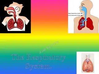
The Respiratory System: An Overview


Respiratory System

Prepared by
Ali Malik Tiryag


Anatomic and Physiologic Overview
The respiratory system is composed of the upper and lower respiratory
tracts. Together, the two tracts are responsible for
ventilation
(movement of
air in and out of the airways). The upper respiratory tract, known as the upper
airway, warms and filters inspired air so that the lower respiratory tract (the
lungs) can accomplish gas exchange or diffusion. Gas exchange involves
delivering oxygen to the tissues through the bloodstream and expelling waste
gases, such as carbon dioxide, during expiration. The respiratory system
depends on the cardiovascular system for perfusion, or blood flow through
the pulmonary system
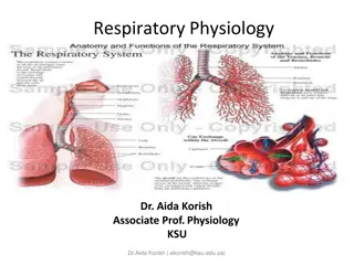
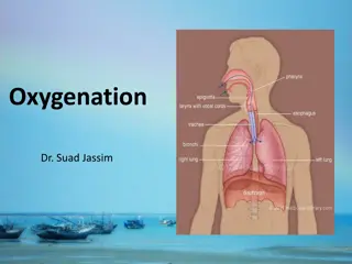
Anatomy of the Respiratory System
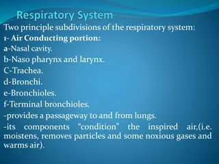
Upper Respiratory Tract
Upper airway structures consist of the nose; paranasal sinuses; pharynx, tonsils, and
adenoids; larynx; and trachea.
Lower Respiratory Tract
The lower respiratory tract consists of the bronchi, bronchioles, alveolar ducts, and
alveoli. Except for the right and left mainstem bronchi, all lower airway structures
are found within the lungs. The right lung is divided into 3 lobes (upper, middle, and
lower) and the left lung into 2 lobes (upper and lower)
Common Symptoms
1- Dyspnea
Dyspnea
(subjective feeling of difficult, breathlessness, shortness of breath) is a
multidimensional symptom common to many pulmonary and cardiac disorders.
Dyspnea may also be associated with allergic reactions, anemia, neurologic or
neuromuscular disorders, trauma, and advanced disease, and is common at the end
of life. Dyspnea can also occur after exercise in people without disease.
In general,
acute diseases of the lungs produce a more severe grade of dyspnea than do chronic
diseases. Sudden dyspnea in a healthy person may indicate pneumothorax (air in the
pleural cavity), acute respiratory obstruction, allergic reaction, or myocardial
infarction.
In immobilized patients, sudden dyspnea may denote pulmonary embolism (PE).
Dyspnea and
tachypnea
(abnormally rapid respirations) accompanied by
progressive
hypoxemia
(low blood oxygen level) in a person who has recently
experienced lung trauma, shock, cardiopulmonary bypass, or multiple blood
transfusions may signal ARDS.
Orthopnea
(shortness of breath when lying flat,
relieved by sitting or standing) may be found in patients with heart disease and
occasionally in patients with chronic obstructive pulmonary disease (COPD);
dyspnea with an expiratory wheeze occurs with COPD.
2- Cough
Cough is a reflex that protects the lungs from the accumulation of secretions or the
inhalation of foreign bodies. Its presence or absence can be a diagnostic clue
because some disorders cause coughing and others suppress it. The cough reflex
may be impaired by weakness or paralysis of the respiratory muscles, prolonged
inactivity, the presence of a nasogastric tube, or depressed function of the brain’s
medullary centers (e.g., anesthesia, brain disorders). Cough results from irritation or
inflammation of the mucous membranes anywhere in the respiratory tract and is
associated with multiple pulmonary disorders. Mucus, pus, blood, or an airborne
irritant, such as smoke or a gas, may stimulate the cough reflex.
Common causes of cough include asthma, gastrointestinal reflux disease, infection,
and side effects of medications, such as angiotensin-converting enzyme (ACE)
inhibitors. To help determine the cause of the cough, the nurse inquires about the
onset and time of coughing. Coughing at night may indicate the onset of left-sided
heart failure or bronchial asthma. A cough in the morning with sputum production
may indicate bronchitis. A cough that worsens when the patient is supine suggests
postnasal drip (rhinosinusitis). Coughing after food intake may indicate aspiration
of material into the tracheobronchial tree. A cough of recent onset is usually from
an acute infection.
The nurse assesses the character of the cough and associated symptoms. A
dry, irritative cough is characteristic of an upper respiratory tract infection
of viral origin, or it may be a side effect of ACE inhibitor therapy. An
irritative, high-pitched cough can be caused by laryngotracheitis. A brassy
cough is the result of a tracheal lesion, and a severe or changing cough may
indicate bronchogenic carcinoma. Pleuritic chest pain that accompanies
coughing may indicate pleural or chest wall (musculoskeletal) involvement.
Violent coughing causes bronchial spasm, obstruction, and further irritation
of the bronchi and may result in syncope (fainting).
3- Sputum Production
Sputum production is the reaction of the lungs to any constantly recurring irritant and often
results from persistent coughing. It may also be associated with a nasal discharge. The
nature of the sputum is often indicative of its cause. A profuse amount of purulent sputum
(thick and yellow, green, or rust colored) or a change in color of the sputum is a common
sign of a bacterial infection. Thin, mucoid sputum frequently results from viral bronchitis.
A gradual increase of sputum over time may occur with chronic bronchitis or bronchiectasis.
Pink-tinged mucoid sputum suggests a lung tumor. Profuse, frothy, pink material, often
welling up into the throat, may indicate pulmonary edema. Foul-smelling sputum and bad
breath point to the presence of a lung abscess, bronchiectasis, or an infection caused by
fusospirochetal or other anaerobic organisms.
4-
Chest Pain
Chest pain or discomfort may be associated with pulmonary, cardiac, gastrointestinal, or
musculoskeletal disease or anxiety. Chest pain associated with pulmonary conditions may
be sharp, stabbing, and intermittent, or it may be dull, aching, and persistent. The pain
usually is felt on the side where the pathologic process is located, although it may be
referred elsewhere—for example, to the neck, back, or abdomen. Chest pain may occur
with pneumonia, pulmonary infarction, or pleurisy, or as a late symptom of bronchogenic
carcinoma. In carcinoma, the pain may be dull and persistent because the cancer has
invaded the chest wall, mediastinum, or spine.
the parietal pleura have a rich supply of sensory nerves that are stimulated by
inflammation and stretching of the membrane. Pleuritic pain from irritation of
the parietal pleura is sharp and seems to “catch” on inspiration; patients often
describe it as being “like the stabbing of a knife.” Patients are more comfortable
when they lay on the affected side because this position splints the chest wall,
limits expansion and contraction of the lung, and reduces the friction between
the injured or diseased pleurae on that side. Pain associated with cough may be
reduced manually by splinting the rib cage.
5- Wheezing
Wheezing is a high-pitched, musical sound heard on either expiration (asthma) or inspiration
(bronchitis). It is often the major finding in a patient with bronchoconstriction or airway
narrowing. Rhonchi are low-pitched continuous sounds heard over the lungs in partial airway
obstruction. Depending on their location and severity, these sounds may be heard with or
without a stethoscope.
6- Hemoptysis
Hemoptysis
is the expectoration of blood from the respiratory tract. It can present as small to
moderate blood-stained sputum to a large hemorrhage and always warrants further
investigation. The onset of hemoptysis is usually sudden, and it may be intermittent or
continuous.
The most common causes are: Pulmonary infection, Carcinoma of the lung, Abnormalities of
the heart or blood vessels, Pulmonary artery or vein abnormalities, PE or infarction. Potential
sources of bleeding include the gums, nasopharynx, lungs, or stomach.
•
Bloody sputum from the nose or the nasopharynx is usually preceded by considerable
sniffing, with blood possibly appearing in the nose.
•
Blood from the lung is usually bright red, frothy, and mixed with sputum. Initial symptoms
include a tickling sensation in the throat, a salty taste, a burning or bubbling sensation in
the chest, and perhaps chest pain, in which case, the patient tends to splint the bleeding
side. This blood has an alkaline pH (greater than 7).
•
Blood from the stomach is vomited rather than expectorated, may be mixed with food, and
is usually much darker and often referred to as “coffee ground emesis.” This blood has an
acid pH (less than 7).
The respiratory system, consisting of the upper and lower respiratory tracts, is responsible for ventilation and gas exchange. Learn about the anatomy, common symptoms like dyspnea and cough, and how the system functions anatomically and physiologically. Understanding these aspects can help in recognizing respiratory issues and seeking timely medical attention.

Download Presentation

Please find below an Image/Link to download the presentation.
The content on the website is provided AS IS for your information and personal use only. It may not be sold, licensed, or shared on other websites without obtaining consent from the author.If you encounter any issues during the download, it is possible that the publisher has removed the file from their server.
You are allowed to download the files provided on this website for personal or commercial use, subject to the condition that they are used lawfully. All files are the property of their respective owners.
The content on the website is provided AS IS for your information and personal use only. It may not be sold, licensed, or shared on other websites without obtaining consent from the author.
E N D
Presentation Transcript
Respiratory System Prepared by Ali Malik Tiryag
Anatomic and Physiologic Overview The respiratory system is composed of the upper and lower respiratory tracts. Together, the two tracts are responsible for ventilation (movement of air in and out of the airways). The upper respiratory tract, known as the upper airway, warms and filters inspired air so that the lower respiratory tract (the lungs) can accomplish gas exchange or diffusion. Gas exchange involves delivering oxygen to the tissues through the bloodstream and expelling waste gases, such as carbon dioxide, during expiration. The respiratory system depends on the cardiovascular system for perfusion, or blood flow through the pulmonary system
Anatomy of the Respiratory System Upper Respiratory Tract Upper airway structures consist of the nose; paranasal sinuses; pharynx, tonsils, and adenoids; larynx; and trachea. Lower Respiratory Tract The lower respiratory tract consists of the bronchi, bronchioles, alveolar ducts, and alveoli. Except for the right and left mainstem bronchi, all lower airway structures are found within the lungs. The right lung is divided into 3 lobes (upper, middle, and lower) and the left lung into 2 lobes (upper and lower)
Common Symptoms 1- Dyspnea Dyspnea (subjective feeling of difficult, breathlessness, shortness of breath) is a multidimensional symptom common to many pulmonary and cardiac disorders. Dyspnea may also be associated with allergic reactions, anemia, neurologic or neuromuscular disorders, trauma, and advanced disease, and is common at the end of life. Dyspnea can also occur after exercise in people without disease. In general, acute diseases of the lungs produce a more severe grade of dyspnea than do chronic diseases. Sudden dyspnea in a healthy person may indicate pneumothorax (air in the pleural cavity), acute respiratory obstruction, allergic reaction, or myocardial infarction.
In immobilized patients, sudden dyspnea may denote pulmonary embolism (PE). Dyspnea and tachypnea (abnormally rapid respirations) accompanied by progressive hypoxemia (low blood oxygen level) in a person who has recently experienced lung trauma, shock, cardiopulmonary bypass, or multiple blood transfusions may signal ARDS. Orthopnea (shortness of breath when lying flat, relieved by sitting or standing) may be found in patients with heart disease and occasionally in patients with chronic obstructive pulmonary disease (COPD); dyspnea with an expiratory wheeze occurs with COPD.
2- Cough Cough is a reflex that protects the lungs from the accumulation of secretions or the inhalation of foreign bodies. Its presence or absence can be a diagnostic clue because some disorders cause coughing and others suppress it. The cough reflex may be impaired by weakness or paralysis of the respiratory muscles, prolonged inactivity, the presence of a nasogastric tube, or depressed function of the brain s medullary centers (e.g., anesthesia, brain disorders). Cough results from irritation or inflammation of the mucous membranes anywhere in the respiratory tract and is associated with multiple pulmonary disorders. Mucus, pus, blood, or an airborne irritant, such as smoke or a gas, may stimulate the cough reflex.
Common causes of cough include asthma, gastrointestinal reflux disease, infection, and side effects of medications, such as angiotensin-converting enzyme (ACE) inhibitors. To help determine the cause of the cough, the nurse inquires about the onset and time of coughing. Coughing at night may indicate the onset of left-sided heart failure or bronchial asthma. A cough in the morning with sputum production may indicate bronchitis. A cough that worsens when the patient is supine suggests postnasal drip (rhinosinusitis). Coughing after food intake may indicate aspiration of material into the tracheobronchial tree. A cough of recent onset is usually from an acute infection.
The nurse assesses the character of the cough and associated symptoms. A dry, irritative cough is characteristic of an upper respiratory tract infection of viral origin, or it may be a side effect of ACE inhibitor therapy. An irritative, high-pitched cough can be caused by laryngotracheitis. A brassy cough is the result of a tracheal lesion, and a severe or changing cough may indicate bronchogenic carcinoma. Pleuritic chest pain that accompanies coughing may indicate pleural or chest wall (musculoskeletal) involvement. Violent coughing causes bronchial spasm, obstruction, and further irritation of the bronchi and may result in syncope (fainting).
3- Sputum Production Sputum production is the reaction of the lungs to any constantly recurring irritant and often results from persistent coughing. It may also be associated with a nasal discharge. The nature of the sputum is often indicative of its cause. A profuse amount of purulent sputum (thick and yellow, green, or rust colored) or a change in color of the sputum is a common sign of a bacterial infection. Thin, mucoid sputum frequently results from viral bronchitis. A gradual increase of sputum over time may occur with chronic bronchitis or bronchiectasis. Pink-tinged mucoid sputum suggests a lung tumor. Profuse, frothy, pink material, often welling up into the throat, may indicate pulmonary edema. Foul-smelling sputum and bad breath point to the presence of a lung abscess, bronchiectasis, or an infection caused by fusospirochetal or other anaerobic organisms.
4- Chest Pain Chest pain or discomfort may be associated with pulmonary, cardiac, gastrointestinal, or musculoskeletal disease or anxiety. Chest pain associated with pulmonary conditions may be sharp, stabbing, and intermittent, or it may be dull, aching, and persistent. The pain usually is felt on the side where the pathologic process is located, although it may be referred elsewhere for example, to the neck, back, or abdomen. Chest pain may occur with pneumonia, pulmonary infarction, or pleurisy, or as a late symptom of bronchogenic carcinoma. In carcinoma, the pain may be dull and persistent because the cancer has invaded the chest wall, mediastinum, or spine.
the parietal pleura have a rich supply of sensory nerves that are stimulated by inflammation and stretching of the membrane. Pleuritic pain from irritation of the parietal pleura is sharp and seems to catch on inspiration; patients often describe it as being like the stabbing of a knife. Patients are more comfortable when they lay on the affected side because this position splints the chest wall, limits expansion and contraction of the lung, and reduces the friction between the injured or diseased pleurae on that side. Pain associated with cough may be reduced manually by splinting the rib cage.
5- Wheezing Wheezing is a high-pitched, musical sound heard on either expiration (asthma) or inspiration (bronchitis). It is often the major finding in a patient with bronchoconstriction or airway narrowing. Rhonchi are low-pitched continuous sounds heard over the lungs in partial airway obstruction. Depending on their location and severity, these sounds may be heard with or without a stethoscope. 6- Hemoptysis Hemoptysis is the expectoration of blood from the respiratory tract. It can present as small to moderate blood-stained sputum to a large hemorrhage and always warrants further investigation. The onset of hemoptysis is usually sudden, and it may be intermittent or continuous.
The most common causes are: Pulmonary infection, Carcinoma of the lung, Abnormalities of the heart or blood vessels, Pulmonary artery or vein abnormalities, PE or infarction. Potential sources of bleeding include the gums, nasopharynx, lungs, or stomach. Bloody sputum from the nose or the nasopharynx is usually preceded by considerable sniffing, with blood possibly appearing in the nose. Blood from the lung is usually bright red, frothy, and mixed with sputum. Initial symptoms include a tickling sensation in the throat, a salty taste, a burning or bubbling sensation in the chest, and perhaps chest pain, in which case, the patient tends to splint the bleeding side. This blood has an alkaline pH (greater than 7). Blood from the stomach is vomited rather than expectorated, may be mixed with food, and is usually much darker and often referred to as coffee ground emesis. This blood has an acid pH (less than 7).
PH 7.35 7.45 PCO2 35 45 mmHg PO2 75 100 mmHg HCO3 22 26 mEq/L SaO2 Greater than 95%