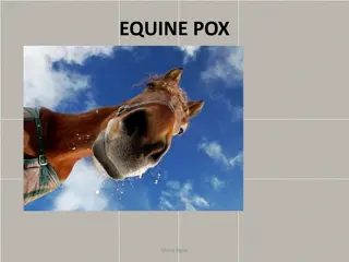
Equine Influenza: An Overview of Respiratory Tract Infections in Horses
Equine influenza is a contagious upper respiratory infection in horses caused by influenza viruses. This article covers the anatomy of the equine respiratory tract, signs, diagnosis, treatment, prevention, and prognosis of equine influenza. It also discusses the histological structure of the respiratory system and different types of infections such as granulomatous rhinitis and mycotic infections.

Download Presentation

Please find below an Image/Link to download the presentation.
The content on the website is provided AS IS for your information and personal use only. It may not be sold, licensed, or shared on other websites without obtaining consent from the author.If you encounter any issues during the download, it is possible that the publisher has removed the file from their server.
You are allowed to download the files provided on this website for personal or commercial use, subject to the condition that they are used lawfully. All files are the property of their respective owners.
The content on the website is provided AS IS for your information and personal use only. It may not be sold, licensed, or shared on other websites without obtaining consent from the author.
E N D
Presentation Transcript
Content: Introduction Anatomic and histological organization Definition Etiology Types Clinical signs Pathogenesis Diagnosis Treatment Prevention Prognosis Equine influenza Conclusions References
INTRODUCTION Equine influenza-epizootic disease of the equine upper respiratory tract.
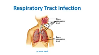
EQUINE RESPIRATORY TRACT To facilitate the understanding of the structure and function, it is convenient to arbitrarily divide the respiratory system into conducting, transitional, and gas exchange systems. Conductive system The conducting system includes nostrils, nasal cavity, paranasal sinuses, nasopharynx, larynx, trachea, and extrapulmonary and intrapulmonary bronchi, all of which are largely lined by pseudostratified, ciliated columnar cells, plus a variable proportion of secretory goblet (mucous) and serous cells. Transitional System The transitional system of the respiratory tract is composed of bronchioles,which are microscopic structures that serve as a transition zone between the conducting system (ciliated) and the gas exchange (alveolar) system Exchange System The gas exchange system of the respiratory tract in all mammals is formed by alveolar ducts and millions of alveoli. The surface of the alveoli is lined by two distinct types of epithelial cells known as type I (membranous) pneumonocytes and type II (granular) pneumonocytes .
EQUINE RESPIRATORY TRACT Airways From the Trachea to the Alveoli. Conducting, transitional, and exchange components of the respiratory system. The transitional zone(bronchioles) is not as equally well developed in all species.
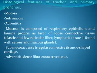
Nasal Mucosa, Granulomatous Rhinitis, Mycotic Infection, The granuloma is composed of three distinct layers: an outer layer of fibroblasts, lymphocytes, and plasma cells (asterisks); a middle thick layer of epithelioid macrophages (double-headed arrow); and a necrotic center of cellular debris containing a fungal hypha (arrow).
WHAT IS EQUINE INFLUENZA? Equine influenza is a common, highly contagious, and self-limiting upper respiratory infection of horses caused by aerogenous exposure to type A strains of influenza virus (H7N7[A/equi-1] and H3N8 [A/equi-2]). It occurs mainly in 2- to 3-year-old horses at the racetrack. As with human influenza, equine influenza is usually a mild disease, but occasionally it can cause severe bronchointerstitial pneumonia with pulmonary edema. In some horses, impaired defense mechanisms caused by the viral infection are complicated by a secondary bacterial bronchopneumonia caused by opportunistic organisms (Streptococcus zooepidemicus, Staphylococcus aureus, or Bacteroides sp.) found in the normal flora of the upper respiratory tract
WHAT CAUSES EQUINE INFLUENZA? There are several strains or subtypes of the equine influenza virus, but one of the most common flu viruses affecting horses is the Type A influenza. All flu viruses are airborne spreading from horse to horse, although some horses are only carriers of the virus, never showing any symptoms and appearing healthy. These horses, however, are still contagious. The virus acts destructively on the cells that line the upper respiratory tract as it replicates inside these cells.
ETIOLOGY OF EQUINE INFLUENZA Equine influenza A virus Equine influenza virus - an orthomyxovirus. Two main strains: H7N7 (formerly subtype 1) and H3N8 (formerly subtype2). There have been very few reports of H7N7 subtype virus infections in the last 20 years, H3N8 infections predominate. There are two main lineages of H3N8 viruses: American and Eurasian.
SYMPTOMS OF EQUINE INFLUENZA A horse with equine influenza has an abnormally high body temperature and nasal discharge, which is usually clear in color. Pneumonia is also a frequent secondary infection for horses with this condition, as their immune systems are compromised. This secondary infection can be deadly in foals. In severe cases of equine influenza, the horse may develop heart and liver complications. Other clinical signs include: fever (up to 106 F [41.1 C]) nasal discharge have a dry, hacking cough occasionally muscle pain occasionally enlarged lymph nodes loss of appetite and weakness distressed coat hair
HOW IS EQUINE INFLUENZA DIAGNOSED? The symptoms of equine influenza are usually enough to form at least a presumptive diagnosis, although signs of influenza are similar to other respiratory viral infections as well. This doesn t matter, as all viral respiratory diseases will be treated the same. Differential diagnosis : Equine rhinopneumonitis Equine viral arteritis Equine rhinovirus and adenovirus infection Pasteurellosis Strangles
HOW IS EQUINE INFLUENZA TREATED? There is no treatment to kill the virus itself, so management of this virus revolves around supportive care. Rest is of the utmost importance; at least six weeks is recommended in order to allow the damaged tissues from the illness to fully heal. It is also important that influenza infected horses be stabled in clean and well-ventilated areas. There is some evidence that the dust found in stables can affect horses suffering or recovering from equine influenza.
VACCINES Vaccines (ATCvet codes: 1.QI05AA01 (WHO) inactivated, 2.QI05AD02 (WHO) live, plus various combinations) are a major defense against the disease. Vaccination schedules generally require a primary course of vaccines, followed by booster shots. It is recommended that horses be vaccinated against equine influenza annually, and competition horses that travel frequently be given a booster every six months as they are at higher risk of contracting the virus. Foals are initially vaccinated at six months of age with a booster 3-6 weeks later and again between 10-12 months. Standard schedules may not maintain absolutely foolproof levels of protection, and more frequent administration is advised in high-risk situations.
PREVENTION OF EQUINE INFLUENZA Prevention of equine influenza outbreaks is maintained through vaccines and hygiene procedures.Isolation of horses for two weeks is common practice when they are being moved to a new environment. The only thing that has prevented the horse flu from becoming a full-blown epidemic has been an equine influenza vaccine. Horses that are at high-risk for contracting this virus, such as racehorses or show horses, should be vaccinated multiple times a year.
CONCLUSIONS Diseases of the respiratory systems have not changed much in the last few decades, but our understanding of many of the processes has progressed dramatically and many of the accepted dogmas were challenged and modified.
REFERENCES: James F. Zachary DVM PhD-Pathologic Basis of Veterinary Disease Expert Consult, 6e-Mosby (2016) Pathologic basis of veterinary disease. Fifth edition. James F. Zachary, M. Donald McGavin https://m.petmd.com/dog/conditions/digestive/cdginflammato ryboweldisease https://ahdc.vet.cornell.edu/docs/equine-influenza.pdf