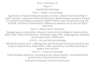
The Diversity of Natural Waters in Water Microbiology
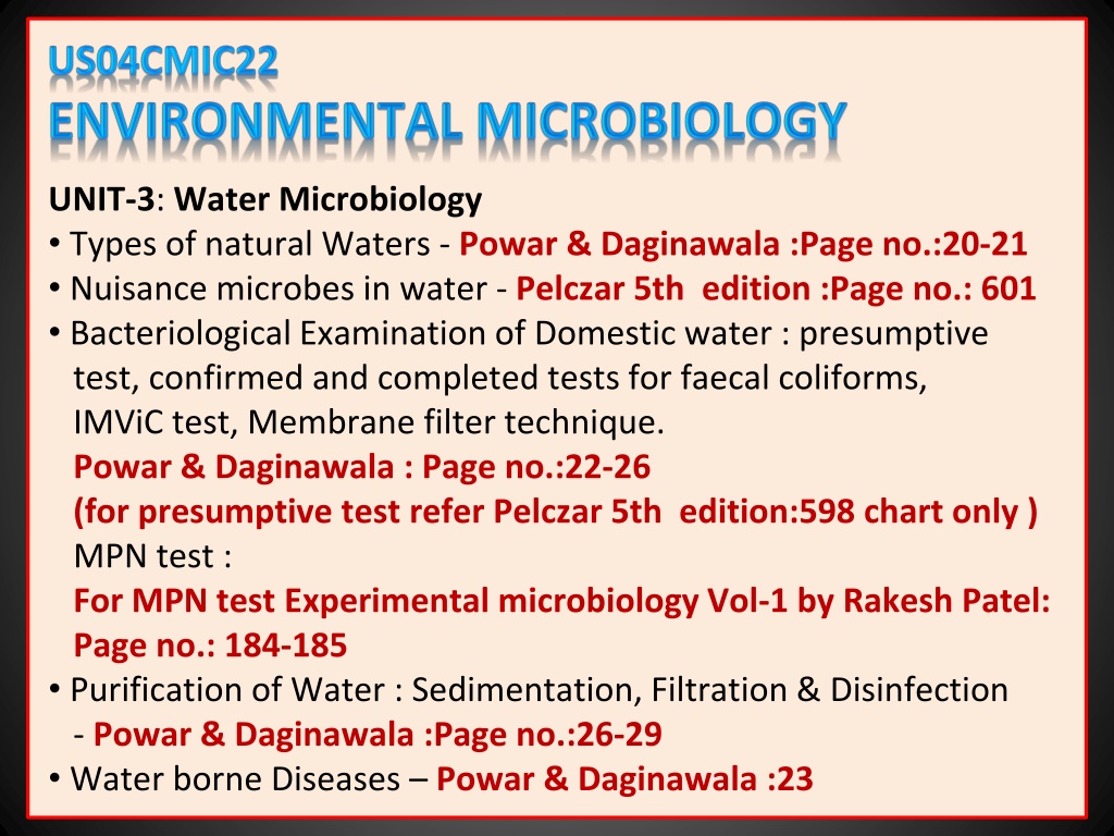
UNIT-3
:
Water Microbiology
•
Types of natural Waters -
Powar & Daginawala :Page no.:20-21
•
Nuisance microbes in water -
Pelczar 5th edition :Page no.: 601
•
Bacteriological Examination of Domestic water : presumptive
test, confirmed and completed tests for faecal coliforms,
IMViC test, Membrane filter technique.
Powar & Daginawala : Page no.:22-26
(for presumptive test refer Pelczar 5th edition:598 chart only )
MPN test :
For MPN test Experimental microbiology Vol-1 by Rakesh Patel:
Page no.: 184-185
•
Purification of Water : Sedimentation, Filtration & Disinfection
-
Powar & Daginawala :Page no.:26-29
•
Water borne Diseases –
Powar & Daginawala :23
Types of natural Waters
•
Water has curious and unusual properties, and plays an
important role in living system. This, "No life without water" is
a common saying It is a master solvent, and all metabolic
reactions of living organism depend on the presence of water.
•
Nearly three fourths of the earth surface is covered by water,
mainly ocean, and to lesser degree by rivers, lakes, and
streams.
•
This water is in continuous circulation, the process is known as
the water cycle or hydrologic cycle.
•
Water is lost from the earth by the way of evaporation,
transpiration and exhalation, and is returned to the earth by
the way of precipitation.
•
Microorganisms get into natural waters from air, soil, sewage,
organic wastes, dead plants and animals, etc. Thus almost any
type of organisms may be found in water.
NATURAL WATERS
Natural waters are commonly grouped into four classes:
(1) atmospheric waters
(2) surface waters
(3) stored waters and
(4) ground waters
(1)
Atmospheric waters
•
Rain, snow and hail which fall on land tend to carry down
particles of dust, soot, and other materials suspended in the
air.
•
These often bear bacteria and other microorganisms on
their surface.
•
The number of organisms depends upon local conditions. After
heavy rain or snow the atmosphere is washed free of organisms.
(2)
Surface waters
•
As soon as rain or snow reaches the earth and flows over the
soil, some of the soil organisms are gathered up by the water.
Bodies of water such as streams, rivers, and oceans represent
surface water.
•
Microbial populations depend upon their numbers in the soil
and, also, upon the kinds and quantities of food material
dissolved out of the soil by water.
•
Climatic, geographical and biological conditions bring about
great variations in microbial populations of surface waters.
•
Rivers and streams show their highest count during the rainy
period. Dust blowing into rivers and streams also contributes
many microorganisms.
•
Animals also make considerable contribution to the microbial
flora of the surface Waters. They bath and often drop their
excreta in the water.
(3)
Stored waters
•
Inland waters held in ponds, lakes, or reservoirs represent
stored waters.
•
Storage generally reduces the numbers of organisms in water.
•
A certain degree of purity and stability is established. Several
factors affect the microbial flora of stored waters.
These are as follows :
Sedimentation
Microorganisms have a specific gravity slightly greater than that
of water, and therefore slowly settle down. However, the most
important factor is their attachment to suspended particles.
Microorganisms are removed from the upper layers of the
water as the suspended particles settle down.
Activities of other organisms
Predatory Protozoa engulf living or dead bacteria for food,
provided the water contains sufficient dissolved oxygen.
Light rays
Direct sunlight is toxic to both vegetative cells and spores of
microorganisms. The toxicity of ultraviolet rays is inversely
proportional to the turbidity of water. In tropical countries direct
sun light is a very effective sterilizing agent.
Temperature
.
Temperature has variable effects. It may kill some organisms and
may stimulate the growth of others. During colder months the
multiplication rate of microorganisms is considerably reduced.
Food supply
If there is considerable vegetation or suspended food particles in
the body of water, it is likely to increase the number of
organisms. On the other hand, certain toxic substances may
bring about marked reduction in the number of organisms.
(4)
Ground waters
•
As water seeps through the earth, microorganisms as well as
suspended particles are removed by filtration in varying
degrees.
•
This depends on the permeability characteristics of the
soil and the depth to which the water penetrates.
•
Ground water brought to the surface by springs or deep wells
contains very few organisms.
•
To construct a well the nature of the soil and underlying porous
strata, the nature of the water table, and the nature, distance,
and direction of the local sources of pollution must be taken
into consideration.
Nuisance microbes in water
Microorganisms Other Than Coliform Bacteria
•
Some microorganisms besides coliform bacteria are of Intestinal
origin and could also be used as indicators of fecal contamination
of water, such as the fecal streptococci.
•
Other intestinal microorganisms, such as intestinal viruses are
frank pathogens and can cause serious diseases.
•
Still other microorganisms are regarded mainly as nuisance
organisms, because they create problems of odor, color, and
taste, or cause obstruction of water flow.
Fecal Streptococci
Fecal streptococci are enteric bacteria found in the intestines of
warm-blooded animals, including humans.
Streptococcus faecalis
is representative of this group: other species are
S. faecium
,
S.
bovis
and
S.equinus
. Because fecal streptococci, particularly,
S.
faecalis
, are abundantly present in the large intestines of humans,
their occurrence in water is indicative of fecal pollution.
Slime-Forming Bacteria
Many bacteria are capable of elaborating gummy or mucilaginous
materials, either as capsular structures or as extracellular excretion
products. The organic and inorganic constituents of the water,
which provide nutrients for the bacteria. help to determine
whether slime is produced and what organisms are responsible
for its production.
Iron Bacteria
The iron bacteria are one of the most important types of nuisance
organisms in water. They transform soluble compounds of iron to
insoluble compounds of iron (ferric hydroxide) which may be
deposited in a sheath around the organism (Sphaerotilus) or
secreted so as to form stalks or ribbons attached to the cell
(Gallionella). This deposition and accumulation of insoluble
material in the piping system may eventually have a significant
effect on the rate of water flow. Iron bacteria can also produce
slime, discolor water, and cause undesirable odors and tastes.
Sulfur Bacteria
Some of the sulfur bacteria are capable of producing and
tolerating extreme acidity, Organisms of the genus
Thiobacillus
oxidize elemental sulfur to sulfuric acid and can produce an acidity
in the range of pH 1; thus they may be responsible for the
corrosion of pipes.
Desulfovibrio desulfuricans
reduces sulfates and
other sulfur compounds to hydrogen sulfide.
Algae
When water is exposed to sunlight, algal growth often results; the
occurrence of algae in water is much like the growth of weeds in a
garden. Algae are present in all natural aquatic environments.
Their nuisance characteristics involve production of turbidity,
discoloration, odor, and taste in water. Algae are frequently the
primary cause for the clogging of filters during water purification.
The diatoms are the most important in this respect, although
green and yellow algae are also involved.
Aside from these
nuisance characteristics, some algae are capable of producing
substances toxic to humans and animals.
Viruses
Many viruses are known to be excreted from humans through the
intestinal tract, and these may find their way via sewage into
sources of drinking water. The enteroviruses are the ones most
commonly found in sewage; they include the polio, coxsackie, and
echo viruses. The virus that causes infectious hepatitis has been
isolated from polluted water and shellfish; occurrence of this
disease has been traced to these sources. Rotaviruses are also of
major importance. The possibility that virus diseases, particularly
the enteric virus diseases, may be waterborne indicates that
methods for evaluating the potability of a water supply from a
virological standpoint should be developed.
Considerable research is underway for the development of a
routine test method for the detection of viruses in water and
wastewater. At the same time more attention is being given to the
assessment of the effectiveness of water treatment processes for
the removal and/or inactivation of viruses.
Bacteriological Examination of Domestic water
:
(
Presumptive test, Confirmed and Completed tests for
faecal coliforms, IMViC test, Membrane filter technique
)
Introduction
:
•
Natural water supplies such as rivers, lakes, and streams contain
sufficient nutrients to support growth of various organisms.
•
Microorganisms enter the water supply in several different ways.
•
In congested centers water supplies get polluted by domestic and
industrial wastes.
•
As a potential carrier of pathogenic microorganisms, water can
endanger health and life.
•
From the standpoint of transmitting human diseases, polluting
waters with soil, rubbish, industrial wastes, and even animal
manure is comparatively harmless. These sources rarely contain
pathogens capable of producing human diseases when swallowed
with drinking water.
•
Sewage containing human excreta, however, is the most
dangerous material that pollutes water. People with
communicable diseases of many kinds eliminate the causative
organisms in their excreta.
•
The most important microbial diseases transmitted through
water are typhoid fever, paratyphoid fever, amoebic dysentery,
bacillary dysentery, cholera, tularem, poliomyelitis, and infectious
hepatitis.
•
Majority of bacteria found in water belong to the groups of
coliforms,
Pseudomonas
and
Proteus
group, plant pathogens, and
the spore formers of the genus
Bacillus
and
Clostridium
.
Potable water or drinking water
, is defined as the
”water which is free from pathogenic microorganisms and
chemicals that are deleterious to human health”
However, other factors such as taste. odor and color must be
absent if water is to be potable.
•
Water contaminated with either domestic or industrial waste is
called non-potable or polluted water.
•
To determine the potability of water quantitative bacteriological
examination may be undertaken. However, there is no single test
or, even combination of tests, that is wholly satisfactory, because
it will give only a fraction of the total count. Theoretically it
would be better to examine water for the presence of the
specific pathogenic microorganisms.
This is also impracticable because of the following reasons
:
1.
The methods are expensive, tedious, and slow, and by that
time the water has already been consumed.
2.
The number of pathogenic organisms may be quite small
compared to non-pathogenic organisms and would be
overlooked.
3. Non-pathogenic organisms may interfere with the examination
of pathogens.
•
The direct examination for pathogens, therefore, is not used in
routine water analysis.
•
Methods commonly used for the bacteriological examination of
water, are based on:
1.
The examination of presence or absence of the more common
organisms of intestinal or sewage origin.(
Qualitative method
)
2.
The approximate determination of total numbers of bacteria
present in the water sample.(
Quantitative method
)
MICROBIAL INDICATORS OF FAECAL CONTAMINATION
•
Drinking water should be free of any pathogenic organisms.
Hence, detection of a specific pathogen would constitute the
most direct evidence of faecal contamination. However, this is
impractical because of following reasons.
1. There are numerous gastrointestinal pathogens, each requiring
different method for isolation
2. Numbers of pathogens are less as compared to non-pathogens
and may escape detection.
3. Non-pathogenic organisms may interfere with examination of
pathogens.
•
Due to the above limitations, sanitary microbiologists use
indicator organisms as an index of possible contamination by
human pathogens.
•
Several organisms which are commonly found in intestinal tract
of humans and animals are considered as indicator organisms.
•
That includes faecal coliforms (Escherichia coli), faecal group D
streptococci (
Streptococcus faecalls
), and
Clostridium
perfringens
.
•
Recently, some other members of the anaerobic intestinal flora,
notably
Bifidobacterium
spp. have been proposed as an
additional indicator bacteria
COLIFORM BACTERIA
•
Coliforms are the members of the family Enterobacteriaceae, it
includes several genera like
Escherichia, Citrobacter, Klebsiella
and
Enterobacter
.
•
Coliforms are defined as facultatively anaerobic, gram-negative,
non sporing, rod shaped bacteria that ferment lactose with acid
and gas formation within 24-48 hours at 37 ̊C. Coliforms include
number of different organisms.
1. Those referred to as typical or faecal (
E.coli
) are commensal of
the intestine and are derived almost exclusively from this
habitat.
2. Others, known as atypical (
Enterobacter, Klebsiella
and
Citrobacter
) may also grow in soil and on vegetation and hence
may often be present in water which is not fecaly contaminated.
•
Thus in carrying out the test for coliform bacilli in water it is
necessary to determine whether the strains present are typical
or atypical.
Escherichia coli
as an indicator organism
•
Among so many indicator organisms, faecal coliforms i.e.
E.coli
has found the most wide spread use as an indicator organism
due to the following reasons.
1.
E.coli
is a normal flora of intestinal tract of healthy humans.
2.
E. coli
is excreted in large numbers in human faeces (~ 5 x 10
7
organisms/gram ). As a result there are high chances of
detecting the organism even after high dilutions.
3.
E.coli
are derived almost exclusively, from intestine of humans.
4. Survival rate of
E.coli
is longer than that of any gastro-intestinal
pathogen.
5. Isolation of
E.coli
is relatively very easy as well as it is non-
pathogenic and harmless.
TESTS FOR DIFFERENTIATION OF COLIFORMS
(1)
IMVIC test
As stated earlier it is essential to differentiate between typical
and atypical coliforms. The differentiation is based on the basis
of four biochemical tests, known as IMVIC tests.
The letter
I
stands for indole test,
M
for methyl red test.
V
for
Voges-Proskauer test and
C
for citrate test.
The letter i between V and C is added solely for euphony. On the
basis of these four tests, the coliforms are differentiated as follows.
Tests
E.coli
Enterobacter aerogens
Indol
+ -
Methyl red
+ -
Voges-Proskauer - +
Citrate
-
+
Test
E. coli
E.aerogenes
Indole Indole is produced Not produced(-)
from tryptophan (+)
Methyl Red Methyl red is turned red, Methyl red remains
which means a pH of yellow, which means
below4.5 is produced(+) less acid produced(-)
Voges-Proskauer Acetyl methyl carbinol Acetyl methyl
is not produced in carbinol is
glucose peptone produced(+)
medium(-)
Citrate Citrate as the sole Supports the
source carbon does growth(+)
not support growth(-)
(2)
Elevated temperature (Eijkman's) test
Another test used for the differentiation between typical and
atypical coliforms is on the basis of the growth at elevated
temperature.
E. coli
is able to grow at a temperature of 44 °C
while the growth of
En. aerogenes
is inhibited at this
temperature.
STANDARDS FOR DRINKING WATER QUALITY
(a)
Qualitative
:
The most stringent standards are imposed on
drinking water. According to the World Health Organization
(WHO), water should be condemned if it is found to contain
more than 10 coliforms or 1
E. coli
per 100 ml. Hence, if there
are 2
E. coli
per 100 ml water becomes unsuitable for drinking
purposes. Thus minimum amount of water sample needed for
analysis is 50 ml (which will theoretically contain at least 1
E.coli.
(b)
Quantitative
:
100 microorganisms / 1ml of water is suitable for
drinking purpose and more than 100 microorganisms/1ml is
unsuitable for drinking purposes.
BACTERIOLOGICAL ANALYSIS OF WATER
•
American Public Health Association (APHA) in their book, titled,
"Standard Methods for Examination of Water and Waste
Water", have described specific techniques for routine
bacteriological analysis of water.
•
All these methods are widely recognized and followed by all
the
sanitary agencies and health laboratories throughout the world.
•
Accordingly, the routine bacteriological procedures consist of:
1.
Standard plate count (SPC) or total viable count (TVC).
(
Quantitative
)
1.
Test for coliforms.
(Qualitative)
2.
Enumeration of coliforms.
(Qualitative)(MPN)
STANDARD PLATE COUNT (SPC) OR TOTAL VIABLE COUNT (TVC)
•
It is a quantitative bacteriological analysis which enumerates
total viable population capable of growing under a given set of
conditions.
•
Plate counts are useful in determining the efficiency of
water/waste water treatment. It is assumed that water of a
good quality properly treated) should give counts less than 100
colonies per milliliter.
Principle
TVC / SPC is based on the assumption that each viable bacterium
develops into a distinct colony. Hence, original number of
microorganisms in the sample can be calculated from number of
colonies and then multiplying it with aliquot factor.
Limitations of viable count
:
1.
There is not a single set of incubation conditions and a
medium composition that would permit growth of all
bacterial types.
2.
Several organisms if stuck together in a clump (in chains or in
clusters) will give rise to a single colony. Because of above
limitations it is not possible to be absolutely certain that each
colony arose from an individual cell, hence results are often
expressed in terms of colony forming units (CFU), rather than
the number of microorganisms.
Requirements
1.
Water sample.
2.
Sterile distilled water dilution tubes (4.5 ml or 9.0 ml).
3.
Sterile melted nutrient agar tubes.
4.
Sterile Petri dishes and sterile 1 ml pipettes.
Procedure
1.
Prepare 10
-1
,10
-2
and 10
-3
.... dilutions of the water sample (if
necessary).
2.
From each of the dilution transfer a fixed amount (e. 0.1 ml)
into sterile melted nutrient agar tube (previously cooled to
50 ̊C), mix it well and pour immediately in sterile Petri dishes.
3.
Label plates (e.g. as 10
-1
/ 0.1 or as the case may be), clearly
indicating the dilution and the volume plated respectively.
Incubate all plates at 37 ̊C for 24 hours.
4.
Count total number of colonies that has developed on each of
the plate. If necessary use colony counter to help counting of
colonies.
5.
Calculate final number of organisms present in the water
sample as follows.
Calculations & interpretation of results
1.
For accuracy of results, countable plates are those which have
colonies in between 30-300. Fewer than 30 colonies are not
acceptable for statistical reasons, and more than 300 colonies
on a plate is likely to produce colonies too close to each other
to be distinguished as individual CFUs.
2.
Lower dilutions (e.g. 10
-1
,10
-2
) at times may show confluent
(lawn) growth, due to high load of organisms in the sample,
these results are represented as "too numerous to count"
(TNTC).
3.
Final CFUs / ml can be calculated by multiplying the average
number of colonies per countable plate by the reciprocal of
the dilution and the reciprocal of the volume plated.
CFUs /ml
=
Average number of colonies
x Volume plated
Dilution
TEST FOR COLIFORMS
To
detect coliforms, a three stage procedure (the presumptive test,
confirmed test, and completed test) is carried out in systematic
order according to the results of each step.
PRESUMPTIVE TEST
Principle
It is based on the principle that coliforms if present in water, will
ferment lactose to produce acid and gas within 24-48 hours.
Production of acid is indicated by pH Indicator and gas is collected
in Durham's vial, both of which are present in the medium.
Media used are highly selective for coliforms, which inhibit growth
of gram-positive organisms. MacConkey's lactose bile broth
(MLBB) or laurel tryptose broth or brilliant green lactose bile broth
(BGLB) can be used for presumptive test.
Different volumes of water are inoculated in MLBB or BGLB which
permits growth of coliforms only. Coliforms will ferment lactose to
produce acid and gas within 24-48 hours and test is considered as
positive. Hence, it may be presumed that coliforms may be present
in water hence the name presumptive test.
Requirements
1.
Water sample.
2.
Sterile 1 ml and 10 ml pipettes
3.
5 MLBB tubes, each with 10 ml double strength medium (2X)
4.
3 MLBB tubes, each with 5 ml single strength medium (X).
Procedure
1.
Shake the water sample vigorously to ensure uniform
distribution of organisms.
2.
With sterile graduated pipettes inoculate the water sample as
follows.
•
5 MLBB double strength tubes with 10 ml water.
•
1 MLBB single strength tube with 1.0 ml water.
•
1 MLBB single strength tube with 0.1 ml water
3. One tube of MLBB single strength is not inoculated and hence,
serves as control.
4.
Incubate all tubes at 37 °C for 24 hours.
5.
Examine tubes for the presence of acid and gas after 24 hours.
6.
If no gas has formed, reincubate all the tubes for another 24
hours (total 48 hours).
7.
Record the presence or absence of acid & gas at each
examination, & interprete as follows:
Interpretations
1.
Absence of gas even after 48 hours indicate a negative test and the
presumptive test is terminated. The water sample is assumed to be potable.
2.
Presumptive test is considered positive if any one or more of tubes show acid
and gas. Positive tubes are retained for confirmed test.
Inoculate 9 Tubes
: 3 MLBB (
2X
) with 10 ml, 3 MLBB (
X
) with 1 ml and 3 MLBB (
X
) with 0.1 ml water sample
Incubate
all tubes
at 37 ̊C
for 24
hours
Negative
:
All tubes show no
color change and
absent of gas in
Durham's vial
Positive
:
Any or More tubes
show color change and
present of gas in
Durham's vial
Note
:
1.
Double strength broth: (contains double the concentration of ingredients except
water) is used when large volumes of water are to be inoculated, because the
medium would otherwise be too diluted and may not support the growth of
bacteria.
2.
False positive presumptive test may be produced due to:
(a) Presence of lactose fermenting organisms other than coliforms.
(b) A synergistic association where the joint action of two organisms on a
carbohydrate re suits into production of gas which will not be formed by either
species when grown separately. Synergism is frequently caused by a joint action of
gram-positive and gram-negative organisms growing together, e.g.
Staphylococcus
aureus
and
Proteus vulgaris
False positive presumptive test can be overcome by adding bile salts and
triphenylmethane dyes which inhibit growth of gram-positive bacteria and thereby
eliminate synergistic effect.
3. Over fermentation: this kind of results are obtained when ratio of number of
organisms to the amount sugar is comparatively very high. Due to less amount of
sugar, acid produced is not sufficient enough to lower the pH at which growth
organism is inhibited. Thus. growth continues and organisms switch over to
peptones when sugars are depleted. Since end products of protein metabolism are
alkaline in nature the basicity of medium increases. Hence, MLBB appears yellow
even though the test is positive
.
CONFIRMED TEST
This test is named so because, positive presumptive tubes having
acid and gas are subjected to further confirmation that positive
results were due to coliforms only. Test involves, streaking of
Eosin methylene blue (EMB) agar or Endo's agar plate and looking
for the growth of typical &/or atypical colonies of coliforms.
Requirements
1.
Positive presumptive tube(s).
2.
EMB agar plate, BGLB
Procedure
1.
Streak EMB agar plate with a loopful of suspension from a
positive presumptive tube (which shows the highest amount
of gas production), so as to get well isolated colonies.
2.
Inoculate one drop in BGLB.
3.
Incubate the plate at 37 ̊C for 24 hours,
4.
Record results and interprete them as follows.
Streak EMB
agar plate
Inoculate
BGLB
Incubate EMB agar
plate and BGLB at 37 ̊C
for 24 hours
BGLB show gas
production in
Durham's vial
indicate positive
confirm test
Growth with
greenish
metallic sheen
indicate positive
confirm test
Interpretations
EMB agar plate permits three types of colonies to develop:
1.
Typical
: Small nucleated with or without greenish metallic
sheen.
2.
Atypical
: large, opaque, pink, non-nucleated, mucoid which
tend to merge with each other.
3.
Negative
: all other types of colonies developing on the plate.
Growth of typical colonies indicate confirmed test positive and
has to proceed for completed test.
If only atypical colonies develop, the test can't be considered
negative since time, coliforms fail to form typical colonies, or
colonies develop slowly, Hence the test should be completed
However, if only negative (others) colonies develop on the
plate: the confirmed test is recorded as negative and further
tests are not necessary.
COMPLETED TEST
In this test the typical &/or atypical colonies growing on EMB agar
plate are subjected to morphological and biochemical verification
so as to prove that they are coliforms. Since this test completes
and finishes the presumptive test for coliform s referred to as
Completed test.
Requirements
1.
EMB/Endo's agar plate having typical/atypical colonies
isolated from positive presumptive tube.
2.
Lactose broth tube (other than the one used in presumptive
test e.g.. Brilliant green lactose bile broth (BGLB) or nutrient
lactose broth with Andrade's indicator and Durham's vial.
3. Nutrient agar slant.
Procedure
1.
Select and mark a well isolated typical atypical colony on EMB
or Endo's agar plate
2.
With the help of Nicrome wire loop, pick up half of the
previously marked typical / atypical colony and transfer it to
BGLB or nutrient lactose broth tube.
3.
From the remaining half of the same colony streak over the
surface of a nutrient agar slant.
4.
Incubate slant and broth at 37 ̊C for 24 hours.
5.
Check Lactose broth for presence of acid and gas.
6.
Prepare Gram's stain of the growth from the surface of agar
slant and observe the slide. Look for the presence of gram-
negative non-spore forming short rods.
7.
Record results and interprete as follows:
EMB with growth
showing greenish
metallic sheen
Inoculate
Lactose
broth
Streak N-agar
slant
Incubate Lactose
broth and N-agar
slant at 37 ̊C for 24
hours
Color change and
present of gas in
Durham's vial of
Lactose broth
indicate positive test
After 24 hours perform gram
staining from N agar slant. If
bacteria are gram negative
non spore forming and show
acid and gas in lactose broth
completed test is positive
Interpretation
1.
Presence of gram-negative, non-spore forming short rods
capable of producing acid and gas from lactose indicates the
completed test positive.
2.
Absence of gram-negative non-spore forming short rods and
the absence of acid and gas from the lactose constitutes a
negative completed test
Conclusion of presumptive test
Positive presumptive test indicates presence of coliforms in water
sample, which points out the faecal contamination of water,
hence
the water is non-potable, as it may carry potentially pathogenic
microorganisms, However, as stated earlier conforms include wide
range of bacteria whose primary source may not be the intestinal
tract of human beings: so further dirferentiation of coliforms is
recommended.
MULTIPLE TUBE (MOST PROBABLE NUMBER (MPN)
TECHNIQUE
Principle
It is a statistical method based on the probability theory. In this
technique, the sample is serially diluted till the number of
organisms reach the point of extinsion. From each of these dilutions
several multiple tubes of a specific medium are inoculated,
Presence of organism is indicated by acid or gas in the medium.
Pattern of positive and negative test results are then used to
estimate the number of bacteria in the original sample. Since the
test gives the most probable number of organisms present in the
sample it is also known as MPN test.
Requirements
1.
3 MLBB tubes each having 10 ml double strength (2x) medium,
2.
7 MLBB tubes each having 5 ml single strength (X) medium.
3.
Sterile 10 ml and I ml pipettes.
4.
Water sample to be tested.
Procedure
1.
Shake the water sample vigorously to ensure uniform
distribution of organisms.
2.
Dilute the sample if necessary.
3.
With the sterile graduated pipettes inoculate the water sample
(diluted sample, if the dilution is done) as follows.
(a) 3 Tubes of MLBB having I0 ml (2X) medium with 10 ml of
sample each.
(b) 3 Tubes of MLBB having 5 ml (X) medium with 1 ml of
sample each.
(c) 3 Tubes of MLBB having 5 ml (X) medium with 0.1 ml of
sample each
4. One tube of MLBB having 5 ml (x) medium is left uninoculated,
which serves as control.
5.
Incubate all tubes at 37 ̊C for 24 hours.
6.
Examine tubes for acid and gas after 24 hours.
7.
If no tube shows acid and gas reincubate all tubes for another
24 hours.
8.
At the end of the incubation period, record the number of
positive tubes in each of three sets (i.e. 10 ml, 1 ml and 0.1
ml). and interprete results as follows.
Interpretation
MeCrady in 1918 computed tables regarding the most probable
number of organisms present in 100 ml of water, on the basis of
various combinations of positive and negative results in the
amounts used for tests Number of organisms per 100 ml is read
from the McCrady's table, and the number is multiplied by the
dilution factor (if any), to come to the final number.
The Membrane Filter Method
•
A filtration technique for enumerating, coliform bacteria in water
was developed in Germany during World War II, and has been
accepted as a standard method.
•
The filtering apparatus is constructed of a glass or stainless steel
funnel and a suction flask.
•
A filter disk composed of cellulose derivative is placed in the
filtering apparatus. After the sterilized filter apparatus is
assembled, a volume of water is passed through the filter disk.
•
The bacteria from the water sample are retained on the
membrane filter. The filter disk is transferred with a sterile
forceps
to a sterile Petridis containing an absorbent pad saturated with an
appropriate medium.
•
The medium diffuses trough the pores of the membrane and
brings nutrient to the bacteria entrapped during filtration. Upon
incubation, colonies of organisms develop upon the filter disk and
can be easily counted.
The Membrane Filter Method
This method possesses distinct advantages, some of which are as
follows:
1.
It permits the examination of bacteria from a large volume of
water. No dilutions are, therefore, required. Colony count is
more accurate and reliable.
2.
The filter disc can be transferred to any appropriate medium
This permits the isolation of any organism on a differential
medium.
3.
It provides a more rapid examination of water than the
standard procedure.
4.
It requires much less equipment and therefore, the
examination can be done in the field.
•
This method cannot be used if water contains considerable
amount of algae, colloidal, or other materials which are likely
to
clog the filter Secondly, if water samples are heavily
contaminated with non coliforms, the growth of coliforms will
be inhibited.
PURIFICATION OF WATER
•
Various methods of water purification been have developed
which depend on the amount and character of water, that is
whether the water is for a single household or a town or city.
•
Water is purified to make it satisfactory in appearance, taste,
and odor as well as safe by removing harmful organisms.
•
Disinfection is the only treatment required for water from
properly constructed wells.
•
Municipal water supplies, however, require a number of
treatments. Three principal methods for the purification of water
are:
1.
Sedimentation
2.
Filtration and
3.
Disinfection.
Sedimentation
•
Water usually undergoes some degree of purification during
storage in ponds or reservoirs.
•
Suspended particles settle and carry down most of the
microorganisms.
•
The rate of purification by sedimentation depends upon the
kind and amount of suspended matter as the well as physical,
chemical and biological conditions of the stored water.
•
The rate of sedimentation is enhanced by adding alum, iron
salts, colloidal silicate etc. which produce flocculent
precipitates.
•
Microorganisms and suspended particles are entrapped and
settle rapidly.
•
Sometimes activated carbon is also added. This adsorbs the
compounds responsible for objectionable color and taste of
water.
•
Microorganisms remain viable for a considerable time, even
though visible evidence of pollution has disappeared.
•
Sedimentation, therefore, reduces the microbial population but
does not sterilize polluted water.
•
To produce potable water further treatment is necessary. Thus
sedimentation is often used as a first stage in purification.
.
Filtration
•
Filtration is an effective means of removing microorganisms and
other suspended matter from water.
•
Many waters require some type of filtration, because of turbidity
and color, presence of a large amount of organic matter, or
sewage pollution.
•
Two types of sand filters are used to purify the clarified water
after sedimentation.
Slow sand filter
•
Slow sand filtration plants require considerable area because the
rate of filtration is slow. A concrete floor with drainage tiles to
collect the filtered water is constructed. The tile is covered with
coarse gravel, fine gravel, coarse sand and finally 2 to 1 feet of
sand at the top.
•
Water seeps through the filter slowly, is collected by tile drain
pipes at the bottom, and is pumped into a reservoir. At best five
million gallons of water per acre, per day, can be filtered.
•
Slow sand filters are clogged by turbid water. Water to be
filtered is, therefore, clarified by sedimentation with or without
coagulation.
•
The purification of water is accomplished not by the screening
action of the sand, for the spaces are much to large, but by a
different principle.
•
A colloidal, flocculent material composed of bacteria, algae, and
Protozoa accumulates in the surface layers of fine sand. This
slimy, gelatinous film closes up the pores between the sand
grains and makes the filter bed more and more effective.
•
Since bacteria have a negative electrical charge and colloidal
material on the sand grains has a positive charge, bacteria are
thus adsorbed on the particles. Bacteria are also ingested by
Protozoa that inhabit the upper layer of the film.
Slow sand filter
Rapid sand filter
•
Rapid sand filters are constructed in a manner similar to that of
slow sand filters. They also consist of layers of sand, gravel, and
rock.
•
Water is pre-treated before filtration by a coagulant such as alum
or ferrous sulphate. The water passes through a settling tank in
which most of the precipitate settles out, and the remainder is
pumped on to the filter.
•
Rapid sand filters soon become clogged and are cleaned by
forcing cleaned water backward (back washed) through the bed
of gravel and sand, and bubbling air through them.
•
The back water rises through the filter and carries the
accumulated material to the sewer. The wash water is thus
wasted.
•
Care is taken in this backwashing procedure to see that the fine
sand on the surface is not lost. Rapid sand filters are usually
operated in batteries, so that some may be in operation while
others are being cleaned.
•
Metabolic activity of microorganisms also greatly reduces the
chemical content of the water. When the gelatinous film finally
become too thick, the efficiency of the filter gradually decreases.
•
The filter is taken out of service and the surface layer is
cleaned.
•
They are nearly as effective as slow sand filters but operate 50
times faster than slow sand filters, Rapid sand filters are capable
of delivering 150 to 200 million gallons of water per acre, per
day.
•
They require a much smaller area of land for more water
filtration and cost much less to install and maintain.
•
Many other filtration devices such as pressure filters, diatomite
filters, membrane filter, reverse osmosis etc., are employed to
remove various impurities in water.
•
Recovery of potable water from the sea and from domestic and
industrial sewage is also undertaken by the use of filtration
techniques.
Rapid sand filter
Disinfection
•
Water purified by sedimentation or filtration cannot be
considered safe for human consumption.
•
Disinfection of public water supply is a final step in water
purification before it reaches the consumer.
•
A number of chemicals have been recommended for the
disinfection of water supplies.
•
Solutions of calcium or sodium hypochlorite are satisfactory for
treating water in small towns. In recent years chlorination of the
public water supply is widely practiced.
•
Chlorine released as gas readily mixes with water. The amount
of
chlorine required depends on the organic matter present, more
chlorine being required if there are more bacteria, more organic
matter, and a shorter time to act.
•
The amount of chlorine taken up is termed chlorine demand.
•
The point at which the available chlorine becomes proportional
to the added chlorine is called the break point.
•
Water is usually treated to contain 0.1 to 0.2 parts per million of
residual chlorine. Residual chlorine is the available chlorine
remaining 20 minutes after its addition to the water.
•
An over dose of chlorine gives peculiar odors and tastes,
because of its action upon various compounds present in water.
Frequently it is due to the formation of chlorophenols.
•
At times chlorine action may be prolonged, particularly in
waters containing considerable organic matter, by the
simultaneous addition of liquid ammonia, with the formation of
chloramines.
•
Chlorine reacts with water to produce hypochlorous acid, which
in turn quickly decomposes and releases oxygen. This nascent
oxygen oxidizes cellular components and the organic matter.
•
Another gas, ozone behaves in a similar manner, as it also
releases oxygen.
•
Chlorine kills most of the microorganisms but does not kill
spores.
•
Chlorinated water is, therefore, not always sterile, but is usually
safe for human consumption.
•
In small communities, where cost is not an important factor,
chlorine is replaced by other purification agents.
•
Germicidal ultraviolet rays are used to disinfect water supplies.
•
Objectionable taste and odor which accompany chlorination are,
therefore, avoided by this process.
•
But the simplest and the best method to make water safe for
human consumption is to boil it for 10 minutes.
•
This practice is often recommended for household use during
floods or other disasters that disrupt the normal water
purification system.
Water borne Diseases
•
Microbial diseases transmitted through water are typhoid fever,
Paratyphoid fever, amoebic dysentery, bacillary dysentery,
cholera, tularemia, poliomyelitis, and infectious hepatitis.
.
.
Water is essential for life and plays a vital role in the ecosystem. This article explores the various types of natural waters, including atmospheric waters, surface waters, stored waters, and ground waters. Each type harbors different microbial populations influenced by factors such as climate, geography, and biological activities. The presence of microorganisms in water poses challenges, leading to the need for bacteriological examinations and purification techniques to prevent waterborne diseases.

Download Presentation

Please find below an Image/Link to download the presentation.
The content on the website is provided AS IS for your information and personal use only. It may not be sold, licensed, or shared on other websites without obtaining consent from the author.If you encounter any issues during the download, it is possible that the publisher has removed the file from their server.
You are allowed to download the files provided on this website for personal or commercial use, subject to the condition that they are used lawfully. All files are the property of their respective owners.
The content on the website is provided AS IS for your information and personal use only. It may not be sold, licensed, or shared on other websites without obtaining consent from the author.
E N D
Presentation Transcript
UNIT-3: Water Microbiology Types of natural Waters - Powar & Daginawala :Page no.:20-21 Nuisance microbes in water - Pelczar 5th edition :Page no.: 601 Bacteriological Examination of Domestic water : presumptive test, confirmed and completed tests for faecal coliforms, IMViC test, Membrane filter technique. Powar & Daginawala : Page no.:22-26 (for presumptive test refer Pelczar 5th edition:598 chart only ) MPN test : For MPN test Experimental microbiology Vol-1 by Rakesh Patel: Page no.: 184-185 Purification of Water : Sedimentation, Filtration & Disinfection - Powar & Daginawala :Page no.:26-29 Water borne Diseases Powar & Daginawala :23
Types of natural Waters Water has curious and unusual properties, and plays an important role in living system. This, "No life without water" is a common saying It is a master solvent, and all metabolic reactions of living organism depend on the presence of water. Nearly three fourths of the earth surface is covered by water, mainly ocean, and to lesser degree by rivers, lakes, and streams. This water is in continuous circulation, the process is known as the water cycle or hydrologic cycle. Water is lost from the earth by the way of evaporation, transpiration and exhalation, and is returned to the earth by the way of precipitation. Microorganisms get into natural waters from air, soil, sewage, organic wastes, dead plants and animals, etc. Thus almost any type of organisms may be found in water.
NATURAL WATERS Natural waters are commonly grouped into four classes: (1) atmospheric waters (2) surface waters (3) stored waters and (4) ground waters (1) Atmospheric waters Rain, snow and hail which fall on land tend to carry down particles of dust, soot, and other materials suspended in the air. These often bear bacteria and other microorganisms on their surface. The number of organisms depends upon local conditions. After heavy rain or snow the atmosphere is washed free of organisms.
(2) Surface waters As soon as rain or snow reaches the earth and flows over the soil, some of the soil organisms are gathered up by the water. Bodies of water such as streams, rivers, and oceans represent surface water. Microbial populations depend upon their numbers in the soil and, also, upon the kinds and quantities of food material dissolved out of the soil by water. Climatic, geographical and biological conditions bring about great variations in microbial populations of surface waters. Rivers and streams show their highest count during the rainy period. Dust blowing into rivers and streams also contributes many microorganisms. Animals also make considerable contribution to the microbial flora of the surface Waters. They bath and often drop their excreta in the water.
(3) Stored waters Inland waters held in ponds, lakes, or reservoirs represent stored waters. Storage generally reduces the numbers of organisms in water. A certain degree of purity and stability is established. Several factors affect the microbial flora of stored waters. These are as follows : Sedimentation Microorganisms have a specific gravity slightly greater than that of water, and therefore slowly settle down. However, the most important factor is their attachment to suspended particles. Microorganisms are removed from the upper layers of the water as the suspended particles settle down. Activities of other organisms Predatory Protozoa engulf living or dead bacteria for food, provided the water contains sufficient dissolved oxygen.
Light rays Direct sunlight is toxic to both vegetative cells and spores of microorganisms. The toxicity of ultraviolet rays is inversely proportional to the turbidity of water. In tropical countries direct sun light is a very effective sterilizing agent. Temperature. Temperature has variable effects. It may kill some organisms and may stimulate the growth of others. During colder months the multiplication rate of microorganisms is considerably reduced. Food supply If there is considerable vegetation or suspended food particles in the body of water, it is likely to increase the number of organisms. On the other hand, certain toxic substances may bring about marked reduction in the number of organisms.
(4) Ground waters As water seeps through the earth, microorganisms as well as suspended particles are removed by filtration in varying degrees. This depends on the permeability characteristics of the soil and the depth to which the water penetrates. Ground water brought to the surface by springs or deep wells contains very few organisms. To construct a well the nature of the soil and underlying porous strata, the nature of the water table, and the nature, distance, and direction of the local sources of pollution must be taken into consideration.
Nuisance microbes in water Microorganisms Other Than Coliform Bacteria Some microorganisms besides coliform bacteria are of Intestinal origin and could also be used as indicators of fecal contamination of water, such as the fecal streptococci. Other intestinal microorganisms, such as intestinal viruses are frank pathogens and can cause serious diseases. Still other microorganisms are regarded mainly as nuisance organisms, because they create problems of odor, color, and taste, or cause obstruction of water flow. Fecal Streptococci Fecal streptococci are enteric bacteria found in the intestines of warm-blooded animals, including humans. Streptococcus faecalis is representative of this group: other species are S. faecium, S. bovis and S.equinus. Because fecal streptococci, particularly, S. faecalis, are abundantly present in the large intestines of humans, their occurrence in water is indicative of fecal pollution.
Slime-Forming Bacteria Many bacteria are capable of elaborating gummy or mucilaginous materials, either as capsular structures or as extracellular excretion products. The organic and inorganic constituents of the water, which provide nutrients for the bacteria. help to determine whether slime is produced and what organisms are responsible for its production. Iron Bacteria The iron bacteria are one of the most important types of nuisance organisms in water. They transform soluble compounds of iron to insoluble compounds of iron (ferric hydroxide) which may be deposited in a sheath around the organism (Sphaerotilus) or secreted so as to form stalks or ribbons attached to the cell (Gallionella). This deposition and accumulation of insoluble material in the piping system may eventually have a significant effect on the rate of water flow. Iron bacteria can also produce slime, discolor water, and cause undesirable odors and tastes.
Sulfur Bacteria Some of the sulfur bacteria are capable of producing and tolerating extreme acidity, Organisms of the genus Thiobacillus oxidize elemental sulfur to sulfuric acid and can produce an acidity in the range of pH 1; thus they may be responsible for the corrosion of pipes. Desulfovibrio desulfuricans reduces sulfates other sulfur compounds to hydrogen sulfide. Algae When water is exposed to sunlight, algal growth often results; the occurrence of algae in water is much like the growth of weeds in a garden. Algae are present in all natural aquatic environments. Their nuisance characteristics involve production of turbidity, discoloration, odor, and taste in water. Algae are frequently the primary cause for the clogging of filters during water purification. The diatoms are the most important in this respect, although green and yellow algae are also involved. Aside from these nuisance characteristics, some algae are capable of producing substances toxic to humans and animals.
Viruses Many viruses are known to be excreted from humans through the intestinal tract, and these may find their way via sewage into sources of drinking water. The enteroviruses are the ones most commonly found in sewage; they include the polio, coxsackie, and echo viruses. The virus that causes infectious hepatitis has been isolated from polluted water and shellfish; occurrence of this disease has been traced to these sources. Rotaviruses are also of major importance. The possibility that virus diseases, particularly the enteric virus diseases, may be waterborne indicates that methods for evaluating the potability of a water supply from a virological standpoint should be developed. Considerable research is underway for the development of a routine test method for the detection of viruses in water and wastewater. At the same time more attention is being given to the assessment of the effectiveness of water treatment processes for the removal and/or inactivation of viruses.
Bacteriological Examination of Domestic water : (Presumptive test, Confirmed and Completed tests for faecal coliforms, IMViC test, Membrane filter technique) Introduction: Natural water supplies such as rivers, lakes, and streams contain sufficient nutrients to support growth of various organisms. Microorganisms enter the water supply in several different ways. In congested centers water supplies get polluted by domestic and industrial wastes. As a potential carrier of pathogenic microorganisms, water can endanger health and life. From the standpoint of transmitting human diseases, polluting waters with soil, rubbish, industrial wastes, and even animal manure is comparatively harmless. These sources rarely contain pathogens capable of producing human diseases when swallowed with drinking water.
Sewage containing human excreta, however, is the most dangerous material that pollutes water. People with communicable diseases of many kinds eliminate the causative organisms in their excreta. The most important microbial diseases transmitted through water are typhoid fever, paratyphoid fever, amoebic dysentery, bacillary dysentery, cholera, tularem, poliomyelitis, and infectious hepatitis. Majority of bacteria found in water belong to the groups of coliforms, Pseudomonas and Proteus group, plant pathogens, and the spore formers of the genus Bacillus and Clostridium. Potable water or drinking water, is defined as the water which is free from pathogenic microorganisms and chemicals that are deleterious to human health However, other factors such as taste. odor and color must be absent if water is to be potable.
Water contaminated with either domestic or industrial waste is called non-potable or polluted water. To determine the potability of water quantitative bacteriological examination may be undertaken. However, there is no single test or, even combination of tests, that is wholly satisfactory, because it will give only a fraction of the total count. Theoretically it would be better to examine water for the presence of the specific pathogenic microorganisms. This is also impracticable because of the following reasons : 1. The methods are expensive, tedious, and slow, and by that time the water has already been consumed. 2. The number of pathogenic organisms may be quite small compared to non-pathogenic organisms and would be overlooked. 3. Non-pathogenic organisms may interfere with the examination of pathogens.
The direct examination for pathogens, therefore, is not used in routine water analysis. Methods commonly used for the bacteriological examination of water, are based on: 1. The examination of presence or absence of the more common organisms of intestinal or sewage origin.(Qualitative method) 2. The approximate determination of total numbers of bacteria present in the water sample.(Quantitative method)
MICROBIAL INDICATORS OF FAECAL CONTAMINATION Hence, detection of a specific pathogen would constitute the most direct evidence of faecal contamination. However, this is impractical because of following reasons. Drinking water should be free of any pathogenic organisms. 1. There are numerous gastrointestinal pathogens, each requiring different method for isolation 2. Numbers of pathogens are less as compared to non-pathogens and may escape detection. 3. Non-pathogenic organisms may interfere with examination of pathogens. indicator organisms as an index of possible contamination by human pathogens. Due to the above limitations, sanitary microbiologists use
tract of humans and animals are considered as indicator organisms. That includes faecal coliforms (Escherichia coli), faecal group D streptococci (Streptococcus faecalls), and Clostridium perfringens. Recently, some other members of the anaerobic intestinal flora, notably Bifidobacterium spp. have been proposed as an additional indicator bacteria Several organisms which are commonly found in intestinal
COLIFORM BACTERIA Coliforms are the members of the family Enterobacteriaceae, it includes several genera like Escherichia, Citrobacter, Klebsiella and Enterobacter. Coliforms are defined as facultatively anaerobic, gram-negative, non sporing, rod shaped bacteria that ferment lactose with acid and gas formation within 24-48 hours at 37 C. Coliforms include number of different organisms. 1. Those referred to as typical or faecal (E.coli) are commensal of the intestine and are derived almost exclusively from this habitat. 2. Others, known as atypical (Enterobacter, Klebsiella and Citrobacter) may also grow in soil and on vegetation and hence may often be present in water which is not fecaly contaminated. Thus in carrying out the test for coliform bacilli in water it is necessary to determine whether the strains present are typical or atypical.
Escherichia coli as an indicator organism Among so many indicator organisms, faecal coliforms i.e. E.coli has found the most wide spread use as an indicator organism due to the following reasons. 1. E.coli is a normal flora of intestinal tract of healthy humans. 2. E. coliis excreted in large numbers in human faeces (~ 5 x 10⁷ organisms/gram ). As a result there are high chances of detecting the organism even after high dilutions. 3.E.coli are derived almost exclusively, from intestine of humans. 4. Survival rate of E.coli is longer than that of any gastro-intestinal pathogen. 5. Isolation of E.coli is relatively very easy as well as it is non- pathogenic and harmless.
TESTS FOR DIFFERENTIATION OF COLIFORMS (1) IMVIC test As stated earlier it is essential to differentiate between typical and atypical coliforms. The differentiation is based on the basis of four biochemical tests, known as IMVIC tests. The letter I stands for indole test, M for methyl red test. V for Voges-Proskauer test and C for citrate test. The letter i between V and C is added solely for euphony. On the basis of these four tests, the coliforms are differentiated as follows. Tests E.coli Enterobacter aerogens Indol + - Methyl red + - Voges-Proskauer - + Citrate - +
Test E. coli E.aerogenes Indole Indole is produced Not produced(-) from tryptophan (+) Methyl Red Methyl red is turned red, Methyl red remains which means a pH of yellow, which means below4.5 is produced(+) less acid produced(-) Voges-Proskauer Acetyl methyl carbinol Acetyl methyl is not produced in carbinol is glucose peptone produced(+) medium(-) Citrate Citrate as the sole Supports the source carbon does growth(+) not support growth(-)
(2)Elevated temperature (Eijkman's) test Another test used for the differentiation between typical and atypical coliforms is on the basis of the growth at elevated temperature. E. coli is able to grow at a temperature of 44 C while the growth of En. aerogenes is inhibited at this temperature. STANDARDS FOR DRINKING WATER QUALITY (a) Qualitative: The most stringent standards are imposed on drinking water. According to the World Health Organization (WHO), water should be condemned if it is found to contain more than 10 coliforms or 1 E. coli per 100 ml. Hence, if there are 2 E. coli per 100 ml water becomes unsuitable for drinking purposes. Thus minimum amount of water sample needed for analysis is 50 ml (which will theoretically contain at least 1 E.coli. (b) Quantitative: 100 microorganisms / 1ml of water is suitable for drinking purpose and more than 100 microorganisms/1ml is unsuitable for drinking purposes.
BACTERIOLOGICAL ANALYSIS OF WATER American Public Health Association (APHA) in their book, titled, "Standard Methods for Examination of Water and Waste Water", have described specific techniques for routine bacteriological analysis of water. All these methods are widely recognized and followed by all the sanitary agencies and health laboratories throughout the world. Accordingly, the routine bacteriological procedures consist of: 1. Standard plate count (SPC) or total viable count (TVC). (Quantitative) 1. Test for coliforms. (Qualitative) 2. Enumeration of coliforms. (Qualitative)(MPN)
STANDARD PLATE COUNT (SPC) OR TOTAL VIABLE COUNT (TVC) It is a quantitative bacteriological analysis which enumerates total viable population capable of growing under a given set of conditions. Plate counts are useful in determining the efficiency of water/waste water treatment. It is assumed that water of a good quality properly treated) should give counts less than 100 colonies per milliliter. Principle TVC / SPC is based on the assumption that each viable bacterium develops into a distinct colony. Hence, original number of microorganisms in the sample can be calculated from number of colonies and then multiplying it with aliquot factor.
Limitations of viable count: 1. There is not a single set of incubation conditions and a medium composition that would permit growth of all bacterial types. 2. Several organisms if stuck together in a clump (in chains or in clusters) will give rise to a single colony. Because of above limitations it is not possible to be absolutely certain that each colony arose from an individual cell, hence results are often expressed in terms of colony forming units (CFU), rather than the number of microorganisms. Requirements 1. Water sample. 2. Sterile distilled water dilution tubes (4.5 ml or 9.0 ml). 3. Sterile melted nutrient agar tubes. 4. Sterile Petri dishes and sterile 1 ml pipettes.
Procedure 1. Prepare 10-1,10-2 and 10-3.... dilutions of the water sample (if necessary). 2. From each of the dilution transfer a fixed amount (e. 0.1 ml) into sterile melted nutrient agar tube (previously cooled to 50 C), mix it well and pour immediately in sterile Petri dishes. 3. Label plates (e.g. as 10-1 / 0.1 or as the case may be), clearly indicating the dilution and the volume plated respectively. Incubate all plates at 37 C for 24 hours. 4. Count total number of colonies that has developed on each of the plate. If necessary use colony counter to help counting of colonies. 5. Calculate final number of organisms present in the water sample as follows.
Calculations & interpretation of results 1. For accuracy of results, countable plates are those which have colonies in between 30-300. Fewer than 30 colonies are not acceptable for statistical reasons, and more than 300 colonies on a plate is likely to produce colonies too close to each other to be distinguished as individual CFUs. 2. Lower dilutions (e.g. 10-1,10-2) at times may show confluent (lawn) growth, due to high load of organisms in the sample, these results are represented as "too numerous to count" (TNTC). 3. Final CFUs / ml can be calculated by multiplying the average number of colonies per countable plate by the reciprocal of the dilution and the reciprocal of the volume plated. CFUs /ml = Average number of colonies x Volume plated Dilution
TEST FOR COLIFORMS Todetect coliforms, a three stage procedure (the presumptive test, confirmed test, and completed test) is carried out in systematic order according to the results of each step. PRESUMPTIVE TEST Principle It is based on the principle that coliforms if present in water, will ferment lactose to produce acid and gas within 24-48 hours. Production of acid is indicated by pH Indicator and gas is collected in Durham's vial, both of which are present in the medium. Media used are highly selective for coliforms, which inhibit growth of gram-positive organisms. MacConkey's lactose bile broth (MLBB) or laurel tryptose broth or brilliant green lactose bile broth (BGLB) can be used for presumptive test.
Different volumes of water are inoculated in MLBB or BGLB which permits growth of coliforms only. Coliforms will ferment lactose to produce acid and gas within 24-48 hours and test is considered as positive. Hence, it may be presumed that coliforms may be present in water hence the name presumptive test. Requirements 1. Water sample. 2. Sterile 1 ml and 10 ml pipettes 3. 5 MLBB tubes, each with 10 ml double strength medium (2X) 4. 3 MLBB tubes, each with 5 ml single strength medium (X). Procedure 1. Shake the water sample vigorously to ensure uniform distribution of organisms. 2. With sterile graduated pipettes inoculate the water sample as follows.
5 MLBB double strength tubes with 10 ml water. 1 MLBB single strength tube with 1.0 ml water. 1 MLBB single strength tube with 0.1 ml water 3. One tube of MLBB single strength is not inoculated and hence, serves as control. 4. Incubate all tubes at 37 C for 24 hours. 5. Examine tubes for the presence of acid and gas after 24 hours. 6. If no gas has formed, reincubate all the tubes for another 24 hours (total 48 hours). 7. Record the presence or absence of acid & gas at each examination, & interprete as follows: Interpretations 1. Absence of gas even after 48 hours indicate a negative test and the presumptive test is terminated. The water sample is assumed to be potable. 2. Presumptive test is considered positive if any one or more of tubes show acid and gas. Positive tubes are retained for confirmed test.
Flow chart of Presumptive Test Inoculate 9 Tubes : 3 MLBB (2X) with 10 ml, 3 MLBB (X) with 1 ml and 3 MLBB (X) with 0.1 ml water sample Incubate all tubes at 37 C for 24 hours Negative: All tubes show no color change and absent of gas in Durham's vial Positive: Any or More tubes show color change and present of gas in Durham's vial
Note: 1. Double strength broth: (contains double the concentration of ingredients except water) is used when large volumes of water are to be inoculated, because the medium would otherwise be too diluted and may not support the growth of bacteria. 2. False positive presumptive test may be produced due to: (a) Presence of lactose fermenting organisms other than coliforms. (b) A synergistic association where the joint action of two organisms on a carbohydrate re suits into production of gas which will not be formed by either species when grown separately. Synergism is frequently caused by a joint action of gram-positive and gram-negative organisms growing together, e.g. Staphylococcus aureus and Proteus vulgaris False positive presumptive test can be overcome by adding bile salts and triphenylmethane dyes which inhibit growth of gram-positive bacteria and thereby eliminate synergistic effect. 3. Over fermentation: this kind of results are obtained when ratio of number of organisms to the amount sugar is comparatively very high. Due to less amount of sugar, acid produced is not sufficient enough to lower the pH at which growth organism is inhibited. Thus. growth continues and organisms switch over to peptones when sugars are depleted. Since end products of protein metabolism are alkaline in nature the basicity of medium increases. Hence, MLBB appears yellow even though the test is positive.
CONFIRMED TEST This test is named so because, positive presumptive tubes having acid and gas are subjected to further confirmation that positive results were due to coliforms only. Test involves, streaking of Eosin methylene blue (EMB) agar or Endo's agar plate and looking for the growth of typical &/or atypical colonies of coliforms. Requirements 1. Positive presumptive tube(s). 2. EMB agar plate, BGLB Procedure 1. Streak EMB agar plate with a loopful of suspension from a positive presumptive tube (which shows the highest amount of gas production), so as to get well isolated colonies. 2. Inoculate one drop in BGLB. 3. Incubate the plate at 37 C for 24 hours, 4. Record results and interprete them as follows.
Flow chart of Confirm test Streak EMB agar plate Inoculate BGLB Incubate EMB agar plate and BGLB at 37 C for 24 hours Growth with greenish metallic sheen indicate positive confirm test BGLB show gas production in Durham's vial indicate positive confirm test
Interpretations EMB agar plate permits three types of colonies to develop: 1. Typical: Small nucleated with or without greenish metallic sheen. 2. Atypical: large, opaque, pink, non-nucleated, mucoid which tend to merge with each other. 3. Negative: all other types of colonies developing on the plate. Growth of typical colonies indicate confirmed test positive and has to proceed for completed test. If only atypical colonies develop, the test can't be considered negative since time, coliforms fail to form typical colonies, or colonies develop slowly, Hence the test should be completed However, if only negative (others) colonies develop on the plate: the confirmed test is recorded as negative and further tests are not necessary.
COMPLETED TEST In this test the typical &/or atypical colonies growing on EMB agar plate are subjected to morphological and biochemical verification so as to prove that they are coliforms. Since this test completes and finishes the presumptive test for coliform s referred to as Completed test. Requirements 1. EMB/Endo's agar plate having typical/atypical colonies isolated from positive presumptive tube. 2. Lactose broth tube (other than the one used in presumptive test e.g.. Brilliant green lactose bile broth (BGLB) or nutrient lactose broth with Andrade's indicator and Durham's vial. 3. Nutrient agar slant.
Procedure 1. Select and mark a well isolated typical atypical colony on EMB or Endo's agar plate 2. With the help of Nicrome wire loop, pick up half of the previously marked typical / atypical colony and transfer it to BGLB or nutrient lactose broth tube. 3. From the remaining half of the same colony streak over the surface of a nutrient agar slant. 4. Incubate slant and broth at 37 C for 24 hours. 5. Check Lactose broth for presence of acid and gas. 6. Prepare Gram's stain of the growth from the surface of agar slant and observe the slide. Look for the presence of gram- negative non-spore forming short rods. 7. Record results and interprete as follows:
Flow chart of Completed test EMB with growth showing greenish metallic sheen Inoculate Lactose broth Streak N-agar slant Incubate Lactose broth and N-agar slant at 37 C for 24 hours After 24 hours perform gram staining from N agar slant. If bacteria are gram negative non spore forming and show acid and gas in lactose broth completed test is positive Color change and present of gas in Durham's vial of Lactose broth indicate positive test
Interpretation 1. Presence of gram-negative, non-spore forming short rods capable of producing acid and gas from lactose indicates the completed test positive. 2. Absence of gram-negative non-spore forming short rods and the absence of acid and gas from the lactose constitutes a negative completed test Conclusion of presumptive test Positive presumptive test indicates presence of coliforms in water sample, which points out the faecal contamination of water, hence the water is non-potable, as it may carry potentially pathogenic microorganisms, However, as stated earlier conforms include wide range of bacteria whose primary source may not be the intestinal tract of human beings: so further dirferentiation of coliforms is recommended.
MULTIPLE TUBE (MOST PROBABLE NUMBER (MPN)TECHNIQUE Principle It is a statistical method based on the probability theory. In this technique, the sample is serially diluted till the number of organisms reach the point of extinsion. From each of these dilutions several multiple tubes of a specific medium are inoculated, Presence of organism is indicated by acid or gas in the medium. Pattern of positive and negative test results are then used to estimate the number of bacteria in the original sample. Since the test gives the most probable number of organisms present in the sample it is also known as MPN test. Requirements 1. 3 MLBB tubes each having 10 ml double strength (2x) medium, 2. 7 MLBB tubes each having 5 ml single strength (X) medium. 3. Sterile 10 ml and I ml pipettes. 4. Water sample to be tested.
Procedure 1. Shake the water sample vigorously to ensure uniform distribution of organisms. 2. Dilute the sample if necessary. 3. With the sterile graduated pipettes inoculate the water sample (diluted sample, if the dilution is done) as follows. (a) 3 Tubes of MLBB having I0 ml (2X) medium with 10 ml of sample each. (b) 3 Tubes of MLBB having 5 ml (X) medium with 1 ml of sample each. (c) 3 Tubes of MLBB having 5 ml (X) medium with 0.1 ml of sample each 4. One tube of MLBB having 5 ml (x) medium is left uninoculated, which serves as control. 5. Incubate all tubes at 37 C for 24 hours. 6. Examine tubes for acid and gas after 24 hours.
7. If no tube shows acid and gas reincubate all tubes for another 24 hours. 8. At the end of the incubation period, record the number of positive tubes in each of three sets (i.e. 10 ml, 1 ml and 0.1 ml). and interprete results as follows. Interpretation MeCrady in 1918 computed tables regarding the most probable number of organisms present in 100 ml of water, on the basis of various combinations of positive and negative results in the amounts used for tests Number of organisms per 100 ml is read from the McCrady's table, and the number is multiplied by the dilution factor (if any), to come to the final number.
The Membrane Filter Method A filtration technique for enumerating, coliform bacteria in water was developed in Germany during World War II, and has been accepted as a standard method. The filtering apparatus is constructed of a glass or stainless steel funnel and a suction flask. A filter disk composed of cellulose derivative is placed in the filtering apparatus. After the sterilized filter apparatus is assembled, a volume of water is passed through the filter disk. The bacteria from the water sample are retained on the membrane filter. The filter disk is transferred with a sterile forceps to a sterile Petridis containing an absorbent pad saturated with an appropriate medium. The medium diffuses trough the pores of the membrane and brings nutrient to the bacteria entrapped during filtration. Upon incubation, colonies of organisms develop upon the filter disk and can be easily counted.
This method possesses distinct advantages, some of which are as follows: 1. It permits the examination of bacteria from a large volume of water. No dilutions are, therefore, required. Colony count is more accurate and reliable. 2. The filter disc can be transferred to any appropriate medium This permits the isolation of any organism on a differential medium. 3. It provides a more rapid examination of water than the standard procedure. 4. It requires much less equipment and therefore, the examination can be done in the field. This method cannot be used if water contains considerable amount of algae, colloidal, or other materials which are likely to clog the filter Secondly, if water samples are heavily contaminated with non coliforms, the growth of coliforms will be inhibited.
PURIFICATION OF WATER Various methods of water purification been have developed which depend on the amount and character of water, that is whether the water is for a single household or a town or city. Water is purified to make it satisfactory in appearance, taste, and odor as well as safe by removing harmful organisms. Disinfection is the only treatment required for water from properly constructed wells. Municipal water supplies, however, require a number of treatments. Three principal methods for the purification of water are: 1. Sedimentation 2. Filtration and 3. Disinfection.
Sedimentation Water usually undergoes some degree of purification during storage in ponds or reservoirs. Suspended particles settle and carry down most of the microorganisms. The rate of purification by sedimentation depends upon the kind and amount of suspended matter as the well as physical, chemical and biological conditions of the stored water. The rate of sedimentation is enhanced by adding alum, iron salts, colloidal silicate etc. which produce flocculent precipitates. Microorganisms and suspended particles are entrapped and settle rapidly. Sometimes activated carbon is also added. This adsorbs the compounds responsible for objectionable color and taste of water.
Microorganisms remain viable for a considerable time, even though visible evidence of pollution has disappeared. Sedimentation, therefore, reduces the microbial population but does not sterilize polluted water. To produce potable water further treatment is necessary. Thus sedimentation is often used as a first stage in purification..


 Surface waters")
 Stored waters")

 Ground waters")


Elevated temperature (Eijkman's) test")

 OR TOTAL VIABLE COUNT (TVC)")


TECHNIQUE")