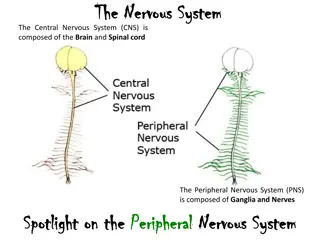
The Central Nervous System

I MSC -V-UNIT
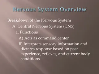
NERVOUS SYSTEM –BRAIM AND SPINAL CORD
Dr.M.Deivanayaki

The central
nervous system
(
CNS
) controls most functions of the body and mind. It
consists of two parts: the
brain
and the
spinal cord
. The
brain
is the center of our
thoughts, the interpreter of our external environment, and the origin of control over
body movement.
The
central nervous system
(
CNS
) is the part of the
consisting
primarily of the
and
. The CNS is so named because it integrates the
received information and coordinates and influences the activity of all parts of the
bodies of
—bilaterally symmetric animalsspinal cordbrainnervous system
The brain and spinal cord
The brain is a complex organ made up of specialized nerve and supportive tissues.
It’s surrounded by many bones that together form the skull. The part of the skull
where the brain sits is called the cranium. The base, or lower part, of the brain is
connected to the spinal cord. Together, the brain and spinal cord are known as the
central nervous system (CNS). Many nerves send electrical signals to and from the
brain and spinal cord.
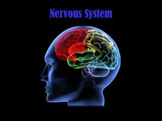
Structure and function of the brain
The brain is the body’s control centre. It constantly receives and interprets nerve
signals from the body and sends new signals based on this information. Different
parts of the brain control movement, speech, emotions, consciousness and internal
body functions, such as heart rate, breathing and body temperature.
Types of cells in the brain
The brain is made up of 2 main types of cells:
Nerve cells
(neurons) are cells that carry the electrical signals that make the nervous system work. They cannot be replaced or repaired if they are damaged. They are the longest cells in the body.
Glial cells
(neuroglial cells) are cells that support, feed and protect the nerve cells. The different types of glial cells are:
•
astrocytes
•
oligodendrocytes
•
ependymal cells
•
microglial cells
Brain regions
Next, we will look at some specific brain regions in a little more detail:
Basal ganglia:
involved in the control of voluntary motor movements, procedural learning, and decisions about which motor activities to
carry out. Diseases that affect this area include
Parkinson’s disease
and
Huntington’s disease
.
Cerebellum:
mostly involved in precise motor control, but also in language and attention. If the cerebellum is damaged, the primary
symptom is disrupted motor control, known as ataxia.
Broca’s area:
this small area on the left side of the brain (sometimes on the right in left-handed individuals) is important in language
processing. When damaged, an individual finds it difficult to speak but can still understand speech. Stuttering is
sometimes associated
with
an underactive Broca’s area.
Corpus callosum:
a broad band of nerve fibers that join the left and right hemispheres. It is the largest white matter structure in the brain
and allows the two hemispheres to communicate.
Dyslexic
children have smaller corpus callosums; left-handed people, ambidextrous
people, and musicians typically have
larger ones
.
Medulla oblongata:
extending below the skull, it is involved in involuntary functions, such as vomiting, breathing, sneezing, and maintaining
the correct
blood pressure
.
Hypothalamus:
sitting just above the brain stem and roughly the size of an almond, the hypothalamus secretes a number of neurohormones
and influences body temperature control, thirst, and hunger.
Thalamus:
positioned in the center of the brain, the thalamus receives sensory and motor input and relays it to the rest of the cerebral
cortex. It is involved in the regulation of consciousness, sleep, awareness, and alertness.
Amygdala:
two almond-shaped nuclei deep within the temporal lobe.
Amygdala:
two almond-shaped nuclei deep within the temporal lobe. They are involved in decision-making, memory, and emotional
responses; particularly negative emotions.
Spinal cord
The spinal cord, running almost the full length of the back, carries information between the brain and body, but also carries out
other tasks.
From the brainstem, where the spinal cord meets the brain, 31 spinal nerves enter the cord.
Along its length, it connects with the nerves of the peripheral nervous system (PNS) that run in from the skin, muscles, and joints.
Motor commands from the brain travel from the spine to the muscles and sensory information travels from the sensory tissues — such as
the skin — toward the spinal cord and finally up to the brain.
The spinal cord contains circuits that control certain reflexive responses, such as the involuntary movement your arm might make if your
finger was to touch a flame.
The circuits within the spine can also generate more complex movements such as walking. Even without input from the brain, the spinal
nerves can coordinate all of the muscles necessary to walk. For instance, if the brain of a cat is separated from its spine so that its brain has
no contact with its body, it will start spontaneously walking when placed on a treadmill.
The brain is only required
to stop and start the
process, or make changes if, for instance, an object appears in your path.
Central glial cells
Also called neuroglia, glial cells are often called support cells for neurons. In the brain,
they outnumber nerve cells 10 to 1.
Without glial cells, developing nerves often lose their way and struggle to form
functioning synapses.
Glial cells are found in both the CNS and PNS but each system has different types. The
following are brief descriptions of the CNS glial cell types:
Astrocytes:
these cells have numerous projections and anchor neurons to their blood
supply. They also regulate the local environment by removing excess ions and recycling
neurotransmitters.
Oligodendrocytes:
responsible for creating the myelin sheath — this thin layer coats
nerve cells, allowing them to send signals quickly and efficiently.
Ependymal cells:
lining the spinal cord and the brain’s ventricles (fluid-filled spaces),
these create and secrete cerebrospinal fluid (CSF) and keep it circulating using their
whip-like cilia.
Radial glia:
act as scaffolding for new nerve cells during the creation of the embryo’s
nervous system.
Visceral
afferent neurons are unipolar neurons that enter the
spinal cord
through
the dorsal root & their cell bodies are located in the dorsal root
ganglia.
Visceral
efferent neurons are motor neurons that conduct impulses to
smooth muscle, cardiac muscle, & glands. These neurons make up the
Autonomic
Nervous System
.
The
viscera
l
(or autonomic) motor
system
controls involuntary functions mediated
by the activity of smooth muscle fibers, cardiac muscle fibers, and glands. autonomic
(
visceral
motor) division of
nervous system
[TA] that part of the
nervous system
that represents the motor innervation of
smooth muscle, cardiac muscle, and gland cells. It consists of two physiologically and
anatomically distinct, mutually antagonistic components: the sympathetic and
parasympathetic parts.
A
spinal nerve
is a mixed
nerve
, which carries motor, sensory, and autonomic signals
between the
spinal
cord and the body. In the human body there are 31 pairs
of
spinal nerves
, one on each side of the vertebral column.
The
cranial nerves
are considered components of the
peripheral nervous
system
(
PNS
), although on a structural level the olfactory (I), optic (II), and
trigeminal (V)
nerves
are more accurately considered part of the central
nervous
system
(CNS).
spinal nerve
The central nervous system, comprising the brain and spinal cord, controls various body and mind functions through intricate neural pathways. Explore the brain regions, such as the basal ganglia and cerebellum, and learn about oligodendrocytes, ependymal cells, and microglial cells.

Download Presentation

Please find below an Image/Link to download the presentation.
The content on the website is provided AS IS for your information and personal use only. It may not be sold, licensed, or shared on other websites without obtaining consent from the author.If you encounter any issues during the download, it is possible that the publisher has removed the file from their server.
You are allowed to download the files provided on this website for personal or commercial use, subject to the condition that they are used lawfully. All files are the property of their respective owners.
The content on the website is provided AS IS for your information and personal use only. It may not be sold, licensed, or shared on other websites without obtaining consent from the author.
E N D
Presentation Transcript
I MSC -V-UNIT NERVOUS SYSTEM BRAIM AND SPINAL CORD Dr.M.Deivanayaki
The central nervous system (CNS) controls most functions of the body and mind. It consists of two parts: the brain and the spinal cord. The brain is the center of our thoughts, the interpreter of our external environment, and the origin of control over body movement. The central nervous system (CNS) is the part of the nervous system consisting primarily of the brain and spinal cord. The CNS is so named because it integrates the received information and coordinates and influences the activity of all parts of the bodies of bilaterally symmetric animals The brain and spinal cord The brain is a complex organ made up of specialized nerve and supportive tissues. It s surrounded by many bones that together form the skull. The part of the skull where the brain sits is called the cranium. The base, or lower part, of the brain is connected to the spinal cord. Together, the brain and spinal cord are known as the central nervous system (CNS). Many nerves send electrical signals to and from the brain and spinal cord. Structure and function of the brain The brain is the body s control centre. It constantly receives and interprets nerve signals from the body and sends new signals based on this information. Different parts of the brain control movement, speech, emotions, consciousness and internal body functions, such as heart rate, breathing and body temperature.
oligodendrocytes ependymal cells microglial cells
Brain regions Next, we will look at some specific brain regions in a little more detail: Basal ganglia: involved in the control of voluntary motor movements, procedural learning, and decisions about which motor activities to carry out. Diseases that affect this area include Parkinson s disease and Huntington s disease. Cerebellum: mostly involved in precise motor control, but also in language and attention. If the cerebellum is damaged, the primary symptom is disrupted motor control, known as ataxia. Broca s area: this small area on the left side of the brain (sometimes on the right in left-handed individuals) is important in language processing. When damaged, an individual finds it difficult to speak but can still understand speech. Stuttering is sometimes associated with an underactive Broca s area. Corpus callosum: a broad band of nerve fibers that join the left and right hemispheres. It is the largest white matter structure in the brain and allows the two hemispheres to communicate. Dyslexic children have smaller corpus callosums; left-handed people, ambidextrous people, and musicians typically have larger ones. Medulla oblongata: extending below the skull, it is involved in involuntary functions, such as vomiting, breathing, sneezing, and maintaining the correct blood pressure. Hypothalamus: sitting just above the brain stem and roughly the size of an almond, the hypothalamus secretes a number of neurohormones and influences body temperature control, thirst, and hunger. Thalamus: positioned in the center of the brain, the thalamus receives sensory and motor input and relays it to the rest of the cerebral cortex. It is involved in the regulation of consciousness, sleep, awareness, and alertness. Amygdala: two almond-shaped nuclei deep within the temporal lobe. Amygdala: two almond-shaped nuclei deep within the temporal lobe. They are involved in decision-making, memory, and emotional responses; particularly negative emotions. Spinal cordThe spinal cord, running almost the full length of the back, carries information between the brain and body, but also carries out other tasks. From the brainstem, where the spinal cord meets the brain, 31 spinal nerves enter the cord. Along its length, it connects with the nerves of the peripheral nervous system (PNS) that run in from the skin, muscles, and joints. Motor commands from the brain travel from the spine to the muscles and sensory information travels from the sensory tissues such as the skin toward the spinal cord and finally up to the brain. The spinal cord contains circuits that control certain reflexive responses, such as the involuntary movement your arm might make if your finger was to touch a flame. The circuits within the spine can also generate more complex movements such as walking. Even without input from the brain, the spinal nerves can coordinate all of the muscles necessary to walk. For instance, if the brain of a cat is separated from its spine so that its brain has no contact with its body, it will start spontaneously walking when placed on a treadmill. The brain is only required to stop and start the process, or make changes if, for instance, an object appears in your path.
Central glial cells Also called neuroglia, glial cells are often called support cells for neurons. In the brain, they outnumber nerve cells 10 to 1. Without glial cells, developing nerves often lose their way and struggle to form functioning synapses. Glial cells are found in both the CNS and PNS but each system has different types. The following are brief descriptions of the CNS glial cell types: Astrocytes: these cells have numerous projections and anchor neurons to their blood supply. They also regulate the local environment by removing excess ions and recycling neurotransmitters. Oligodendrocytes: responsible for creating the myelin sheath this thin layer coats nerve cells, allowing them to send signals quickly and efficiently. Ependymal cells: lining the spinal cord and the brain s ventricles (fluid-filled spaces), these create and secrete cerebrospinal fluid (CSF) and keep it circulating using their whip-like cilia. Radial glia: act as scaffolding for new nerve cells during the creation of the embryo s nervous system. Visceral afferent neurons are unipolar neurons that enter the spinal cord through the dorsal root & their cell bodies are located in the dorsal root ganglia. Visceral efferent neurons are motor neurons that conduct impulses to smooth muscle, cardiac muscle, & glands. These neurons make up the Autonomic Nervous System.
The visceral (or autonomic) motor system controls involuntary functions mediated by the activity of smooth muscle fibers, cardiac muscle fibers, and glands. autonomic (visceral motor) division of nervous system [TA] that part of the nervous system that represents the motor innervation of smooth muscle, cardiac muscle, and gland cells. It consists of two physiologically and anatomically distinct, mutually antagonistic components: the sympathetic and parasympathetic parts. A spinal nerve is a mixed nerve, which carries motor, sensory, and autonomic signals between the spinal cord and the body. In the human body there are 31 pairs of spinal nerves, one on each side of the vertebral column. The cranial nerves are considered components of the peripheral nervous system (PNS), although on a structural level the olfactory (I), optic (II), and trigeminal (V) nerves are more accurately considered part of the central nervous system (CNS).

 controls most functions of")


 motor system controls")