Structure and Function of the Cerebrum


CEREBRUM
CEREBRUM

Dr. Jamila EL Medany

Objectives
Objectives

At the end of the lecture, the student should be able to:
List the parts of the cerebral hemisphere (cortex, medulla, basal
nuclei, lateral ventricle).
Describe the subdivision of a cerebral hemisphere into lobes.
List the important sulci and gyri of each lobe.
Describe different types of fibers in cerebral medulla
(association, projection and commissural) and give example of
each type.
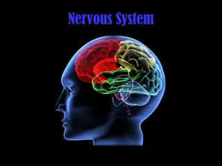
Cerebrum
Cerebrum
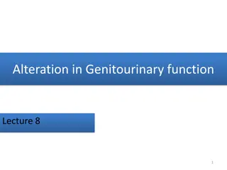
Largest part of the forebrain.
Divided into two halves, the
(
cerebral hemipheres
)
,
which
are separated by a
deep
median longitudinal
fissure
which lodges the
falx
cerebri.
In the depth of the fissure,
the hemispheres are
connected by a bundle of
fibers called the
corpus
callosum
.
Median longitudinal fissure
Corpus callosum
Right
hemisphere
Left
hemisphere
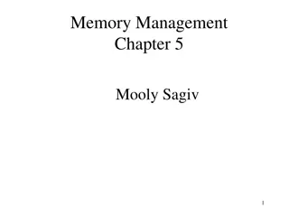
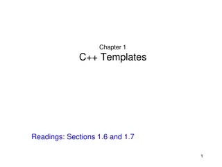
Cerebral cortex
:
Superficial layer
of grey matter
Medulla (White matter) :
Deeper
to the cortex, contains axons to and
from the cells of the cortex
Basal ganglia
:
Number of nuclear
masses buried within the white
matter
Lateral ventricle
:
The cavity of
hemisphere
Cortex
Basal
ganglia
WM
WM
WM
WM
Lateral
ventricle
Structure of Cerebrum
Superolateral
Superolateral
Medial
Medial
Inferior
(tentorial
)
Surfaces(3)
Surfaces(3)
Lobes of Cerebrum
T
h
e
s
u
p
e
r
f
i
c
i
a
l
l
a
y
e
r
o
f
g
r
e
y
m
a
t
t
e
r
i
s
h
i
g
h
l
y
c
o
n
v
o
l
u
t
e
d
t
o
f
o
r
m
a
c
o
m
p
l
e
x
p
a
t
t
e
r
n
o
f
r
i
d
g
e
s
(
g
y
r
i
)
a
n
d
g
r
o
o
v
e
s
(
s
u
l
c
i
)
.
T
h
i
s
a
r
r
a
n
g
e
m
e
n
t
m
a
x
i
m
i
z
e
s
t
h
e
s
u
r
f
a
c
e
a
r
e
a
o
f
t
h
e
c
e
r
e
b
r
a
l
c
o
r
t
e
x
(
a
b
o
u
t
7
0
%
i
s
h
i
d
d
e
n
w
i
t
h
i
n
t
h
e
d
e
p
t
h
s
o
f
s
u
l
c
i
)
.
S
g
•
Three sulci
, consistent in
position, named
central,
lateral
(
sylvian) &
parieto-
occipital
,
divide each
hemisphere into
FOUR
lobes:
Frontal
,
Parietal
,
Temporal
&
Occipital
(
named after
overlying bones) Functionally
each hemisphere contains a
‘
limbic lobe
limbic lobe
’
’
on the medial
surface.
motor function,
motivation,
aggression,
smell and
mood
emotions,
memory
storage &
Linking
conscious
intellectual
functions with
the unconscious
autonomic
functions,
smell, hearing,
memory and
abstract
thought
visual processing
reception
and
evaluation
of sensory
information
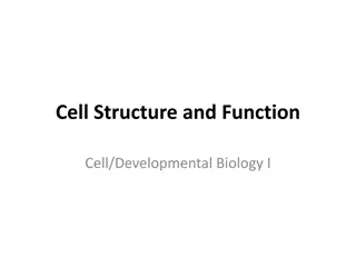
Frontal lobe:
Precentral gyrus.
Superior & inferior
frontal sulci
divide
the lobe into
superior, middle &
inferior frontal gyri.
Superior , middle &
Superior , middle &
inferior frontal gyri
inferior frontal gyri
Precentral
Precentral
gyrus
Superior
parietal lobule
Inferior
parietal
lobule
Postcentral
Postcentral
gyrus
gyrus
Intraparietal
sulcus
sfs
ifs
Parietal lobe:
Postcentral gyrus.
Intraparietal sulcus
divide
the lobe into superior &
inferior parietal lobules.
MAIN GYRI IN
SUPEROLATERAL
SURFACE
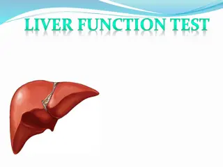
Temporal lobe:
Superior & inferior
temporal sulci
giving
rise to superior,
middle & inferior
temporal gyri.
Insula
: the gyrus in
the depth of lateral
sulcus, covered by
parts of frontal,
parietal & temporal
lobes called the
opercula
(removed in
lower picture.).
Superior, middle &
inferior temporal
gyri
insula
sts
its
Medial Surface
Sulci: Parietooccipital, Calcarine, Cingulate
Gyri: Cingulate, Parahippocampal
•
Brodmann
produced a
numbered, cytological
map
of cerebral cortex
based upon its regional
histological
characteristics.
•
Subdivisions with
similar
cellular and laminar
structure
a
re called
'areas'
'areas'
•
Brodmann's numbering of
these cortical locations
has become one of the
standard ways to identify
brain areas.
Functional Areas
Functional Areas
of the
of the
Cerebral Cortex
Cerebral Cortex
Frontal Lobe
Primary motor cortex:
Located
in precentral gyrus
(Brodmann
area 4).
Premotor cortex
:
Located in the
region immediately anterior to
the precentral gyrus
(Brodmann’s
area 6).
Frontal eye field:
Located in the middle frontal
gyrus immediately in front of motor cortex
(Brodmann’s area 8).
Broca’s (motor
speech) area:
Located in the
inferior frontal
gyrus of the
dominant
hemisphere, usually
left
(Brodmann’s
area 44 & 45).
Prefrontal cortex:
Extensive region
of the frontal lobe
anterior to
premotor area.
Parietal
Parietal
lobe
lobe
Primary visual cortex
:
located
on the medial surface of the
hemisphere, in the gyri
surrounding the calcarine
sulcus
(Brodmann’s area 17).
Occipital lobe
Occipital lobe
Visual association cortex
:
l
ocated
around the primary visual
cortex. Area 19
Parietal association cortex:
located posterior to primary
somatosensory cortex.
P
rimary somatosensory cortex
:
located in postcentral gyrus
(Brodmann’s area 1, 2, 3).
Temporal Lobe
Temporal Lobe
Auditory association cortex
:
located immediately around
the primary auditory cortex
(also includes
Wernick’s area)
P
a
r
a
h
i
p
p
o
c
a
m
p
a
l
g
y
r
u
s
:
l
o
c
a
t
e
d
i
n
t
h
e
i
n
f
e
r
o
m
e
d
i
a
l
p
a
r
t
o
f
t
e
m
p
o
r
a
l
l
o
b
e
.
D
e
e
p
t
o
t
h
i
s
g
y
r
u
s
l
i
e
s
t
h
e
h
i
p
p
o
c
a
m
p
u
s
a
n
d
t
h
e
a
m
y
g
d
a
l
a
,
w
h
i
c
h
a
r
e
p
a
r
t
s
o
f
l
i
m
b
i
c
s
y
s
t
e
m
Primary auditory cortex
: located
in the superior surface of the
superior temporal gyrus
(
Brodmann’s area 41, 42)
Language AreaS
Language AreaS
Organized around the lateral
Sulcus.
Broca’s area
:
concerned with
expressive aspects of language.
Wernick’s area:
responsible for
comprehension of the spoken
words.
Angular gyrus & Supramarginal
gyrus:
nearby regions of
temporal lobe and parietal lobe
o fthe inferior parietal lobule)
are important in naming,
reading, writing, and
calculation.
The localization of
Speech centers
& Mathematical ability
is the
criterion for defining the dominant
cerebral hemisphere.
In 96% of normal
right-handed
individuals and 70% of normal
left-
handed
individuals, the Left
hemisphere contains the language
centers. These are
Left Hemisphere
Dominant.
Cerebral dominance becomes
established during the
first few
years after birth.
Verbal
Memory
Shape
Memory
Hemispheres communicate
via the corpus
callosum
White Matter
Underlies the cortex, contains nerve fibers, neuroglia cells and blood vessels.
The nerve fibers originate, terminate or sometimes both, within the cortex.
Depending on their origin & termination
,
these nerve fibers are classified
into three types:
Association
,
Projection
&
Commissural
Association fibers:
Unite
different parts of the same
hemisphere, are of two
types: long & short
Commissural fibers:
Connect
the corresponding regions of
the two hemispheres
Projection fibers
:
Consist of
afferent and efferent fibers
of the cerebral cortex
Dive into the intricate details of the cerebrum, the largest part of the forebrain. Explore its parts including the cortex, medulla, basal ganglia, and lateral ventricle. Learn about the lobes, important sulci, and gyri. Understand the types of fibers in the cerebral medulla and their functions. Unravel the mysteries of sensory information reception, motor functions, emotions, memory, and more associated with the cerebral hemisphere.

Download Presentation

Please find below an Image/Link to download the presentation.
The content on the website is provided AS IS for your information and personal use only. It may not be sold, licensed, or shared on other websites without obtaining consent from the author.If you encounter any issues during the download, it is possible that the publisher has removed the file from their server.
You are allowed to download the files provided on this website for personal or commercial use, subject to the condition that they are used lawfully. All files are the property of their respective owners.
The content on the website is provided AS IS for your information and personal use only. It may not be sold, licensed, or shared on other websites without obtaining consent from the author.
E N D
Presentation Transcript
CEREBRUM CEREBRUM Dr. Jamila EL Medany Dr. Jamila EL Medany
Objectives At the end of the lecture, the student should be able to: List the parts of the cerebral hemisphere (cortex, medulla, basal nuclei, lateral ventricle). Describe the subdivision of a cerebral hemisphere into lobes. List the important sulci and gyri of each lobe. Describe different types of fibers in cerebral medulla (association, projection and commissural) and give example of each type.
Cerebrum Cerebrum Corpus callosum Largest part of the forebrain. Divided into two halves, the (cerebral hemipheres), which are separated by a deep median longitudinal fissure which lodges the falx cerebri. In the depth of the fissure, the hemispheres are connected by a bundle of fibers called the corpus callosum. Left Right hemisphere hemisphere Median longitudinal fissure
Structure of Cerebrum Structure of Cerebrum Cortex Basal ganglia Cerebral cortex: Superficial layer of grey matter Medulla (White matter) : Deeper to the cortex, contains axons to and from the cells of the cortex Basal ganglia: Number of nuclear masses buried within the white matter Lateral ventricle: The cavity of hemisphere WM WM Lateral ventricle
Surfaces( Surfaces(3 3) ) Medial Superolateral Inferior (tentorial)
Lobes of Cerebrum Lobes of Cerebrum The superficial layer of grey matter is highly convoluted to form a complex pattern of ridges (gyri) and grooves (sulci). This arrangement maximizes the surface area of the cerebral cortex (about 70% is hidden within the depths of sulci). Three sulci, consistent in position, named central, lateral (sylvian) & parieto- occipital, divide each hemisphere into FOUR lobes: Frontal, Parietal, Temporal & Occipital (named after overlying bones) Functionally each hemisphere contains a limbic lobe on the medial surface. S g
Function of Lobes Function of Lobes reception and evaluation of sensory information motor function, motivation, aggression, smell and mood visual processing emotions, memory storage & Linking conscious intellectual functions with the unconscious autonomic functions, smell, hearing, memory and abstract thought
Frontal lobe: Precentral gyrus. Superior & inferior frontal sulci divide the lobe into superior, middle & inferior frontal gyri. Parietal lobe: Postcentral gyrus. Intraparietal sulcus divide the lobe into superior & inferior parietal lobules. Precentral gyrus Postcentral gyrus Superior parietal lobule sfs ifs Inferior parietal lobule Intraparietal sulcus Superior , middle & inferior frontal gyri MAIN GYRI IN SUPEROLATERAL SURFACE
Superior, middle & inferior temporal gyri Temporal lobe: Superior & inferior temporal sulci giving rise to superior, middle & inferior temporal gyri. Insula: the gyrus in the depth of lateral sulcus, covered by parts of frontal, parietal & temporal lobes called the opercula (removed in lower picture.). sts its insula
Medial Surface Medial Surface Sulci: Parietooccipital, Calcarine, Cingulate Gyri: Cingulate, Parahippocampal
Brodmann Brodmann s s Map Map Brodmann produced a numbered, cytological map of cerebral cortex based upon its regional histological characteristics. Subdivisions with similar cellular and laminar structure are called 'areas' Brodmann's numbering of these cortical locations has become one of the standard ways to identify brain areas.
Functional Areas Functional Areas of the of the Cerebral Cortex Cerebral Cortex
Frontal Lobe Frontal Lobe Premotor cortex: Located in the region immediately anterior to the precentral gyrus (Brodmann s area 6). Primary motor cortex: Located in precentral gyrus (Brodmann area 4). Prefrontal cortex: Extensive region of the frontal lobe anterior to premotor area. Broca s (motor speech) area: Located in the inferior frontal gyrus of the dominant hemisphere, usually left (Brodmann s area 44 & 45). Frontal eye field: Located in the middle frontal gyrus immediately in front of motor cortex (Brodmann s area 8).
Parietal Parietal lobe lobe Primary somatosensory cortex: located in postcentral gyrus (Brodmann s area 1, 2, 3). Parietal association cortex: located posterior to primary somatosensory cortex. Occipital lobe Primary visual cortex: located on the medial surface of the hemisphere, in the gyri surrounding the calcarine sulcus (Brodmann s area 17). Visual association cortex: located around the primary visual cortex. Area 19
Temporal Lobe Temporal Lobe Primary auditory cortex: located in the superior surface of the superior temporal gyrus (Brodmann s area 41, 42) Auditory association cortex: located immediately around the primary auditory cortex (also includes Wernick s area) Parahippocampal gyrus: located in the inferomedial part of temporal lobe. Deep to this gyrus lies the hippocampus and the amygdala, which are parts of limbic system
Language Language AreaS AreaS Organized around the lateral Sulcus. Broca s area: concerned with expressive aspects of language. Wernick s area: responsible for comprehension of the spoken words. Angular gyrus & Supramarginal gyrus: nearby regions of temporal lobe and parietal lobe o fthe inferior parietal lobule) are important in naming, reading, writing, and calculation.
Hemispheric Dominance Hemispheric Dominance The localization of Speech centers & Mathematical ability is the criterion for defining the dominant cerebral hemisphere. In 96% of normal right-handed individuals and 70% of normal left- handed individuals, the Left hemisphere contains the language centers. These are Left Hemisphere Dominant. Cerebral dominance becomes established during the first few years after birth. Verbal Memory Shape Memory Hemispheres communicate via the corpus callosum
White Matter White Matter Underlies the cortex, contains nerve fibers, neuroglia cells and blood vessels. The nerve fibers originate, terminate or sometimes both, within the cortex. Depending on their origin & termination, these nerve fibers are classified into three types: Association, Projection & Commissural Association fibers: Unite different parts of the same hemisphere, are of two types: long & short Commissural fibers: Connect the corresponding regions of the two hemispheres Projection fibers: Consist of afferent and efferent fibers of the cerebral cortex