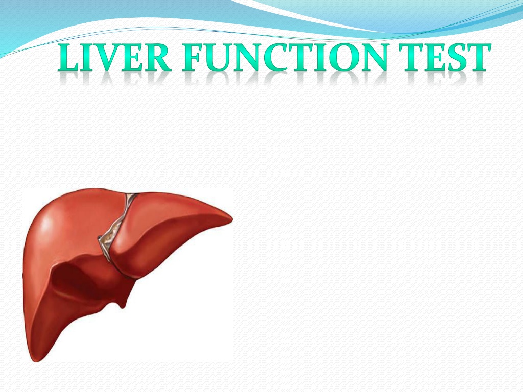
Liver Function Tests and Their Significance
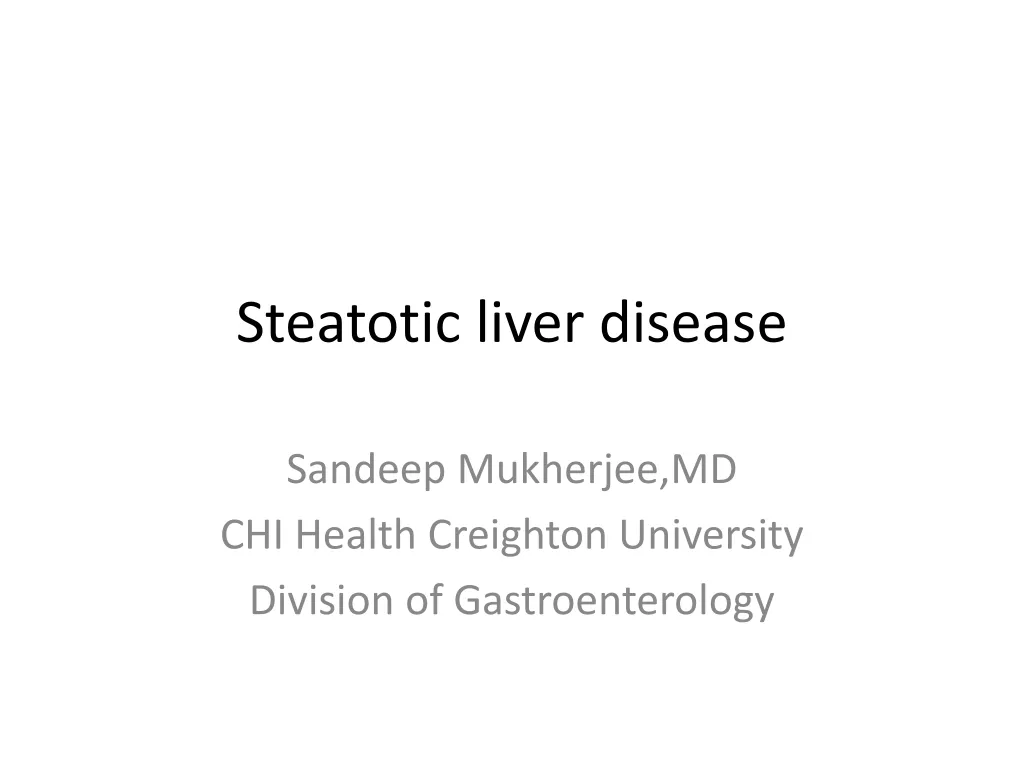
Function of Liver
1
.Metabolic Functions:
Urea cycle
Glycogen synthesis
Vitamin Metabolism
Minral Metabolism
Lipid Metabolism
Glycolysis
2
.Excretory Functions:
Cholesterol
Bile Pigments
Bile salts
3
.Protective Functions & detoxification:
Ammonia
Clearance of insulin, PTH, estrogens, cortisol.
4
.Hematological function:
Formation of Blood
Destruction of erythrocytes
5
. Synthetic functions:
Protein, Albumin, Prothrombin, Hormones
6
.Storage Functions:
Glycogen, Vitamin A, D and B12
USES
Screening
of liver dysfunction
To recognize
Pattern
of liver disease
To Assess
Prognosis of patient
Follow up
of disease
To evaluate the response to therapy
Liver function tests include:
SGPT (ALT-Alkaline Transaminase)
SGOT (AST-Aspartate Transaminase)
GGT (Gama glutamic traspeptidase)
ALP (Alkaline Phosphatase)
Bilirubin
Total Protein
Serum Albumin
Serum Globulin
PT/BT /CT (Prothombin, Bledding, Clotting
Time)
5’ nucleotide
Other test include:
Blood ammonia
LDH (Lactate Dehydrate)
AFP (Alfa feto protein)
Ceruloplasmin
Leucine aminopeptidase
Alpha - 1 antitrypsin
Procollagen III peptide
Cholesterol
Glycoprotein
SGPT(ALT)
Method: L- Alanine LDH UV Kinetic (IFCC
kinetic)
Measuring the rate of decrease in absorbance of
NADH at 340 nm due to the oxidation of NADH to
NAD.
Pyruvate+NADH+H+
Ldh
Lactate +NAD+
Significance
•
Serum Glutamic Pyruvate Transaminase (SGPT)
•
Alanine aminotransferase (ALT)
•
Enzyme present in hepatocytes .
•
Significantly elevated levels of ALT (
SGPT
) often
suggest the existence of other medical problems such
as viral hepatitis, diabetes, congestive heart failure,
liver damage, bile duct problems, infectious
mononucleosis, or myopathy.
•
So ALT is commonly used as a way of screening for
liver problems.
INERFARANCE:
Alanine transaminase (ALT) –
10-40 u/L
•
When a cell is damaged, it leaks this enzyme into the
blood, where it is measured.
•
ALT rises in
–
Viral hepatitis
–
Alcoholic Liver disease
–
Hepatic congestion
–
Hepatocellular carcinoma
–
Cholecystitis
–
Paracetamol (acetaminophen) overdose.
SGOT (AST) Principal
Method: L- Alanine LDH UV Kinetic
SGOT (AST) catalyzes the transfer of amino group
between L-Aspartate and
α
Ketoglutarate to form
Oxaloacetate and Glutamate.
The Oxaloacetate formed reacts with NADH in the
presence of Malate Dehydrogenase to form NAD.
The rate of oxidation of NADH to NAD is measured as
a decrease in absorbance which is proportional to the
SGOT (AST) activity in the sample.
L-Aspartate +
α
Ketoglutarate SGOT Oxaloacetate + L-Glutamate
Significance
Serum Glutamic Oxaloacetic Transaminase (SGOT)
The AST is a cellular enzyme is found in highes concentration
in heart muscle, The cells of the liver the cells of the skeletal
muscle & in smaller amounts in other weaves.
The blood SGOT levels are thus elevated with liver damage
or insult to the heart.
Some medication can also raise SGOT level.
Interference
•
Normal Range:-
10-40 U/L
•
It is raised in acute liver damage,
•
But,also present
–
Red blood cells
–
Cardiac and Skeletal muscle
–
And therefore it is not specific to the liver
.
GGT PRINCIPAL
Method: Carboxy Substrate Method
GGT catalyzes the transfer of amino group between L-
γ
-Glutamyl-3-carboxy-4 nitroanilide and Glycylglycine
to form L-
γ
-Glutamylglycylglycine and 5-amino-2-
nitrobenzoate.
The rate of formation of 5-amino-2-nitrobenzoate is
measured as an increase in absorbance which is
proportional to the GGT acitivity in the sample.
L-
γ
-Glutamyl 3-carboxy 4-nitroanilide + Glycylglycine
GGT L-
γ
-Glutamyl glycylglycine+ 5-Amino-2-
nitrobenzoate
.
Significance
The gamma-glutamyl transferase (
GGT
) test may be
used to determine the cause of elevated alkaline
phosphatase (ALP).
Both ALP and
GGT
are elevated in disease of the bile
ducts and in some liver diseases.
Interference
•
Normal Range:
10-30 u/L
.
Although reasonably specific to the liver and a more
sensitive marker for cholestatic damage than ALP.
•
Gamma glutamyl transpeptidase (GGT) may be elevated
with even minor, sub-clinical levels of liver dysfunction.
•
GGT is raised in alcohol toxicity (acute and chronic).
ALP PRINCIPAL
Method: para-nitrophenylphosphate with AMP
buffer
Alkaline phosphatase in the sample catalyzes the
hydrolysis of colourless
p
-nitrophenyl phosphate
(
p-
NPP) to give
p-
nitrophenol and inorganic
phosphate.
At the pH of the of the assay (alkaline), the
p-
nitrophenol is in the yellow phenoxide form.
The rate of absorbance increase at 404 nm is
directly proportional to the alkaline phosphatase
activity in the sample.
Optimized concentrations of zinc and magnesium
ions are present to activate the ALP in the sample.
CLINICAL SIGNIFICANCE
Increased levels of serum ALP;
In growing children
Bone disease like;
Metastasis, rickets, healing fractures, Osteomalacia.
Interference
•
Mild elevations (<500 IU/L)
–
Viral hepatitis
–
Alcoholic cirrhosis
–
Infiltrative liver disease like lymphoma, sarcoidosis
•
Moderate elevation (500 – 1000 IU/L)
–
Cholecystitic
–
Gall bladder stone
•
Severely elevation (>1500 IU/L)
–
Osteomalacia
–
Osteoporosis
–
Ricket
–
Osteosarcoma
–
Bone tumour
–
Paget's disease
Bilirubin PRINCIPAL
Method: Diazo Reaction
Bilirubin glucuronide +diazonium salt azodye
(tan or pink to viotel)
Different between Unconjugated &
Conjugated Bilirubin
Bilirubin Pathway
Type & Cause of Jaundice
Pre-hepatic Jaundice
Neonatal
(Physiological) Jaundice
Malaria
G 6 PD deficiency
Thalassaemia
Sickle cell disease
Mis-match Blood
Transfusion
Auto-immune
Intra-Hepatic Jaundice
Acute Viral hepatitis
Alcohol Cirrhosis
Cirrhosis of Liver
Primray Biliary Cirrhosis,
Haemochromatosis
Wilson Disease
Alpha-1 antitrypsin deficiency
Drug induce – Quinine Group,
NSAID, Chemotherapeutic drugs
Post Hepatic Jaundice
Gall Bladder - Common Bile Duct - Pancreatic duct Stone
Gall Bladder - Hepatic – Pancreatic – Duodenal Carcinoma
OBSTRUCTIVE JAUNDICE
PHYSIOLOGIC JAUNDICE OF THE
NEWBORN
PHOTOTHERAPY
31
Van den bergh Test :Direct &
Indirect Bilirubin
Method: Diazo Reaction
This is specific reaction to identify the increase in
serum bilirubin level.
Normal serum gives a negative Van den Bergh
reaction.
sulfanilic acid + sodium nitrate Diazotized
Diazotized sulfanilic acid + bilirubin Azobilirubin
(purple color).
Give major causes for increase in
blood Bilirubin level:-
Major causes for increase bilirubin levels in blood:
Hemolysis:-
Damage to RBC may cause increased breakdown
of Hb producing Unconjugated Bilirubin, which may
overload liver conjugating system, causing
Hyperbilirubinemia;
Failure of Conjugating system in the liver,
Obstruction in the Biliary system,
Total Protein PRINCIPAL
Method: Biuret
Proteins react with cupric ions in alkaline medium to
form a violet colored complex. The intensity of the
color produced is directly proportional to proteins
present in the specimen and can be measured on a
photometer at 530 nm (or by using a green filter).
Normal Range
Serum Protein: 6-8 g/dl
Albumin Principal
Method: Bromocresol Green (BCG)
Albumin present in serum binds specifically with
bromocresol green at pH 4.1 to form green colored
complex, intensity of which can be measured
colorimetrically by using 640 nm (or a red filter)
Normal Range
3.3-4.8 g/dl
Total Protein & Albumin
•
Both decrease in Hepato-cellular disease.
•
Because it is synthesized & store into liver.
•
It may found decrease in following disease:
•
Malnutrition
•
Chronic disease
•
Nephrotic syndrome
•
Inflammatory bowel disease
•
Chronic infection
•
Tuberculosis
Prothombin time Principal
Method: Capillary tube method
When preformed tissue thromboplastin and calcium
chloride are added to citrated plasma, the plasma
clots.
The time taken for the clot to appear is called
Prothrombin time (PT).
Significance
Prothrombin time
(PT) is a blood test that measures
how long it takes blood to clot.
A
prothrombin time
test can be used to check for
bleeding problems.
PT is also used to check whether medicine to prevent
blood clots is working. A PT test may also be called an
INR test.
Normal Range
11 to 16 second
Interferance
•
Another measure of hepatic synthetic function is the
prothrombin time.
•
Prothrombin time is affected by proteins synthesized by the
liver.
•
Thus, in patients who have prolonged prothrombin times, liver
disease may be present.
•
Since a prolonged PT is not a specific test for liver disease,
confirmation of other abnormal liver tests is essential.
•
Diseases such as malnutrition, in which decreased vitamin
K ingestion is present, may result in a prolonged PT time.
•
An indirect test of hepatic synthetic function includes
administration of vitamin K (10mg) subcutaneously over
three days.
•
Several days later, the prothrombin time may be
measured. If the prothrombin time becomes normal, then
hepatic synthetic function is intact.
•
This test does not indicate that there is no liver disease,
but is suggestive that malnutrition may coexist with (or
without) liver disease.
Bledding Time
Determination of bleeding time helps to detect
vascular defect & platelet disorder.
Prolonged bleeding time is generally absociated
thrombocytopenia.
Principle:-
A 1mm deep prick made on ear lobe or finger
of the patient the length of time required for bleeding
to cease is record.
Normal Range:-
1-5 minutes
Procedure
Sterilize the finger lip or ear lobe with spirit and given
a deep prick to get free flow of blood, immediately,
with a clean, white filter paper and note the time.
Continue to apply the filter paper to pricked site at the
interval 30 seconds, till no blood stain is seen on the
filter paper. The duration of time from the firs sport till
no blood stain on filter paper is known as BLEEDING
TIME.
Clotting Time
Principle:-
Blood is collected in capillary tube after a
finger prick & the stop watch is started. The formation
of fibrin string is noted by breaking the capillary tube
at regular interval, The time is noted at the first
appearance of the fibrin string.
This method is generally useful in severe clotting
disorders.
Normal Range:-
4-9 min.
Procedure
Sterilize the finger top with spirit and given a deep
prick to get free flow of blood. Start the stop watch as
soon as blood starts coming out. Fill a thin capillary
tube (it will be filled by capillary action by just
applying the tip on the blood drop) with blood. After
every 30 seconds, break a small portion of the capillary
tube till a thin line of unbroken coagulam is seen
between the broken ends. Stop the stop watch and
note the time. This is CLOTTING TIME.
Blood Ammonia
Ammonia is a by product of amino acid catabolism.
Ammonia is used as a potential marker of hepatic
encephaophathy, but it is not a good test for liver
function.
High annonia levels have been found with near normal
liver function and vice versa.
Roll of liver
Liver converts blood ammonia into urea and muscles use it
in transmination reaction to produce glutamate or alanine
etc..
Pateints with advanced liver diseases usually also have
muscle wasting which then also contributes, to
hyperammonemia.
Liver biopsy is oftern the last test used to arrive at a final
diagnosis of liver disease.
It is indicated in chronic cases of liver diseases
characterized by unexplained clinical findings pointing to
liver disease.
Coagulation
tests
•
The liver is responsible for the production of coagulation
factors.
•
If it is increased, it means it is taking longer than usual
for blood to clot.
•
It will only be increased if the liver is so damaged that
synthesis of vitamin K-dependent coagulation factors has
been impaired.
•
It is not a sensitive measure of liver function.
Hepatic neoplasm markers
Alpha fetoprotein (AFP): primary hepatocellular
carcinoma
Carcinoembryonic antigen (CEA): increased CEA:
liver metastatic carcinoma or other carcinomas of the
gastrointestinal system
Abnormal prothrombin (APT): increased APT
primary hepatocellular carcinoma
The liver carries out essential metabolic, excretory, protective, synthetic, and storage functions in the body. Liver function tests play a crucial role in screening for liver dysfunction, recognizing patterns of liver disease, assessing patient prognosis, monitoring disease progression, and evaluating therapy responses. These tests measure various enzymes, proteins, and substances in the blood that indicate liver health and function. Elevated levels of certain markers like SGPT (ALT) can point to liver issues such as viral hepatitis, diabetes, or liver damage. Other tests like blood ammonia and AFP are also used to assess liver health. Understanding the significance and interpretation of liver function test results is key in diagnosing and managing a range of liver conditions.

Download Presentation

Please find below an Image/Link to download the presentation.
The content on the website is provided AS IS for your information and personal use only. It may not be sold, licensed, or shared on other websites without obtaining consent from the author.If you encounter any issues during the download, it is possible that the publisher has removed the file from their server.
You are allowed to download the files provided on this website for personal or commercial use, subject to the condition that they are used lawfully. All files are the property of their respective owners.
The content on the website is provided AS IS for your information and personal use only. It may not be sold, licensed, or shared on other websites without obtaining consent from the author.
E N D
Presentation Transcript
Function of Liver 1.Metabolic Functions: Urea cycle Glycogen synthesis Vitamin Metabolism Minral Metabolism Lipid Metabolism Glycolysis 2.Excretory Functions: Cholesterol Bile Pigments Bile salts 3.Protective Functions & detoxification: Ammonia Clearance of insulin, PTH, estrogens, cortisol.
4.Hematological function: Formation of Blood Destruction of erythrocytes 5. Synthetic functions: Protein, Albumin, Prothrombin, Hormones 6.Storage Functions: Glycogen, Vitamin A, D and B12
USES Screening of liver dysfunction To recognize Pattern of liver disease To Assess Prognosis of patient Follow up of disease To evaluate the response to therapy
Liver function tests include: SGPT (ALT-Alkaline Transaminase) SGOT (AST-Aspartate Transaminase) GGT (Gama glutamic traspeptidase) ALP (Alkaline Phosphatase) Bilirubin Total Protein Serum Albumin Serum Globulin PT/BT /CT (Prothombin, Bledding, Clotting Time) 5 nucleotide
Other test include: Blood ammonia LDH (Lactate Dehydrate) AFP (Alfa feto protein) Ceruloplasmin Leucine aminopeptidase Alpha - 1 antitrypsin Procollagen III peptide Cholesterol Glycoprotein
SGPT(ALT) Method: L- Alanine LDH UV Kinetic (IFCC kinetic) Measuring the rate of decrease in absorbance of NADH at 340 nm due to the oxidation of NADH to NAD.
Significance Serum Glutamic Pyruvate Transaminase (SGPT) Alanine aminotransferase (ALT) Enzyme present in hepatocytes . Significantly elevated levels of ALT (SGPT) often suggest the existence of other medical problems such as viral hepatitis, diabetes, congestive heart failure, liver damage, bile duct problems, infectious mononucleosis, or myopathy. So ALT is commonly used as a way of screening for liver problems.
INERFARANCE: Alanine transaminase (ALT) 10-40 u/L When a cell is damaged, it leaks this enzyme into the blood, where it is measured. ALT rises in Viral hepatitis Alcoholic Liver disease Hepatic congestion Hepatocellular carcinoma Cholecystitis Paracetamol (acetaminophen) overdose.
SGOT (AST) Principal Method: L- Alanine LDH UV Kinetic SGOT (AST) catalyzes the transfer of amino group between L-Aspartate and Ketoglutarate to form Oxaloacetate and Glutamate. The Oxaloacetate formed reacts with NADH in the presence of Malate Dehydrogenase to form NAD. The rate of oxidation of NADH to NAD is measured as a decrease in absorbance which is proportional to the SGOT (AST) activity in the sample.
L-Aspartate + Ketoglutarate SGOT Oxaloacetate + L-Glutamate
Significance Serum Glutamic Oxaloacetic Transaminase (SGOT) The AST is a cellular enzyme is found in highes concentration in heart muscle, The cells of the liver the cells of the skeletal muscle & in smaller amounts in other weaves. The blood SGOT levels are thus elevated with liver damage or insult to the heart. Some medication can also raise SGOT level.
Interference Normal Range:- 10-40 U/L It is raised in acute liver damage, But,also present Red blood cells Cardiac and Skeletal muscle And therefore it is not specific to the liver.
GGT PRINCIPAL Method: Carboxy Substrate Method GGT catalyzes the transfer of amino group between L- -Glutamyl-3-carboxy-4 nitroanilide and Glycylglycine to form L- -Glutamylglycylglycine and 5-amino-2- nitrobenzoate. The rate of formation of 5-amino-2-nitrobenzoate is measured as an increase in absorbance which is proportional to the GGT acitivity in the sample.
L- -Glutamyl 3-carboxy 4-nitroanilide + Glycylglycine GGT L- -Glutamyl glycylglycine+ 5-Amino-2- nitrobenzoate.
Significance The gamma-glutamyl transferase (GGT) test may be used to determine the cause of elevated alkaline phosphatase (ALP). Both ALP and GGT are elevated in disease of the bile ducts and in some liver diseases.
Interference Normal Range: 10-30 u/L. Although reasonably specific to the liver and a more sensitive marker for cholestatic damage than ALP. Gamma glutamyl transpeptidase (GGT) may be elevated with even minor, sub-clinical levels of liver dysfunction. GGT is raised in alcohol toxicity (acute and chronic).
ALP PRINCIPAL Method: para-nitrophenylphosphate with AMP buffer Alkaline phosphatase in the sample catalyzes the hydrolysis of colourless p-nitrophenyl phosphate (p-NPP) to give p-nitrophenol and inorganic phosphate. At the pH of the of the assay (alkaline), the p- nitrophenol is in the yellow phenoxide form. The rate of absorbance increase at 404 nm is directly proportional to the alkaline phosphatase activity in the sample. Optimized concentrations of zinc and magnesium ions are present to activate the ALP in the sample.
CLINICAL SIGNIFICANCE Increased levels of serum ALP; In growing children Bone disease like; Metastasis, rickets, healing fractures, Osteomalacia. Sex Age Reference Interval Male-femals 4-15 yr 54-36 U/L Male 20-50 yr 53-128 U/L Male >60 yr 56-119 U/L femals 20-50 yr 42-98 U/L femals >60 yr 53-141 U/L
Interference Mild elevations (<500 IU/L) Viral hepatitis Alcoholic cirrhosis Infiltrative liver disease like lymphoma, sarcoidosis Moderate elevation (500 1000 IU/L) Cholecystitic Gall bladder stone Severely elevation (>1500 IU/L) Osteomalacia Osteoporosis Ricket Osteosarcoma Bone tumour Paget's disease
Bilirubin PRINCIPAL Method: Diazo Reaction Bilirubin glucuronide +diazonium salt azodye (tan or pink to viotel)
Different between Unconjugated & Conjugated Bilirubin UNCONJUGATED CONJUGATED Insoluble Soluble <1.3 In water In alcohol Normal Soluble Soluble <0.4 Present Normally absent Not absorbed In bile In Urine Absorption gut Absent Always absent Absorbed Diffuses-yellow colour Indirect + Diffusion into tissues Van den bergh Doesn t diffuse Direct +
Type & Cause of Jaundice Pre-hepatic Jaundice Intra-Hepatic Jaundice Neonatal (Physiological) Jaundice Acute Viral hepatitis Alcohol Cirrhosis Malaria Cirrhosis of Liver G 6 PD deficiency Primray Biliary Cirrhosis, Thalassaemia Haemochromatosis Sickle cell disease Wilson Disease Mis-match Blood Transfusion Alpha-1 antitrypsin deficiency Drug induce Quinine Group, NSAID, Chemotherapeutic drugs Auto-immune Post Hepatic Jaundice Gall Bladder - Common Bile Duct - Pancreatic duct Stone Gall Bladder - Hepatic Pancreatic Duodenal Carcinoma
PHYSIOLOGIC JAUNDICE OF THE NEWBORN
Heamolytic Obstructive r Blood Examination Total Billirubin Direct Billirubin Indirect Billirubin Normal Normal ALT Normal Normal Alkaline phosphatase Normal / Normal Urine Examination Normal Normal / Bile Pigment Urobillinogen Normal / Absent Absent Normal Normal / Bile Salt Stool Examination Specific Investigation Normal Normal Clay Colour 31 Haemoglobin, LDH Liver Function Test USG Abdomen
Van den bergh Test :Direct & Indirect Bilirubin Method: Diazo Reaction This is specific reaction to identify the increase in serum bilirubin level. Normal serum gives a negative Van den Bergh reaction. sulfanilic acid + sodium nitrate Diazotized Diazotized sulfanilic acid + bilirubin Azobilirubin (purple color).
Give major causes for increase in blood Bilirubin level:- Major causes for increase bilirubin levels in blood: Hemolysis:- Damage to RBC may cause increased breakdown of Hb producing Unconjugated Bilirubin, which may overload liver conjugating system, causing Hyperbilirubinemia; Failure of Conjugating system in the liver, Obstruction in the Biliary system,
Total Protein PRINCIPAL Method: Biuret Proteins react with cupric ions in alkaline medium to form a violet colored complex. The intensity of the color produced is directly proportional to proteins present in the specimen and can be measured on a photometer at 530 nm (or by using a green filter). Normal Range Serum Protein: 6-8 g/dl
Albumin Principal Method: Bromocresol Green (BCG) Albumin present in serum binds specifically with bromocresol green at pH 4.1 to form green colored complex, intensity of which can be measured colorimetrically by using 640 nm (or a red filter) Normal Range 3.3-4.8 g/dl
Total Protein & Albumin Both decrease in Hepato-cellular disease. Because it is synthesized & store into liver. It may found decrease in following disease: Malnutrition Chronic disease Nephrotic syndrome Inflammatory bowel disease Chronic infection Tuberculosis
Prothombin time Principal Method: Capillary tube method When preformed tissue thromboplastin and calcium chloride are added to citrated plasma, the plasma clots. The time taken for the clot to appear is called Prothrombin time (PT).
Significance Prothrombin time (PT) is a blood test that measures how long it takes blood to clot. A prothrombin time test can be used to check for bleeding problems. PT is also used to check whether medicine to prevent blood clots is working. A PT test may also be called an INR test. Normal Range 11 to 16 second
Interferance Another measure of hepatic synthetic function is the prothrombin time. Prothrombin time is affected by proteins synthesized by the liver. Thus, in patients who have prolonged prothrombin times, liver disease may be present. Since a prolonged PT is not a specific test for liver disease, confirmation of other abnormal liver tests is essential.
Diseases such as malnutrition, in which decreased vitamin K ingestion is present, may result in a prolonged PT time. An indirect test of hepatic synthetic function includes administration of vitamin K (10mg) subcutaneously over three days. Several days later, the prothrombin time may be measured. If the prothrombin time becomes normal, then hepatic synthetic function is intact. This test does not indicate that there is no liver disease, but is suggestive that malnutrition may coexist with (or without) liver disease.
Bledding Time Determination of bleeding time helps to detect vascular defect & platelet disorder. Prolonged bleeding time is generally absociated thrombocytopenia. Principle:- A 1mm deep prick made on ear lobe or finger of the patient the length of time required for bleeding to cease is record. Normal Range:- 1-5 minutes
Procedure Sterilize the finger lip or ear lobe with spirit and given a deep prick to get free flow of blood, immediately, with a clean, white filter paper and note the time. Continue to apply the filter paper to pricked site at the interval 30 seconds, till no blood stain is seen on the filter paper. The duration of time from the firs sport till no blood stain on filter paper is known as BLEEDING TIME.
Clotting Time Principle:- Blood is collected in capillary tube after a finger prick & the stop watch is started. The formation of fibrin string is noted by breaking the capillary tube at regular interval, The time is noted at the first appearance of the fibrin string. This method is generally useful in severe clotting disorders. Normal Range:- 4-9 min.
Procedure Sterilize the finger top with spirit and given a deep prick to get free flow of blood. Start the stop watch as soon as blood starts coming out. Fill a thin capillary tube (it will be filled by capillary action by just applying the tip on the blood drop) with blood. After every 30 seconds, break a small portion of the capillary tube till a thin line of unbroken coagulam is seen between the broken ends. Stop the stop watch and note the time. This is CLOTTING TIME.
Blood Ammonia Ammonia is a by product of amino acid catabolism. Ammonia is used as a potential marker of hepatic encephaophathy, but it is not a good test for liver function. High annonia levels have been found with near normal liver function and vice versa.
Roll of liver Liver converts blood ammonia into urea and muscles use it in transmination reaction to produce glutamate or alanine etc.. Pateints with advanced liver diseases usually also have muscle wasting which then also contributes, to hyperammonemia. Liver biopsy is oftern the last test used to arrive at a final diagnosis of liver disease. It is indicated in chronic cases of liver diseases characterized by unexplained clinical findings pointing to liver disease.
Coagulation tests The liver is responsible for the production of coagulation factors. If it is increased, it means it is taking longer than usual for blood to clot. It will only be increased if the liver is so damaged that synthesis of vitamin K-dependent coagulation factors has been impaired. It is not a sensitive measure of liver function.
Hepatic neoplasm markers Alpha fetoprotein (AFP): primary hepatocellular carcinoma Carcinoembryonic antigen (CEA): increased CEA: liver metastatic carcinoma or other carcinomas of the gastrointestinal system Abnormal prothrombin (APT): increased APT primary hepatocellular carcinoma


")


 Principal")