RICKETTSIAL DISEASES


RICKETTSIAL DISEASES
KAUSHAL KUMAR
Assistant Professor & Head
Department of Veterinary Pathology
Bihar Veterinary College
Bihar Animal Sciences University, Patn
a


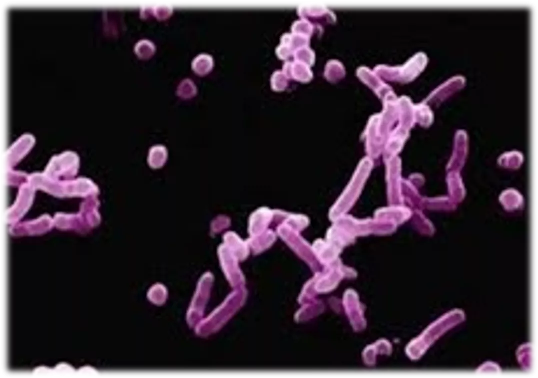
Rickettsia


A genus of nonmotile, Gram-
negative, nonspore-forming,
highly pleomorphic, cocci, bacilli and
thread shaped bacteria.
Genus
Rickettsia
was named
after
Howard Taylor Ricketts
, in
honour of his pioneering work on tick-
borne spotted fever.
Rickettsial Diseases
Rickettsial diseases (rickettsioses) and related
diseases (anaplasmosis, ehrlichiosis, Q fever, scrub
typhus) are caused by a group of gram-negative,
obligately intracellular coccobacilli. All, except
for
Coxiella burnetii
, have an arthropod vector.
Anaplasmosis
Nature of the disease
A form of ‘tick fever’ in cattle,
Caused by: Rickettsia-like-Organism
Anaplasmcentrale (
sometimes
)
Anaplasma marginale (
Usually
)
&
A parasite of a red blood cells
Characterized by
Initial high fever &
Progressive anaemia
Anaplasmosis
Susceptible Species:
Cattle,
Bos indicus
(less susceptible)
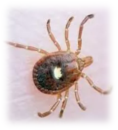
Transmission:
Boophilus microplus
(Cattle tick)-
Major vector
Others
-
Boophilus, Dermacentor, Rhipicephalus, Ixodes, Hyalomma & Argas
Mechanical transmission via veterinary
instruments
Anaplasmosis
Clinical Sign:
Steadily increasing temperatures
Anaemia, weakness and respiratory distress
Depression and anorexia
Jaundice marked
Brown Urine (presence of bile pigments)
Severely affected animals may die
Necropsy Lesion:
Gross findings are due to anemia and resulting anoxia.
Pale and jaundiced tissues
Spleenomegale (soft reddish-brown pulp)
Hepatomegale, (yellow-brown and mottled)
Anaplasmosis
Diagnosis:
Particularly in convalescent animals
Confirmed by microscopic examination of stained blood smears.
Serological testing for antibodies to
A. marginale
Canine Ehrlichiosis
Nature of the disease
A tick-born disease of dogs,
Also known as
Canine Rickettsiosis ; Canine Hemorrhagic Fever,
Canine Typhus ; Tracker Dog Disease &
Tropical Canine Pancytopaenia (TCP)
Caused by: Rickettsia-like-Organism
Ehrlichia canis
A parasite of
a monocyte in the peripheral blood
Characterized by
High fever &
Lowered peripheral blood cell counts(pancytopenia),
Ehrlichiosis
Susceptible Species:
Dogs and some species of wild canidae
German Shepherd-more susceptible
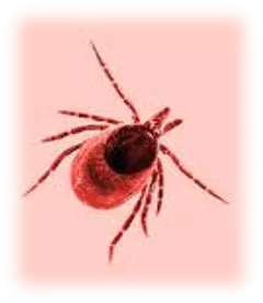
Transmission:
Rhipicephalus sanguineus (
brown dog tick) -Major vector
Others
-
Boophilus, Dermacentor, Ixodes, Hyalomma & Argas
Ehrlichiosis
Clinical Sign:
wide range of clinical syndromes
Acute stage
, -
Fever and epistaxis or other haemorrhages
Lowered peripheral blood cell
Subclinical phase
-No outward signs and
Last for the rest of the dog's life-Carrier states
Chronic phase-
The third and most serious stage
Pancytopenia,
Extensive haemorrhage (bleeding, )with epistaxis
bacterial infection, lameness,
Neurological, ophthalmic disorders, and kidney disease
Necropsy Lesion:
Emaciated carcase
Pale mucous membranes
Oedema of the limbs, ascites and hydro-pericardium
Haemorrhages in GIT, internal organs, s/c tissues and eye
Enlarged lymph nodes and spleen
Ehrlichiosis
Ehrlichiosis
Diagnosis:
1.
Blood smear Examination
(Uncommon):
For the presence of the
ehrlichia
canis, within a white blood cell.
For abnormalities in the No. of RBC, WBC, and most commonly platelets
2.
Serologic test:
for the presence of Ab against the
Ehrlichia
organism
3.
PCR:
To detect genetic material of the bacteria.
But yield a -ve result (subclinical and chronic phases)
Q. Fever
Nature of the disease
A zoonotic disease whose most common animal reservoirs are cattle, sheep,
and goats.
well-recognized cause of abortions in Ruminants and pets
has been developed as a
biological weapon
"Q" stands for "query
First described in slaughterhouse workers at Brisbane, State of Queensland.
Caused by:
Coxilla Burneti
Closely related to the Rickettsiae, but as similar to
Legionella and Francisella.
discovered in 1937, by Frank Macfarlane
Burnet
Cox and Davis
elucidated the transmission when they isolated it from
ticks.
Q. Fever
Susceptible Species:
C
attle, sheep, goats, and other domestic mammals,
Other animals : dogs, cats,
rabbits,
horses, pigs,
camels, buffalo, rodents
, and some
birds.
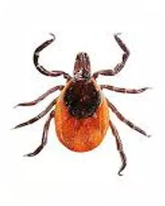
Transmission:
Inhalation of a spore-like small-cell variant,
Contact with the milk, urine, feces, vaginal mucus, or
semen of infected animals.
Rarely
,
the disease is tick-borne.
Q. Fever
C. burneti
resides and reproduces in the acidified
phagolysosomes of host monocytes and macrophages.
Clinical Sign:
Typical clinical signs :
Late Abortion,
Stillbirth,
Weak calves, and
Repeat breeding.
Moreover, experimental inoculation
in cattle
respiratory disorders and
cardiac failures (myocarditis)
frequent abortions and irregular repeat breedings.
Once a animal is infected,
C burnetii
can localize in mammary
glands, placenta, and uterus, from which it may be shed in
subsequent parturitions and lactations.
Q. Fever
Necropsy Lesion:
-
Necrotsing placentitis in ruminants
Diagnosis:
1.
Isolation and identification
of organisms in case of infectious
abortion
2.
Immunohistochemical Test:
for the presence of Ab against the
C. burnetii
3.
Immunofluoresence Test
on Paired sera taken 2 week apart to
detect recent infection
4.
PCR:
To detect
C. burnetii in tissue
.
Thanks
Rickettsial diseases, including anaplasmosis, are caused by gram-negative bacteria and often transmitted via arthropod vectors. Anaplasmosis affects cattle and is characterized by fever, anemia, and other clinical signs. Diagnosis involves blood smear examination and serological testing for antibodies. Canine ehrlichiosis is another tick-borne disease affecting dogs. Understanding these diseases is essential for veterinary professionals.

Download Presentation

Please find below an Image/Link to download the presentation.
The content on the website is provided AS IS for your information and personal use only. It may not be sold, licensed, or shared on other websites without obtaining consent from the author.If you encounter any issues during the download, it is possible that the publisher has removed the file from their server.
You are allowed to download the files provided on this website for personal or commercial use, subject to the condition that they are used lawfully. All files are the property of their respective owners.
The content on the website is provided AS IS for your information and personal use only. It may not be sold, licensed, or shared on other websites without obtaining consent from the author.
E N D
Presentation Transcript
RICKETTSIAL DISEASES KAUSHAL KUMAR Assistant Professor & Head Department of Veterinary Pathology Bihar Veterinary College Bihar Animal Sciences University, Patna
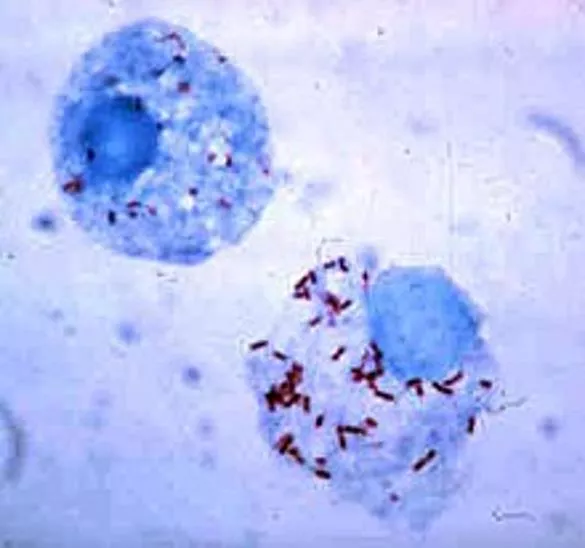
Rickettsia A genus of nonmotile, Gram- negative, nonspore-forming, highly pleomorphic, cocci, bacilli and thread shaped bacteria. Rickettsia Genus was named after Howard Taylor Ricketts, in honour of his pioneering work on tick- Red-stained Rickettsia rickettsii visible in cells of an Ixodid vector tick borne spotted fever.
Rickettsial Diseases Rickettsial diseases (rickettsioses) and related diseases (anaplasmosis, ehrlichiosis, Q fever, scrub typhus) are caused by a group of gram-negative, obligately intracellular coccobacilli. All, except for Coxiella burnetii, have an arthropod vector.
Anaplasmosis Nature of the disease A form of tick fever in cattle, Caused by: Rickettsia-like-Organism Anaplasmcentrale (sometimes) Anaplasma marginale (Usually) & A parasite of a red blood cells Characterized by Initial high fever & Progressive anaemia
Anaplasmosis Susceptible Species: Cattle, Bos indicus (less susceptible) Transmission: Boophilus microplus (Cattle tick)-Major vector Others -Boophilus, Dermacentor, Rhipicephalus, Ixodes, Hyalomma & Argas Mechanical transmission via veterinaryinstruments
Anaplasmosis Clinical Sign: Steadily increasing temperatures Anaemia, weakness and respiratory distress Depression and anorexia Jaundice marked Brown Urine (presence of bile pigments) Severely affected animals may die Necropsy Lesion: Gross findings are due to anemia and resulting anoxia. Pale and jaundiced tissues Spleenomegale (soft reddish-brown pulp) Hepatomegale, (yellow-brown and mottled)
Anaplasmosis Diagnosis: Particularly in convalescent animals Confirmed by microscopic examination of stained blood smears. Serological testing for antibodies to A. marginale
Canine Ehrlichiosis Nature of the disease A tick-born disease of dogs, Also known as Canine Rickettsiosis ; Canine Hemorrhagic Fever, Canine Typhus ; Tracker Dog Disease & Tropical Canine Pancytopaenia (TCP) Caused by: Rickettsia-like-Organism Ehrlichia canis A parasite of a monocyte in the peripheral blood Characterized by High fever & Lowered peripheral blood cell counts(pancytopenia),
Ehrlichiosis Susceptible Species: Dogs and some species of wild canidae German Shepherd-more susceptible Transmission: Rhipicephalus sanguineus (brown dog tick) -Major vector Others -Boophilus, Dermacentor, Ixodes, Hyalomma & Argas
Ehrlichiosis Clinical Sign:wide range of clinical syndromes Acute stage, - Fever and epistaxis or other haemorrhages Lowered peripheral blood cell Subclinical phase-No outward signs and Last for the rest of the dog's life-Carrier states Chronic phase-The third and most serious stage Pancytopenia, Extensive haemorrhage (bleeding, )with epistaxis bacterial infection, lameness, Neurological, ophthalmic disorders, and kidney disease Necropsy Lesion: Emaciated carcase Pale mucous membranes Oedema of the limbs, ascites and hydro-pericardium Haemorrhages in GIT, internal organs, s/c tissues and eye Enlarged lymph nodes and spleen
Ehrlichiosis Diagnosis: Blood smear Examination (Uncommon): For the presence of the ehrlichia canis, within a white blood cell. For abnormalities in the No. of RBC, WBC, and most commonly platelets 2. Serologic test: for the presence of Ab against the Ehrlichia organism 3. PCR: 1. To detect genetic material of the bacteria. But yield a -ve result (subclinical and chronic phases)
Q. Fever Nature of the disease A zoonotic disease whose most common animal reservoirs are cattle, sheep, and goats. well-recognized cause of abortions in Ruminants and pets has been developed as a biological weapon "Q" stands for "query First described in slaughterhouse workers at Brisbane, State of Queensland. Caused by: Coxilla Burneti Closely related to the Rickettsiae, but as similar to Legionella and Francisella. discovered in 1937, by Frank Macfarlane Burnet Cox and Davis elucidated the transmission when they isolated it from ticks.
Q. Fever Susceptible Species: Cattle, sheep, goats, and other domestic mammals, Other animals : dogs, cats, rabbits, horses, pigs, camels, buffalo, rodents, and some birds. Transmission: Inhalation of a spore-like small-cell variant, Contact with the milk, urine, feces, vaginal mucus, or semen of infected animals. Rarely, the disease is tick-borne.
Q. Fever C. burneti resides and reproduces in the acidified phagolysosomes of host monocytes and macrophages. Clinical Sign: Typical clinical signs : Late Abortion, Stillbirth, Weak calves, and Repeat breeding. Moreover, experimental inoculation in cattle respiratory disorders and cardiac failures (myocarditis) frequent abortions and irregular repeat breedings. Once a animal is infected, C burnetii can localize in mammary glands, placenta, and uterus, from which it may be shed in subsequent parturitions and lactations.
Q. Fever Necropsy Lesion: - Necrotsing placentitis in ruminants Diagnosis: 1. Isolation and identification of organisms in case of infectious abortion 2. Immunohistochemical Test: for the presence of Ab against the C. burnetii 3. Immunofluoresence Test on Paired sera taken 2 week apart to detect recent infection 4. PCR: To detect C. burnetii in tissue.