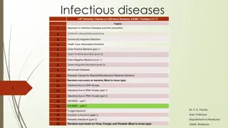
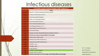
High Consequence Infectious Diseases (HCID)
High Consequence Infectious Diseases (HCID) encompass serious illnesses like Middle East Respiratory Syndrome (MERS), Ebola Virus Disease (EVD), and more. Patient screening for symptoms is crucial, and HCID is defined as diseases with high mortality rates and potential risks to healthcare personnel. Diseases like Ebola, Lassa fever, and Nipah virus fall under HCID with no routine vaccines available for some. Understanding HCID helps in early detection and prevention of potential outbreaks.

Download Presentation

Please find below an Image/Link to download the presentation.
The content on the website is provided AS IS for your information and personal use only. It may not be sold, licensed, or shared on other websites without obtaining consent from the author.If you encounter any issues during the download, it is possible that the publisher has removed the file from their server.
You are allowed to download the files provided on this website for personal or commercial use, subject to the condition that they are used lawfully. All files are the property of their respective owners.
The content on the website is provided AS IS for your information and personal use only. It may not be sold, licensed, or shared on other websites without obtaining consent from the author.
E N D
Presentation Transcript
High Consequence Infectious Diseases (HCID) Disease Specifics
High Consequence Infectious Diseases Middle East Respiratory Syndrome (MERS) Ebola Virus Disease (EVD) Marburg hemorrhagic fever (Marburg HF) Lassa Fever Crimean-Congo Hemorrhagic Fever (CCHF) Nipha Virus (NiV) Monkeypox
Patient Screening All patients should be screened for Respiratory symptoms Fever Rash Travel history in last 30 days Screening all patients will aid in identifying a high consequence infectious disease (HCID) or other contagious illnesses such as measles, chickenpox, and influenza
HCID Definition A high consequence infectious disease (HCID) is defined by the Minnesota HCID Collaborative* as a disease that: All forms of medical waste are classified as Category A infectious substances (UN2814) by the U.S. Department of Transportation or Has potential to cause a high mortality among otherwise healthy people and no routine vaccine exists and some types of direct clinical specimens pose generalized risks to laboratory personnel or risk of secondary airborne spread or unknown mode of transmission * MN HCID Collaborative: MN Department of Health, Mayo Clinic, University of Minnesota Medical Center, Minnesota Hospital Association, Minnesota Health Care Coalitions, Minnesota HCID-Ready EMS services
HCID for which no routine vaccine currently available HCID For Which No Routine Vaccine is Currently Available person-to-person spread Category A waste Generalized laboratory risk from direct clinical specimens Risk of airborne spread in healthcare settings or unknown mode of transmission Syndrome Pathogen Examples Unknown highly fatal disease with evidence of Yes Yes Yes Ebola virus, Marburg virus, Lassa virus, Crimean-Congo virus, Guanarito virus, Machupo virus, Junin virus, Sabia virus, Lujo virus, Chapare virus, Kayasnur Forest Disease, Omsk Hemorrhagic Fever, Hantaviruses causing HFRS Yes/No (none are known to be transmitted via airborne spread, but all potential modes of transmission may be unknown for some rare pathogens) Hemorrhagic fever Yes Yes Poxvirus diseases Variola (smallpox) virus, Monkeypox Yes Yes Yes Febrile neurological or respiratory illness Nipah virus, Hendra virus Yes ? Yes (Nipah virus only) MERS-CoV, SARS-CoV, Pandemic Influenza Febrile respiratory illness No No Yes
HCID Screening Guidance A suggested framework to aid with the Identify, Isolate, and Inform components of HCID preparedness Impact not limited to HCIDs; designed to prevent spread of both common and are rare infections Emphasizes respiratory etiquette 4 short questions for all patients 1 additional question in some circumstances
If HCID suspected do the following Place appropriate isolation signage at the patient s door Evaluate persons accompanying the patient for illness and/or exposure to a HCID Track all health care providers (HCP) who have had contact with the suspected HCID patient for potential exposure Track all the HCP who have entered the patients room for potential exposure Clinical staff should contact the laboratory leadership regarding sending specimens to the facility s clinical laboratory
Middle East Respiratory Syndrome (MERS) CDC: Middle East Respiratory Syndrome (MERS) (https://www.cdc.gov/coronavirus/mers/index.html) WHO: Middle East respiratory syndrome coronavirus (MERS-CoV) (https://www.who.int/emergencies/mers-cov/en/)
History of MERS-CoV Infection Middle East Respiratory Syndrome (MERS) is caused by a virus called Middle East Respiratory Syndrome Coronavirus (MERS-CoV). Most MERS patients develop severe acute respiratory illness with symptoms of fever, cough, and shortness of breath. About 3 to 4 out of every 10 patients reported with MERS have died. Health officials first reported the disease in Saudi Arabia in September 2012. Through retrospective (backward-looking) investigations, health officials later identified that the first known cases of MERS occurred in Jordan in April 2012. So far, all cases of MERS have been linked through travel to, or residence in, countries in and near the Arabian Peninsula. The largest known outbreak of MERS outside the Arabian Peninsula occurred in the Republic of Korea in 2015. The outbreak was associated with a traveler returning from the Arabian Peninsula. There was a total of 186 cases which occurred primarily due to transmission in health care facilities. The case fatality rate was 44%.1 MERS-CoV has spread from ill people to others through close contact, such as caring for or living with an infected person. 1 Park J, Lee K, Lee K, et al. Hospital Outbreaks of Middle East Respiratory Syndrome, Daejeon, South Korea, 2015. Emerg Infect Dis. 2017;23(6):898-905.
About Middle East Respiratory Syndrome (MERS) Screen all patients for: Respiratory symptoms Fever Rash Travel history in last 30 days Screening all patients will aid in identifying an HCID or other contagious illnesses such as measles, chickenpox, and influenza Symptoms Fever, cough, shortness of breath - may have diarrhea and nausea/vomiting, sore throat, coryza, headache, dizziness, abdominal pain In severe cases pneumonia and kidney failure Some have mild illness (like a cold) or no symptoms People with pre-existing conditions may be more likely to be infected or have a severe case Causative agent Coronavirus called Middle East Respiratory Syndrome Coronavirus (MERS-CoV)
About MERS continued Reservoir Humans and camels Source is likely an animal source in the Arabian Peninsula Incubation period Usually about 5-6 days, but can range from 2-14 days Transmission Close contact Thought to spread from an infected person s respiratory secretions such as through coughing The precise ways the virus spreads are not currently well understood Diagnosis For suspect case, contact MDH at 651-201-5414 or 1-877-676-5414 MDH can perform testing for MERS-CoV Specimens for testing: lower respiratory specimen, NP swab and serum
MERS Management and Treatment Lab Specimens Follow standard laboratory practices using Standard Precautions for potential MERS-CoV specimens Specimens are Category B per Department of Transportation. Must package appropriately for transport. Management of contacts Identify persons at risk for contact with patient: staff, other patients, visitors Evaluate persons who accompany the patient for symptoms of MERS Develop plan with the state and federal authorities for monitoring exposed persons and facility staff Monitor exposed persons for 14 days for symptoms of MERS Treatment There is no specific antiviral treatment recommended for MERS-CoV infection. Individuals with MERS often receive medical care to help relieve symptoms. For severe cases, current treatment includes care to support vital organ functions.
MERS Isolation Precautions Isolation Clinical symptoms and epidemiologic risk should be met to designate a patient under investigation (PUI) for MERS CDC: MERS Interim Guidance for Healthcare Professionals (https://www.cdc.gov/coronavirus/mers/interim-guidance.html) Place face mask (not N95) on any patient with respiratory symptoms Place patient in airborne infection isolation room (AIIR) as soon as possible Hand hygiene, personal protective equipment (PPE): gloves, gown, N95 or PAPR, eye protection Identify others at risk for exposure (persons accompanying patient, other patients, visitors) Limit transport of patient around facility Only essential persons should enter room. Consider using phone or intercom for communication with patient. Length of isolation determined on a case-by-case basis with consult from state and federal health authorities
MERS and Infection Prevention and Control Cleaning Standard cleaning and disinfection procedures are appropriate for MERS-CoV in health care settings, including those patient-care areas in which aerosol-generating procedures are performed. If there are no available EPA-registered products that have a label claim for MERS-CoV, products with label claims against human coronaviruses should be used according to label instructions. Waste Management of laundry, food service utensils, and medical waste should also be performed in accordance with routine procedures Prevention No vaccine Protect from respiratory diseases in general: hand hygiene, respiratory etiquette
Patient Under Investigation (PUI) Definition MERS Fever1 AND pneumonia or acute respiratory distress syndrome (based on clinical or radiologic evidence) AND EITHER: history of travel from countries in or near the Arabian Peninsula2 within 14 days before symptom onset, OR close contact3 with a symptomatic traveler who developed fever and acute respiratory illness (not necessarily pneumonia) within 14 days after traveling from countries in or near the Arabian Peninsula2, OR a member of a cluster of patients with severe acute respiratory illness (e.g., fever1 and pneumonia requiring hospitalization) of unknown etiology in which MERS-CoV is being evaluated, in consultation with state and local health departments, OR A. Fever1 AND symptoms of respiratory illness (not necessarily pneumonia; e.g., cough, shortness of breath) AND being in a healthcare facility (as a patient, worker, or visitor) within 14 days before symptom onset in a country or territory in or near the Arabian Peninsula2 in which recent healthcare-associated cases of MERS have been identified. OR B. Fever1 OR symptoms of respiratory illness (not necessarily pneumonia; e.g. cough, shortness of breath) AND close contact3 with a confirmed MERS case while the case was ill. C. Footnotes are on subsequent slide
Confirmed and Probable Case Definition MERS-CoV Confirmed Case A confirmed case is a person with laboratory confirmation of MERS-CoV infection. Confirmatory laboratory testing requires a positive PCR on at least two specific genomic targets or a single positive target with sequencing on a second. Probable Case A probable case is a PUI with absent or inconclusive laboratory results for MERS-CoV infection who is a close contact3 of a laboratory-confirmed MERS-CoV case. Examples of laboratory results that may be considered inconclusive include a positive test on a single PCR target, a positive test with an assay that has limited performance data available, or a negative test on an inadequate specimen. Footnotes are on subsequent slide
MERS PUI Definition Footnotes 1. Fever may not be present in some patients, such as those who are very young, elderly, immunosuppressed, or taking certain medications. Clinical judgement should be used to guide testing of patients in such situations. 2. Countries considered in the Arabian Peninsula and neighboring include: Bahrain; Iraq; Iran; Israel, the West Bank, and Gaza; Jordan; Kuwait; Lebanon; Oman; Qatar; Saudi Arabia; Syria; the United Arab Emirates (UAE); and Yemen. 3. Close contact is defined as a) being within approximately 6 feet (2 meters), or within the room or care area, of a confirmed MERS case for a prolonged period of time (such as caring for, living with, visiting, or sharing a healthcare waiting area or room with, a confirmed MERS case) while not wearing recommended personal protective equipment or PPE (e.g., gowns, gloves, NIOSH-certified disposable N95 respirator, eye protection); or b) having direct contact with infectious secretions of a confirmed MERS case (e.g., being coughed on) while not wearing recommended personal protective equipment.
Middle East Respiratory Syndrome Coronavirus (MERS-CoV) Overview Disease & Agent Geographic Areas Transmission Incubation period Signs & Symptoms Mortality rate Diagnostic Testing Prevention & Treatment Isolation & PPE Cleaning Specimen transport and waste Middle East Respiratory Syndrome (MERS) is caused by Middle East Respiratory Syndrome Coronavirus (MERS-CoV) Linked to travel in and near the Arabian Peninsula Close contact Usually about 5- 6 days but can range from 2-14 days Fever, cough, shortness of breath - may have diarrhea and nausea/vomiting , sore throat, coryza, headache, dizziness, abdominal pain About 3 to 4 out of every 10 patients reported with MERS have died Specimens for testing: lower respiratory specimen, NP swab and serum There is no specific antiviral treatment recommended for MERS-CoV infection Place facemask (not N95) on any patient with respiratory symptoms Standard cleaning and disinfection procedures Transport specimens as Category B infectious waste Thought to spread from an infected person s respiratory secretions such as though coughing 2015 Korean outbreak traveler returning from the Arabian Peninsula If available EPA - registered products do not have a label claim for MERS- CoV, products with label claims against human coronaviruses should be used according to label instructions Management of laundry, food service utensils, and medical waste should be performed in accordance with routine procedures For suspect case, contact MDH at 651- 201-5414 or 1-877-676-5414 Place patient in airborne infection isolation room (AIIR) as soon as possible Individuals with MERS often receive medical care to help relieve symptoms. For severe cases, current treatment includes care to support vital organ functions The precise ways the virus spreads are not currently well understood Source is likely an animal source in the Arabian Peninsula In severe cases can be followed by pneumonia and kidney failure MDH can perform testing for MERS-CoV Hand hygiene, personal protective equipment (PPE): gloves, gown, N95 or PAPR), eye protection Some have mild illness (like a cold) or no symptoms People with pre- existing conditions may be more likely to be infected or have a severe case References: CDC: Middle East Respiratory Syndrome (MERS) (https://www.cdc.gov/coronavirus/mers/index.html) WHO: Middle East respiratory syndrome coronavirus (MERS-CoV) (https://www.who.int/emergencies/mers-cov/en/)
Ebola Virus Disease (EVD) CDC: Ebola (Ebola Virus Disease) (https://www.cdc.gov/vhf/ebola/index.html) WHO: Ebola virus disease (https://www.who.int/health-topics/ebola)
History of Ebola Virus Disease (EVD) People probably initially infected with Ebola virus from an infected animal, such as a fruit bat or nonhuman primate. The virus then spreads person to person. Mortality rate may be as high as 50%. EVD was discovered in 1976 when two consecutive outbreaks of fatal hemorrhagic fever occurred in different parts of Central Africa. The first outbreak occurred in the Democratic Republic of Congo (formerly Zaire) in a village near the Ebola River, which gave the virus its name. Viral and epidemiologic data suggest that Ebola virus existed long before these recorded outbreaks occurred. Factors like population growth, encroachment into forested areas, and direct interaction with wildlife (such as bushmeat consumption) may have contributed to the spread of the Ebola virus. Occurrences Since 1976, the virus has emerged periodically in several African countries 2014-16 Guinea, Liberia, Sierra Leone - outbreak of 28,610 cases 2018 Democratic Republic of Congo (formerly Zaire)
About Ebola Virus Disease (EVD) Screen all patients for: Respiratory symptoms Fever Rash Travel history in last 30 days Screening all patients will aid in identifying an HCID or other contagious illnesses such as measles, chickenpox, and influenza Symptoms Fever, severe headache, muscle pain, weakness, fatigue, diarrhea, vomiting, abdominal pain, unexplained hemorrhage, potential rash Lab findings may include leukopenia frequently with lymphopenia followed by elevated neutrophils and a left shift. Platelet counts often are decreased in the 50,000 to 100,000 range. Amylase and hepatic transaminases may be elevated Causative agent Ebola virus negative stranded RNA virus in the family of Filoviridae. Five Ebola virus species are known (Zaire, Sudan, Tai Forest, Bundibugyo, Reston) and 4 have been shown to cause human disease. Zaire is the species which has caused recent outbreaks in humans. Reston causes disease in nonhumans.
About Ebola Virus Disease (EVD) continued Reservoir African fruit bats are likely involved in the spread of Ebola virus. Scientists continue to search for conclusive evidence of the bat s role in transmission of Ebola. Incubation period Symptoms may appear from 2-21 days after exposure with an average range of 8-10 days Transmission Direct contact (to broken skin or mucous membranes in the eyes, nose, or mouth) with blood or body fluids of an ill person with EVD Ebola can remain in certain body fluids after a person has recovered from the infection. Semen, breast milk, ocular fluid, and spinal column fluid. Research is underway on this topic. There is no evidence that EVD is spread through mosquitoes or other insects
EVD Management and Treatment Diagnosis see subsequent slide for case definition For suspect case, contact MDH at 651-201-5414 or 1-877-676-5414 MDH can perform testing for EVD from serum Lab Specimens Specimens are Category A per Department of Transportation. Must package appropriately for transport. Management of contacts Evaluate persons who accompany the patient for symptoms of EVD Identify and log persons potentially exposed to patient: staff, other patients, visitors and develop plan with the state and federal authorities for monitoring exposed persons and facility staff Monitor exposed persons for 21 days Treatment No specific antiviral treatment. Some agents continued to be studied (e.g. ZMapp)
EVD Isolation Precautions Isolation Clinical symptoms and epidemiologic risk should be used to designate a person under investigation (PUI) Place face mask (not N95) on any patient with respiratory symptoms Place patient in private room. Airborne infection isolation room (AIIR) preferred. If no private bathroom use commode. Post appropriate isolation signage. Level 1 or Level 2 Full Barrier Isolation. Post personnel at door to ensure PPE is donned and doffed appropriately. Create a doffing area. Dedicate medical equipment and remove all nonessential items from the room Limit transport and perform minimum procedures and blood draws Minimize or avoid aerosol generating procedures (BiPAP, bronchoscopy, sputum induction, intubation and extubation and open suctioning of airway); these procedures require Level 2 Full Barrier HCID PPE. Hand hygiene, Level 1 personal protective equipment (PPE): gloves (2 pairs), gown, face mask, eye protection Level 2 PPE required for any patient with vomiting, diarrhea, bleeding, or clinically unstable Consider using phone or intercom for communication with patient
EVD Infection Prevention and Control Persistence of the virus On dry surfaces, like doorknobs and countertops, the virus can survive for several hours In body fluids like blood, the virus can survive up to several days at room temperature Cleaning Disinfection of Ebola virus should be done using a U.S. Environmental Protection Agency (EPA)- registered hospital disinfectant with a label claim for a non-enveloped virus. Although, Ebola is an enveloped virus and is easier to kill than non-enveloped viruses, as a precaution selection of a disinfectant product with a higher potency than what is normally required for an enveloped virus is being recommended at this time. See List L: EPA s Registered Antimicrobial Products that Meet the CDC Criteria for Use Against the Ebola Virus (https://www.epa.gov/pesticide-registration/list-l-epas-registered-antimicrobial-products-meet- cdc-criteria-use-against) Waste Is Category A infectious waste. Hold waste in the room of a Person Under Investigation (PUI) for EVD until ruled out. Consult with the MDH on management of the waste. Prevention Vaccine trials are underway Protection from body fluids and contaminated environment of persons with EVD
Case Definitions for Ebola Virus Disease (EVD) Current EVD risk factors: Contact with blood or bodily fluids of acutely ill persons with suspected or confirmed EVD such as: providing care in a home or healthcare setting participation in funeral rituals, including preparation of bodies for burial or touching a corpse at a traditional burial ceremony working in a laboratory where human specimens are handled handling wild animals or carcasses that may be infected with Ebola virus (primates, fruit bats, duikers) sexual history, specifically if the patient has had contact with the semen from a man who has recovered from Ebola virus disease (for example, oral, vaginal, or anal sex). Person Under Investigation (PUI) A person who has both consistent signs or symptoms and risk factors as follows should be considered a PUI: 1. Elevated body temperature or subjective fever or symptoms, including severe headache, fatigue, muscle pain, vomiting, diarrhea, abdominal pain, or unexplained hemorrhage; AND 2. An epidemiologic risk factor (as listed above) within the 21 days before the onset of symptoms. Confirmed Case 1. Laboratory-confirmed diagnostic evidence of Ebola virus infection
Personal Protective Equipment (PPE) for Evaluating Clinically Stable PUIs for Ebola Patient is clinically stable AND is not bleeding, vomiting, or having diarrhea, and does not require aerosol-generating procedures Use Level 1 Full Barrier HCID PPE Wear a single use (disposable): Fluid-resistant gown that extends to at least mid-calf or single-use (disposable) fluid- resistant coveralls without integrated hood (ANSI/AAMI Level 3) Disposable face mask Full face shield Gloves with extended cuffs. Two pairs of gloves should be worn. At a minimum, outer gloves should have extended cuffs.
Personal Protective Equipment (PPE) for Evaluating Clinically Unstable PUIs for Ebola Patient meets the definition of a Person Under Investigation (PUI) for Ebola and is exhibiting obvious bleeding, vomiting, or diarrhea; Or is clinically unstable and/or will require invasive or aerosol-generating procedures (e.g., intubation, suctioning, active resuscitation) Or is a person with confirmed Ebola Use Level 2 Full Barrier HCID PPE Cover all skin by wearing a single use (disposable): Impermeable garment: gown or coverall (ANSI/AAMI Level 4) N95 respirator or PAPR preferred (disinfect motor part of PAPR) Gloves (2 pairs), at a minimum outer gloves should have extended cuffs Boot covers Apron
Ebola Virus Disease (EVD) Overview Disease & Agent Geographic areas Transmission Incubation period Signs & Symptoms Mortality rate Diagnostic Testing Prevention & Treatment Isolation and PPE Cleaning Specimen transport and waste Disease & Agent Ebola virus negative stranded RNA virus in the family of Filoviridae Democratic Republic of Congo, Sudan, Cote D Ivore, Gabon, Uganda, Republic of the Congo, Guinea, Liberia, Sierra Leonne Probably initially from an infected animal such as a fruit bat Symptoms may appear from 2- 21 days with an average range of 8-10 days Fever, severe headache, muscle pain, weakness, fatigue, diarrhea, vomiting, abdominal pain, unexplained hemorrhage, potential rash May be as high as 50% Consult with facility s Lab Director before sending any specimens to the facility s general lab Vaccine trials are underway Place facemask (not N95) on any patient with respiratory symptoms EPA registered hospital disinfectant on List L with a label claim for a non- enveloped virus. Specimens are Category A per Department of Transportation Ebola virus negative stranded RNA virus in the family of Filoviridae Protection from body fluids and environment of persons with EVD Must package appropriately for transport Person to person through blood and body fluids Airborne Infection Isolation Room For suspect case, contact MDH at 651- 201-5414 or 1-877-676- 5414 Category A infectious waste. Hold waste in room until ruled in or out. MDH will assist with waste disposal. No specific antiviral treatment Ebola can remain in semen, breast milk, ocular fluid, and spinal column fluid Gloves 2 pairs, gown, N95 or PAPR, eye protection. Leukopenia frequently with lymphopenia followed by elevated neutrophils and a left shift. Platelet counts often are decreased in the 50,000 to 100,000 range. Amylase and hepatic transaminases may be elevated MDH can perform testing for EVD from serum Cover all skin if unstable patient, diarrhea, or bleeding No evidence that EVD is spread through mosquitoes or other insects References: CDC: Ebola (Ebola Virus Disease) (https://www.cdc.gov/vhf/ebola/index.html) WHO: Ebola virus disease (https://www.who.int/health-topics/ebola)
Marburg hemorrhagic fever (Marburg HF) CDC: Marburg hemorrhagic fever (Marburg HF) (https://www.cdc.gov/vhf/marburg/index.html) WHO: Marburg virus disease (https://www.who.int/csr/disease/marburg/en/)
History of Marburg hemorrhagic fever Marburg virus was first recognized in 1967, when outbreaks of hemorrhagic fever occurred simultaneously in laboratories in Marburg and Frankfurt, Germany and in Belgrade, Yugoslavia (now Serbia). Thirty-one people became ill. They were laboratory workers followed by medical personnel and family members who had cared for them. Seven deaths were reported. The lab workers were exposed to imported African green monkeys or their tissues during research. Outbreaks have started with mine workers in bat infested mines. In 2012 there were 15 confirmed cases and 8 probable cases in Uganda. There were 15 deaths. In 2008, U.S. and Dutch travelers who visited caves in Maramagambo Forest in Uganda (home to thousands of bats) acquired Marburg HF. In 2005 there was an outbreak in Angola. The case-fatality rate for Marburg hemorrhagic fever is between 23-90%.
About Marburg hemorrhagic fever (Marburg HF) Screen all patients for: Respiratory symptoms Fever Rash Travel history in last 30 days Screening all patients will aid in identifying an HCID or other contagious illnesses such as measles, chickenpox, and influenza Symptoms Symptom onset is sudden with fever, chills, headache, and myalgia. Around the fifth day after the onset of symptoms, a maculopapular rash, most prominent on the trunk (chest, back, stomach), may occur. Nausea, vomiting, chest pain, a sore throat, abdominal pain, and diarrhea may then appear. Symptoms become increasingly severe and can include jaundice, inflammation of the pancreas, severe weight loss, delirium, shock, liver failure, massive hemorrhaging, and multi-organ dysfunction. Causative agent Marburg virus - a genetically unique zoonotic (or, animal-borne) RNA virus of the Filoviridae family
About Marburg HF continued Reservoir The reservoir host of Marburg virus is the African fruit bat, Rousettus aegyptiacus. Fruit bats do not to show signs of illness. Primates (including humans) can become infected with Marburg virus, and develop serious disease with high mortality. The fruit bat has a wide distribution across Africa which increases the risk of outbreaks in Africa. Incubation period 5-10 days Transmission It is unknown how Marburg virus first transmits from its animal host to humans. Two cases in tourists in Uganda in 2008 most likely had unprotected contact with infected bat feces or aerosols. Person-to-person transmission can occur with exposure to blood and body fluids and contaminated equipment Veterinarians and laboratory or quarantine facility workers who handle non-human primates from Africa, may also be at increased risk of exposure
Marburg HF Diagnosis, Isolation, and Management Diagnosis Difficult due the non-specific symptoms. Fever and travel history are important. Consult with MDH. Call 651-201-4515 or 1-877-676-5414 for assistance. Antigen-capture enzyme-linked immunosorbent assay (ELISA) testing, polymerase chain reaction (PCR), and IgM-capture ELISA within a few days of symptom onset. Virus isolation may also be performed. IgG-capture ELISA is appropriate for testing persons later. Lab Specimens Specimens are Category A per Department of Transportation. Must package appropriately for transport. Isolation Airborne Infection Isolation Room Post appropriate isolation signage. Level 1 or Level 2 Full Barrier Isolation. Gloves (2 pairs), gown, N95 or PAPR, eye protection. Cover all skin if unstable patient, diarrhea, or bleeding. Management of contacts Evaluate persons who accompany the patient for symptoms of EVD Identify and log persons potentially exposed to patient: staff, other patients, visitors and develop plan with the state and federal authorities for monitoring exposed persons and facility staff
Marburg HF Treatment and Infection Prevention Treatment Supportive care Cleaning Disinfection of Marburg virus should be done using a U.S. Environmental Protection Agency (EPA)-registered hospital disinfectant with a label claim for a non-enveloped virus. Although, Marburg is an enveloped virus and is easier to kill than non-enveloped viruses, as a precaution selection of a disinfectant product with a higher potency than what is normally required for an enveloped virus is being recommended at this time. See List L: EPA s Registered Antimicrobial Products that Meet the CDC Criteria for Use Against the Ebola Virus (https://www.epa.gov/pesticide-registration/list-l-epas-registered-antimicrobial-products-meet-cdc-criteria- use-against) Waste Is Category A infectious waste. Hold waste in the room of a suspect Marburg case until HCIDs are ruled out. Consult with the MDH on management of the waste. Prevention Avoiding fruit bats Prevent contact with blood or body fluids and contaminated environment from case
Marburg hemorrhagic fever Overview Disease & Agent Geographic areas Transmission Incubation period Signs & Symptoms Mortality rate Diagnostic Testing Prevention & Treatment Isolation & PPE Cleaning Specimen transport and waste Marburg virus - a genetically unique zoonotic (or, animal- borne) RNA virus of the Filoviridae family Areas with fruit bats, especially caves or mines, in Uganda, Kenya, Democratic Republic of the Congo, Angola, South Africa. Unknown how Marburg virus first transmits from its animal host to humans but most likely unprotected contact with infected bat feces or aerosols 5-10 days Sudden onset with fever, chills, headache, and myalgia. Around day 5 a maculopapular rash, most prominent on the chest, back, stomach, may occur. Nausea, vomiting, chest pain, sore throat, abdominal pain, and diarrhea may then appear. Symptoms become increasingly severe and can include jaundice, inflammation of the pancreas, severe weight loss, delirium, shock, liver failure, massive hemorrhaging, and multi- organ dysfunction. 23-90% ELISA and PCR Avoid fruit bats and sick non- human primates in Africa Place facemask (not N95) on any patient with respiratory symptoms EPA registered hospital disinfectant on List L with a label claim for a non- enveloped virus. Category A specimens and waste For suspect case, contact MDH at 651-201-5414 or 1-877-676-5414 Prevent contact with blood or body fluids and contaminated environment from case Airborne Infection Isolation Room Outbreaks associated with imported African monkeys have occurred in Germany and Serbia Person-to-person transmission can occur with exposure to blood and body fluids and contaminated equipment Gloves (2 pairs), gown, N95 or PAPR, eye protection. Cover all skin if unstable patient, diarrhea, or bleeding Supportive care References: CDC: Marburg hemorrhagic fever (Marburg HF) (https://www.cdc.gov/vhf/marburg/index.html) WHO: Marburg virus disease (https://www.who.int/csr/disease/marburg/en/)
Lassa Fever CDC: Lassa Fever (https://www.cdc.gov/vhf/lassa/index.html) WHO: Lassa fever (https://www.who.int/health-topics/lassa- fever/)
History of Lassa Fever Lassa fever is an acute viral illness that occurs in west Africa. Discovered in 1969 when two missionary nurses died in Nigeria. The virus is named after the town in Nigeria where the first cases occurred. Around 15-20% of patients hospitalized die. But overall, the death rate is about 1%. There is a 95% mortality in in fetuses of infected mothers Lassa fever is endemic in parts of west Africa including Sierra Leone, Liberia, Guinea and Nigeria; however, other neighboring countries are also at risk, as the animal vector for Lassa virus, the "multimammate rat" (Mastomys natalensis) is distributed throughout the region. In 2009, the first case from Mali was reported in a traveler living in southern Mali; Ghana reported its first cases in late 2011. Isolated cases have also been reported in C te d Ivoire and Burkina Faso and there is serologic evidence of Lassa virus infection in Togo and Benin. The number of Lassa virus infections per year in west Africa is estimated at 100,000 to 300,000, with approximately 5,000 deaths. These are crude estimates because surveillance for cases of the disease is not uniformly performed. In some areas of Sierra Leone and Liberia, it is known that 10%-16% of people admitted to hospitals every year have Lassa fever, which indicates the serious impact of the disease on the population of this region.
About Lassa Fever Screen all patients for: Respiratory symptoms Fever Rash Travel history in last 30 days Screening all patients will aid in identifying an HCID or other contagious illnesses such as measles, chickenpox, and influenza Symptoms Majority of Lassa infections (80%) are mild and undiagnosed. Fever, general malaise, weakness, headache. In 20% of infected individuals may progress to hemorrhaging (in gums, eyes, or nose, as examples), respiratory distress, repeated vomiting, facial swelling, pain in the chest, back, and abdomen, and shock. Hearing loss, tremors, and encephalitis. Death may occur within two weeks after symptom onset due to multi-organ failure. Causative agent Lassa virus, a member of the virus family Arenaviridae, is a single-stranded RNA virus and is zoonotic, or animal-borne
About Lassa Fever continued Reservoir The reservoir, or host, of Lassa virus is a rodent known as the "multimammate rat" (Mastomys natalensis) Incubation period 1-3 weeks Transmission Contact with urine and feces of rats. Ingestion or inhalation of virus. Rats themselves are sometimes consumed as food. Can be spread from person to person via blood and body fluids or contaminated equipment Casual contact (including skin-to-skin contact without exchange of body fluids) does not spread Lassa virus Diagnosis For suspect case, contact MDH at 651-201-5414 or 1-877-676-5414, Specimens for testing: serum Enzyme-linked immunosorbent serologic assays (ELISA), which detect IgM and IgG antibodies as well as Lassa antigen. Reverse transcription-polymerase chain reaction (RT-PCR) can be used in the early stage of disease. The virus itself may be cultured in 7 to 10 days.
Lassa Fever Isolation and Management Lab Specimens Specimens are Category A per Department of Transportation. Must package appropriately for transport. Isolation Place facemask (not N95) on any patient with respiratory symptoms Airborne Infection Isolation Room Post appropriate isolation signage. Level 1 or Level 2 Full Barrier Isolation. Gloves (2 pairs), gown, N95 or PAPR, eye protection. Cover all skin if unstable patient, diarrhea, or bleeding Management of contacts Evaluate persons who accompany the patient for symptoms of EVD Identify and log persons potentially exposed to patient: staff, other patients, visitors and develop plan with the state and federal authorities for monitoring exposed persons and facility staff
Lassa Fever Treatment and Infection Prevention Treatment Ribavirin, an antiviral drug, has been used with success along with supportive care Cleaning Disinfection of Lassa virus should be done using a U.S. Environmental Protection Agency (EPA)- registered hospital disinfectant with a label claim for a non-enveloped virus. Although, Lassa is an enveloped virus and is easier to kill than non-enveloped viruses, as a precaution selection of a disinfectant product with a higher potency than what is normally required for an enveloped virus is being recommended at this time. See List L: EPA s Registered Antimicrobial Products that Meet the CDC Criteria for Use Against the Ebola Virus (https://www.epa.gov/pesticide-registration/list-l-epas-registered-antimicrobial- products-meet-cdc-criteria-use-against) Waste Is Category A infectious waste. Hold waste in the room of a suspect Lassa Fever case until HCIDs are ruled out. Consult with the MDH on management of the waste. Prevention Avoid rat feces and urine Prevent contact with blood or body fluids from case
Lassa Fever Overview Disease & Agent Geographic area Transmission Incubation period Signs & Symptoms Mortality rate Diagnostic Testing Prevention & Treatment Isolation & PPE Cleaning Specimen transport and waste Lassa Fever Found in rural West Africa. Mainly in Sierra Leone, Liberia, Guinea, and Nigeria. Contact with urine and feces of rats. Ingestion or inhalation of virus. Rats themselves are sometimes consumed as food. 1-3 weeks Majority of Lassa infections (80%) are mild and undiagnosed. 15-20% of patients hospitalized die. But overall, the death rate is about 1%. Serum, ELISA IgM & IgG antibodies and Lassa antigen. RT PCR in early stage. Virus can be cultured by 7- 10 days Avoid rat feces and urine. Place facemask (not N95) on any patient with respiratory symptoms EPA registered hospital disinfectant on List L with a label claim for a non- enveloped virus. Category A specimens and waste Discovered in 1969 in Lassa, Nigeria. Fever, general malaise, weakness, headache. In 20% of infected individuals, may progress to hemorrhaging (in gums, eyes, or nose, as examples), respiratory distress, repeated vomiting, facial swelling, pain in the chest, back, and abdomen, and shock. Hearing loss, tremors, and encephalitis. Death may occur within two weeks after symptom onset due to multi-organ failure. Ribavirin, an antiviral drug, has been used with success along with supportive care The virus, a member of the virus family Arenaviridaeis a single- stranded RNA virus and is zoonotic, or animal-borne. 95% mortality in in fetuses of infected mothers. Airborne Infection Isolation Room Lassa virus infections per year in west Africa is estimated at 100,000 to 300,000, with approximately 5,000 deaths. Can be spread from person to person via blood and body fluids or contaminated equipment. Gloves (2 pairs), gown, N95 or PAPR, eye protection. Cover all skin if unstable patient, diarrhea, or bleeding For suspect case, contact MDH at 651- 201-5414 or 1-877-676- 5414 Casual contact (including skin-to- skin contact without exchange of body fluids) does not spread Lassa virus. References: CDC: Lassa Fever (https://www.cdc.gov/vhf/lassa/index.html) WHO: Lassa fever (https://www.who.int/health-topics/lassa-fever/)
Crimean-Congo Hemorrhagic Fever Virus (CCHF) CDC: Crimean-Congo Hemorrhagic Fever (CCHF) (https://www.cdc.gov/vhf/crimean- congo/index.html) WHO: Crimean-Congo haemorrhagic fever (https://www.who.int/health- topics/crimean-congo-haemorrhagic- fever/)
History of Crimean-Congo Hemorrhagic Fever Virus Crimean-Congo hemorrhagic fever (CCHF) is caused by infection with a tick- borne virus (Nairovirus) in the family Bunyaviridae. The disease was first characterized in the Crimea in 1944 and given the name Crimean hemorrhagic fever. It was then later recognized in 1969 as the cause of illness in the Congo, thus resulting in the current name of the disease. Crimean-Congo hemorrhagic fever is found in Eastern Europe, particularly in the former Soviet Union, throughout the Mediterranean, in northwestern China, central Asia, southern Europe, Africa, the Middle East, and the Indian subcontinent. In documented outbreaks of CCHF, fatality rates in hospitalized patients have ranged from 9% to as high as 50%.
About Crimean-Congo Hemorrhagic Fever Virus (CCHF) Screen all patients for: Respiratory symptoms Fever Rash Travel history in last 30 days Screening all patients will aid in identifying an HCID or other contagious illnesses such as measles, chickenpox, and influenza Symptoms Headache, high fever, back pain, joint pain, stomach pain, and vomiting. Red eyes, a flushed face, a red throat, and petechiae on the palate are common, jaundice. As illness progresses, large areas of severe bruising, severe nosebleeds, and uncontrolled bleeding at injection sites can be seen, beginning on about the fourth day of illness and lasting for about two weeks. Causative agent Nairovirus in the family Bunyaviridae
")


")


")

")


 Definition MERS")


")
")
")
")
 continued")


")
 for Evaluating")
 for Evaluating")
 Overview")
")

")


")