Neonatal Diseases
Neonatal and postnatal diseases in livestock, with a focus on perinatal, neonatal, and postnatal stages. It covers common classifications, causes, and risk factors associated with these diseases, particularly focusing on neonatal diarrhea (Calf Scour). The content delves into noninfectious and infectious causes of these diseases, highlighting the importance of passive immunity transfer and proper nutrition for pregnant dams. Additionally, it discusses specific pathogens such as Escherichia coli and viruses like coronavirus and rotavirus that contribute to calf scours.
Download Presentation

Please find below an Image/Link to download the presentation.
The content on the website is provided AS IS for your information and personal use only. It may not be sold, licensed, or shared on other websites without obtaining consent from the author.If you encounter any issues during the download, it is possible that the publisher has removed the file from their server.
You are allowed to download the files provided on this website for personal or commercial use, subject to the condition that they are used lawfully. All files are the property of their respective owners.
The content on the website is provided AS IS for your information and personal use only. It may not be sold, licensed, or shared on other websites without obtaining consent from the author.
E N D
Presentation Transcript
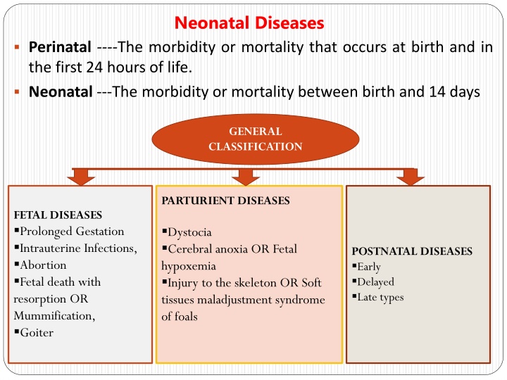
Neonatal Diseases Perinatal ----The morbidity or mortality that occurs at birth and in the first 24 hours of life. Neonatal ---The morbidity or mortality between birth and 14 days GENERAL CLASSIFICATION PARTURIENT DISEASES FETAL DISEASES Prolonged Gestation Intrauterine Infections, Abortion Fetal death with resorption OR Mummification, Goiter Dystocia Cerebral anoxia OR Fetal hypoxemia Injury to the skeleton OR Soft tissues maladjustment syndrome of foals POSTNATAL DISEASES Early Delayed Late types
POSTNATAL DISEASES Early postnatal disease (within 48 hours of birth) Deaths by an infectious (acquired congenitally.) Most diseases occurring are noninfectious and metabolic (e.g., hypoglycemia hypothermia. Congenital commonly manifest during this period but may sometimes manifest later. Infectious diseases, but most manifest clinically at a later age because of their long incubation period; some (e.g., navel infection, septicemic disease, and enterotoxigenic colibacillosis) have a short enough incubation to occur during this period. Delayed postnatal disease (2 to 7 days of age) Desertion by the mother, mammary incompetence resulting in starvation, and diseases associated with increased susceptibility to infection as a result of failure in the transfer of colostral immunoglobulins Examples colibacillosis, joint ill, lamb dysentery, disease, and most of the viral enteric infections in young animals rotavirus and coronavirus). Late postnatal disease (1 to 4 weeks of age) There is still some influence of hypogammaglobulinemia, with late-onset enteric diseases and the development and severity of respiratory disease and disease will Cryptosporidiosis, muscle enterotoxemia (Not directly associated with transfer of immunoglobulins) white and include disease, septicemic (e.g.,
Postnatal Disease Neonatal Diarrhea (Calf Scour) Highest risk for death in the first 2 weeks of life due to Septicemic and enteric diseases (diarrhea) Respiratory disease (pneumonia) being more common after 2 weeks of age Failure of transfer of passive immunity is a major determinant of this mortality Etiology: grouped :1) noninfectious causes and 2) infectious causes predisposing or contributing factors Noninfectious causes: Inadequate nutrition of the pregnant dam, particularly: Especially at last third of gestation. Both the quality and quantity of colostrum Deficiencies in vitamins A and E, and trace minerals
Inadequate environment for the newborn calf Colostrum given to calves >24 to 36 hours old is practically useless; antibodies are seldom absorbed this late in life Infectious causes: Bacteria: Escherichia coli, Salmonella spp., Clostridium perfringens, and other bacteria Viruses: coronavirus, rotavirus, BVD virus, IBR virus Protozoa: Cryptosporidium, coccidia Yeasts and molds A. Bacterial Causes of Calf Scours: Escherichia coli (E. coli): E. Coli affects mostly within first 3 days of life Numerous serotypes (kinds) of E. coli. It first colonize (or adhere to) the calf s gut through as pili or fimbriae 1.
These pili may possess the K99 antigen and called enterotoxigenic E. coli (E.T.E.C.) ETEC produces heat stable toxin leading to secretory diarrhea and severe dehydration. 2. Salmonella: Salmonella produces a potent toxin or an endotoxin (poison) within its own cells. Antibiotic treatment damages the salmonella organism, causing it to release the endotoxin, resulting in endotoxic shock, and severe illness. Therefore, treatment should be designed to combat endotoxic shock. Calves are usually affected at six days of age or older. The source of Salmonella infection in a herd can be from other cattle, birds, cats, rodents, the water supply, or human carrier.
3. Clostridium perfringens: Clostridium perfringens infections are commonly known as enterotoxemia. Enterotoxemia is fatal and is caused by toxins released by various types of C. perfringens. The disease has a sudden onset. Affected calves become listless, and strain or kick at their abdomen. Bloody diarrhea may or may not occur. Infection is usually associated with changing weather conditions, changes in the feed or feeding of the cows, or management practices that cause the calf to not nurse for a longer period of time than usual. The hungry calf may over consume milk, which establishes a media in the gut conducive to growth and production of toxins by clostridial organisms. In many cases, calves may die without any signs being observed.
B. Viral Causes of Calf Scours: 1.Coronavirus and Rotavirus Both of these viruses posses the ability to disrupt the cells which line the small intestine, resulting in diarrhea and dehydration. Coronavirus also damages the cells in the intestinal crypts (where new intestinal cells are produced) and slows the healing process in the intestinal lining. Furthermore, the damage caused by either corona or rotavirus is often compounded by bacterial infections and creating the risk for fatal diarrhea Calves as young as one or two days old may scour from corona or rotavirus, but outbreak mostly occur near a week of age and older 2.Bovine Virus Diarrhea (BVD) Virus Diarrhea begins about 24 hours to three days after exposure and may persist for days or weeks (if the animal survives that long). Erosions and ulcers on the tongue, lips, and in the mouth are the usual lesions found in the live calf.
3. Infectious Bovine Rhinotracheitis (IBR) Virus Mainly causes respiratory disease, abortions, vaginitis, and conjunctivitis in adult , but causes digestive disorders in young calves Affected calves had erosions and ulcers in the esophagus, and complicated by dullness, loss of weight, scours, and death. C. Protozoan Causes of Calf Scours: 1. Cryptosporidium : It is a protozoan parasite that is much smaller than coccidia It has the ability to adhere to the cells that line the small intestine and to damage the microvilli. Calves infected with Cryptosporidium range from one to three weeks in age. 2. Coccidiosis: It caused by one-celled parasites that invade the intestinal tract Two, Eimeria zurnii and Eimeria bovis, were usually associated with clinical infections in cattle.
The disease occur in calves 3 weeks of age and older, usually following stress, poor sanitation, overcrowding or sudden changes of feed. Fecal staining of the tail and perineum will be present. This may contain blood or not. The intestinal hemorrhage may subsequently lead to anemia and hypoproteinemia Clinical signs: Diarrhea, blood and fibrin in the feces, depression, and fever AND severe in young or debilitated calves. Extent of dehydration (percent) is judged by: Sunken eyes, skin tenting for 3-5 secs: 6-7 % dehydration Depression, skin tenting for 8-10 secs, dry mucus membranes: 8- 10% dehydration Recumbent, cool extremities, poor pulse: 11-12% dehydration
Management: Correction of the dehydration, acidosis, and electrolyte loss Withhold milk to calves and feed them whey Oral electrolyte replacement (depends on the extent of dehydration) In mild diarrhea the amount of oral fluids may be 1.1 kg/day whereas depressed calves may require 3 to 4 kg of oral electrolytes Intravenous fluids and glucose(advanced state) Oral fluids (1 teaspoonful of salt+ 1 teaspoonful of soda bicarbonate+2 teaspoonfuls of glucose +a pinch of potassium chloride +1.5 to 2 liters of body temperature water OR 1 tablespoon (15g) baking soda+ 1 teaspoon salt (5g)+ 250 ml (eight ounces) of 50 percent dextrose+ enough warm water to make one gallon and administer up to 1 liter of this material every three to four hours, depending upon the degree of dehydration and fluid loss.
Do not use milk or milk replacers, as milk in the intestinal tract makes an ideal medium for bacteria such as E. coli Antibiotics may be given and Sulfonamides may be used for thereatment of choice for coccidiosis for many years Amprolium as a preventative; this should be supplied at the rate of 5 mg/kg of body weight for a period of 21 days Management: 1. Nutrition Balanced in energy, protein, minerals, and vitamins to pregnant female Particular care must be taken to provide them with sufficient feed energy for maintenance and growth. Failure to meet energy needs will not only result in a weak calf at birth, but also contributes to increased dystocia (difficult calving), delayed return to estrus, and lowered conception rates 2. Environment and sanitation
3. Attention to the newborn calf should receive sufficient colostrum and may scour later in life and so must receive one to two quarts of colostrum during the first two to four hours immediately after birth 4. Vaccination programs (K99 E. coli antigen singly or in combination with coronavirus and rotavirus) CALF DIPHTHERIA Infectious disease involving larynx, buccal cavity and is characterized by fever and ulceration. When larynx involved, called necrotic laryngitis and when oral cavity involved called necrotic stomatitis Etiology:Fusobacterium necrophorum, a gm ve bacteria Epidemiology: World wide Poor and unsanitary management Traumatic injury to M.M of oral cavity Necrotic stomatitis in calves < 3months and necrotic laryngitis occur in claves up to 18 months of age
Pathogenesis: On reaching the site of infection---- causes inflammation, oedema and necrosis (oral mucosa, pharynx and larynx)----It leads to varying degree of closure of rimaglottidis, inspiratory dyspnoea and stridor---the lesion may extend to arytenoid cartilages resulting into laryngeal chondritis in which delayed healing may be observed Formation of abscess in different body organs as been also occur Clinical findings: Differ depending on organ involved Necrotic stomatitis: Difficulty in suckling, depressed and anorectic Temperature-103-104 o F Cheeks posterior to the lip commissures may be swollen, saliva often mixed with food particles drool from mouth Foul odor in breath Some times swelling and protrusion of tongue In severe cases, the lesion may spread to facial tissue, throat, vulva and around coronets Involvement of lung pneumonia and death due to toxaemia
Necrotic Laryngitis: High body temperature (1060F) Anorexia Depression Rapid respiration Salivation Nasal discharge Protrusion of tongue Moist painful coughing with inspiratory dypnoea Dysphagia and halitosis When infection goes to lung Bronchopneumonia Death if not treated due to toxaemia, pneumonia and obstruction of respiratory airways
Diagnosis: Clinical signs and along with inspection of managemenatal and feeding practices Isolation of organism (Swab) Treatment: Debridement of lesions and application of Tincture iodine as local antiseptics (Sulphamethazine @150mg/Kg BW for 3-5 days followed by oral. Sulphonamides and Sulphamerazine) Other antibiotics (Penicillin, Chloramphenicol and combination of penicillin and streptomycin ) may be used about 3 weeks Provision of soft, palatable and nutritious diets In necrotic laryngitis, use antibiotics and Corticosteroids(edema) and in severe cases trachestomy may be required
Calf Pneumonia Pneumonia is an inflammation of the lungs Etiology: Multifactorial disease. Factors leading to pneumonia include:
Clinical Signs: Problems that occur within 5 days of birth usually have their source as the dam or the calving environment. After 7 days of age, problems develop from a source in the calf environment Treatment: Antibiotic therapy is necessary along with non-steroidal anti-inflammatory drugs like aspirin, banamine or ketoprofen
HYPOTHERMIA IN NEWBORNS Homeothermic animals keep their body temperature almost constant to different ambient temperatures (Thermoregulation ) Hypothermia is a lower than normal body temperature, which occurs when excess heat is lost or insufficient heat is produced Etiology: 1. Excessive Loss of Heat: If exposure to excessively cold air temperatures >increased metabolic activity, shivering and sustained muscular contraction, and peripheral vasoconstriction---Heat loss 2. Insufficient Heat Production: Insufficient body reserves of energy and insufficient feed intake result in insufficient heat production 3. Combination of Excessive Heat Loss and Insufficient Heat Production: Insufficient energy intake or starvation of newborn farm animals in a cold environment can be a major cause of hypothermia.
Thermoregulation in Neonatal Farm Animals: Neonatal ruminants are precocial in their development and have well-developed thermoregulatory mechanisms Prolonged exposure to heat or cold induces hormonal and metabolic changes Glucocorticoid hormones, catecholamines , fat, glycogen, and proteins are used for heat production Cold-Induced Thermogenesis : 1. Shivering thermogenesis: involuntary, periodic contractions of skeletal muscle 2. Nonshivering thermogenesis: Involve brown adipose tissue (abdominal cavity , perirenal, around large blood vessels, and in the inguinal and prescapular areas), which is present in neonatal lambs, kids, and calves but not in piglets Norepinephrine- increased blood flow to brown adipose tissue Glucocorticoids mobilization of lipid and glycogen to supply energy substrates Thyroid hormones- regulating cold thermogenesis
In neonatal lambs, approximately 40% of the thermogenic response during summit metabolism( cold-induced peak metabolic rate) is attributed to non-shivering thermogenesis, with the balance of about 60% attributed to shivering thermogenesis. Risk Factors for Neonatal Hypothermia: Calves: Calves born outdoors during cold weather Wind, rain, and snow decrease the level of insulation and increase the lower critical temperature Dystocia-inhibit nonshivering thermogenesis and impair cold tolerance immediately after birth Colostrum Lamb: Bad weather Starvation, low birth weight, birth injury, and sparse hair coat Colostrum intake---lambs require 180 to 210 mL colostrum /Kg BW in the first 18 hours after birth to provide sufficient energy substrate for heat production
Wetness of the fleece--Wet lambs suffer a reduction in coat insulation Piglets: At birth, the newborn piglet experiences a sudden and dramatic 15 to 20 C decrease in its thermal environment, because poorly insulation pig does not possess brown adipose tissue, so rely essentially on muscular thermogenesis for thermoregulatory purposes Newborn pigs shiver vigorously from birth because it is the main heat-producing mechanismduring the first 5 days of life Body reserves (glycogen and fat) are important for the piglet to survive in the first few hours Foals: premature, dysmature, or affected with neonatal maladjustment syndrome cannot maintain their rectal temperatures at normal values during the first few hours after birth
The lower critical temperature for healthy foals is estimated to be about 10 C and for sick foals is about 24 C When wet with amniotic fluid, the lower critical temperature probably will be much higher Covering these foals with rugs and providing thermal radiation using radiant heaters would increase the lower critical temperature Premature foals are the most compromised compared with dysmature and those with neonatal maladjustment syndrome Colostrum intake
Moisture and wind produce Emitting heat in electromagnetic waves Amniotic fluid on the newborns surface Cold surfaces
PATHOGENESIS: Sudden exposure to cold ambient temperature Decreased cardiac output, heart rate, and blood pressure. Subnormal Body temperature Shivering Muscular weakness Mental depression, Respiratory failure Recumbency (have a weak suck reflex) A state of collapse (Bradycardia, weak arterial pulse, and collapse of the veins) coma and death The entire body, especially the extremities, becomes cold and the rectal temperature is below 37 C and may drop to 30 C or lower Convulsions in some cases especially in piglets that have inadequate intake of milk, caused by marked hypoglycemia rather than hypothermia
TREATMENT: Supplemental heat--infrared heat lamps Rectal temperature---taken every 30 minutes during rewarming to assess progress Colostrum or milk should be warmed to 40 C and intubated using an esophageal feeder Intravenous warmed fluids Intravenous dextrose (1 mL of 50% dextrose per kilogram BW) The repeated administration of warm (40 C) 0.9% NaCl enemas via a flexible soft tube Lamb: Twin and triplet lambs are more susceptible to hypothermia than singles because of lower body energy reserves The milk requirement of two or three lambs is higher than that of a single lamb and starvation is more likely
CONTROL: Lambs and Calves: Effective management strategies to limit the risk factors (Malnutrition of the Dam During Late Gestation, Dystocia and Colostrum supplies ) Note: 1. During a normal delivery fetal hypoxemia anaerobic glycolysis compensate for within hours after birth acidosis lactic acid, and a mixed respiratory metabolic In prolonged dystocia metabolic acidosis nonshivering thermogenesis inhibited and impair cold tolerance, immediately after birth Dystocia may result in a weak calf that has a poor suck reflex, and a poor appetite for colostrum, resulting in colostrum deprivation and hypogammaglobulinemia
2. Management strategies to prevent hypothermia from excessive heat loss (changing the calving season to a warmer time of the year, providing a dry, draft-free environment, protective shelter) 3. To provide adequate quantities of colostrum, beginning as soon after birth to provide immunoglobulin and energy sources Fading Puppy Complex The condition in which puppies are born apparently normal but fail to thrive and die, usually, before they reach 14 days of age It constitutes about 50% of the total neonatal deaths in canines Etiology: 1. Environmental 2. Genetic 3. 3. infectious 1. Environmental: Environmental Hypothermia or hyperthermia Maternal factors (Overweight or older dam) Environmental toxins(thinner chemical fumes) skin of neonates/Breathing
2. Genetic or congenital factors: Physical defects: Abnormalities of the mouth, anus, skull, and heart Swimmer (flat) puppies ( flattened and widened chests) Pectus excavatum is a severe deformity resulting from intrusion of the breastbone into the chest cavity Birth weigh: Varies with the breed (Eg: Pomeranian about 120 g and Great Dane about 625 g ) Weight gain should be steady and should gain 5% to 10% of birth weight daily Neonates should be weighed twice a day In toy-breeds, a transient juvenile hypoglycaemia, associated with low body weight.
3. Infectious agents: Bacterial infection: The immature immune systems, keep the risk for infection through the placenta, umbilicus, or gastrointestinal or respiratory tract from contaminated environments. Clinical signs of bacterial infection may vary but usually include vomiting, diarrhoea, constant crying, fever, failure to nurse, and sloughing of the ear and tail tips and toes Viral infection: Many viruses like Canine herpes virus infection, is a very common in puppies. The signs vary from constant crying to abdominal pain. In Canine parvovirus (type 1) infection, there is a rapid onset of crying, failure to nurse, vomiting, diarrhoea, difficult breathing (dyspnoea), and weakness. Canine adenovirus is also implicated rarely as a cause for this condition
Intestinal parasites: Transplacental transmission of roundworm and hookworm Pups can also acquire roundworm infection through the dam s milk. Besides, some protozoan parasites can also cause diarrhoea in the young ones. The condition is rarely fatal but can contribute to illness and put a neonate at higher risk of additional infection Icterus neonatorum: This form of haemolytic anaemia arises in pups as a result of previous blood transfusions of the bitch with a blood group similar to that of the pups. The antibodies are transferred through the colostrum to cause a haemolytic crisis in the pups. Such pups are weak, anaemic and jaundiced
Clinical signs: The clinical signs are vague and insidious It is often too difficult to save a puppy once clinical signs appear. The common findings are a low birth weight or failure to gain weight at the same rate as their siblings Decreased activity and inability to suckle Have a tendency to remain separate from the mother and the rest of the litter. They are often reported to cry weakly in a high pitched tone. Some people refer to this as seagulling due to its similarity to the cry of seagulls. These puppies often quickly progress to severe lethargy, loss of muscle tone and death.
Prevention: Provide adequate warmth to avoid become chilled and temperature of slightly above 103 F (39 C) should be maintained. Chilled puppies should always be warmed slowly to prevent tissue hypoxia Starvation and hypoglycaemia should avoided to limit the risk of hypothermia Due to water turnover rates twice that of the adult as well as immature kidney function, the neonate is particularly at risk from dehydration. The use of 5% dextrose/ Ringer lactate solution by subcutaneous route @ 1ml/30g bodyweight is recommended followed by oral 5-10% glucose @ 0.25 ml/30g until urine flow is normal Provide colostrums within first 12-24 hours In infectious condition, antibiotics may be of help along with good hygiene and management procedures Vaccination of the dam against important infectious diseases and deworming prior to whelping can help to reduce the early neonatal losses Congenital defects may be corrected with surgery and physical therapy
