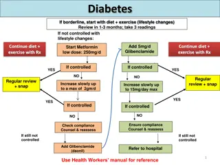
Pulmonary Hypertension
Mr. A, a 71-year-old man with a history of bladder cancer and pulmonary embolism, presents with breathlessness. Various imaging and diagnostic tests reveal findings suggestive of pulmonary hypertension. The case involves evaluation through echocardiography, V/Q scan, CTPA, and right heart catheterization. Differential diagnosis includes chronic thromboembolic pulmonary hypertension and other causes of PH. Interpretation of RHC values indicates significant findings related to PAH and left heart dysfunction. The case underscores the importance of accurate diagnosis in managing pulmonary hypertension.

Download Presentation

Please find below an Image/Link to download the presentation.
The content on the website is provided AS IS for your information and personal use only. It may not be sold, licensed, or shared on other websites without obtaining consent from the author.If you encounter any issues during the download, it is possible that the publisher has removed the file from their server.
You are allowed to download the files provided on this website for personal or commercial use, subject to the condition that they are used lawfully. All files are the property of their respective owners.
The content on the website is provided AS IS for your information and personal use only. It may not be sold, licensed, or shared on other websites without obtaining consent from the author.
E N D
Presentation Transcript
Pulmonary Hypertension 3 short cases Hannah Carlin SpR2 19.1.22
Mr A, 71y man Presented to local service with breathlessness Still able to walk 3-4 miles but subjectively slower and more breathless History of bladder cancer treated with curative surgery 2019/2020: provoked PE during neo-adjuvant chemotherapy treated with 3 months of anticoagulation Further saddle PE following surgery (off anticoagulation) Lifelong apixaban now No other comorbidities
Mr A Referral letter: Slow on inclines, unable to play tennis Echo at time of PE: dilated and impaired function of RV CT 5 months later: residual thrombus in bilateral lower lobe pulmonary arteries. V/Q: large mismatched perfusion defects to right apex and lower lobe as well as left lower lobe Echo 7 months post PE: Normal LA/LV. RV normal size but impaired function. "High probability of PH"
Mr A CTPA: Significant improvement in clot burden compared to index scan. Persisting organized clot both lower lobes with webs and post-thrombotic stenoses Echo: Normal LV size and function. Normal biatrial size. Normal RV. Indeterminate probability of PH. BNP 148 (normal) PFTs: Normal spirometry and lung volumes, TLCO and KCO both approx. 75% predicted 6MWT 480m with no desaturation CPET: reached VO2max of 17ml/min/kg (85% predicted)
MR A Right heart catheter: RA pressure RV systolic pressure PA pressure mPAP PCWP CO CI PVR MvO2 3 39 48/3 24 9 5.4 2.6 2.7 72% 1-7 15-25 15-25 / 4-12 10-20 6-12 4 - 6 2.4 - 4 <3 75-80%
What is the most likely diagnosis? Chronic thromboembolic pulmonary hypertension Chronic thromboembolic disease Pulmonary hypertension secondary to left heart disease Pulmonary hypertension secondary to lung disease No cause for symptoms identified
Important RHC values RA pressure > 10 overloaded RA mPAP 25 PH PA wedge pressure (PCWP) >15 left heart dysfunction PVR 3 elevated (usually >10 PAH, CTD lower / congenital higher
CTED (without PH) Nonresolution of clot AND in-situ thrombosis However the mPAP remains < 25 mmHg and PVR <3. either the number of occluded segments is insufficient to affect resistance at rest or no secondary vasculopathy has developed Functional impairment has been demonstrated on CPET (reduction of maximal work rate) and right heart/PA pressures elevated during exercise (vs PH and controls) Pulmonary endarterectomy high-risk, high morbidity surgery Balloon pulmonary angioplasty a future treatment option?
What is the prevalence of CTEPH following acute PE? 0.01 - 0.5% 0.5 4% 4 7% 7 9% 9 - 15 %
In patients presenting with acute PE, which of the following is not a risk factor for CTEPH? Splenectomy Previous VTE episode Pacemaker Inflammatory bowel disease Obesity hypoventilation
Mr B, 72y man Referred after an echo detected PASP 110mmHg Arranged by cardiology as part of followup for an ACS episode treated medically Complex medical history: IHD with previous MI in 2006 and 2019; recent angio showed two vessel disease with 50-60% stenosis in LAD not amenable to PCI Known mild LVSD EF 45-50% CKD with eGFR around 30 PE 2016 on warfarin Peripheral vascular disease
Mr B Felt much better following recent cardiology treatment WHO functional class 3 but leg pain rather than breathlessness limiting mobility CTPA: possible CTPEH but poor quality, V/Q heterogenous but with possible matched defect in left lower lobe Referred to FH for inpatient assessment INR noted to be 1.5 on admission 6MWT poor 210m but had to stop several times with desaturation to 68%
Mr B Echo: Normal LA size. Normal LV size but regional wall motion artefacts and mildly impaired function EF 45-50%. RV dilated and functionally impaired. Significant TR with PASP >100 BNP 5971 (previously 14800) CTPA: Persisting significant chronic thromboembolic disease and clot burden PFTs: lung volumes and spirometry normal, TLCO 35% / KCO 48%
Mr B Right heart catheter RA pressure RV systolic pressure PA pressure mPAP PCWP CO CI PVR MvO2 4 80 90/20 38 22 3.8 2.1 4.2 62% 1-7 15-25 15-25 / 4-12 10-20 6-12 4 - 6 2.4 - 4 <3 75-80%
According to ESC guidelines how would you classify his pulmonary hypertension? Group 1 Group 2 Group 3 Group 4 Group 5
1. mPAP 25? yes PH present Left heart dysfunction 2. PCWP > 15? yes no Not PH no Precapillary PH 3. CO normal / low yes no ?left to right shunt if CO high
Ms C, 22y woman Fit and well young woman Worsening breathlessness for around 7 months, attributed to asthma by GP (no spirometry or face-to-face review) Syncope episode attributed to heat Admitted acutely after second syncope episode and investigated after BNP found to be >5000 WHO performance status 4 CTPA: grossly dilated right sided cardiac chambers with reflux of contrast into IVC Echo: Severe right heart dilatation and dysfunction with PASP >100
Ms C Right heart catheter RA pressure RV systolic pressure PA pressure mPAP PCWP CO CI PVR MvO2 20 98 100/48 67 11 1.7 1.1 32 - 1-7 15-25 15-25 / 4-12 10-20 6-12 4 - 6 2.4 - 4 <3 75-80%
Which feature is least helpful for risk stratification? Presence of syncope WHO functional class Clinical signs of right heart failure Resting oxygen saturations Right heart catheter haemodynamic measurements
What is the optimal drug treatment for this patient? Dual therapy with PDE1 inhibitor and endothelin antagonist Sequential trials of PDE1 inhibitor or endothelin antagonist followed by dual if nor response Oral prostanoid Immediate triple therapy with IV prostanoid, PDE1 inhibitory, endothelin antagonist Sequential triple therapy
What is the most common gene implicated in familial PAH? ENG1 BMPR2 MUC5B F508del SERPINA1
What is the mode of inheritance of this gene? Autosomal recessive Autosomal dominant with complete penetrance Autosomal dominant with incomplete penetrance X-linked None of the above


")