Protein Domains and Databases
 undefined


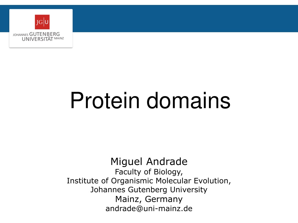
Protein domains
Miguel Andrade
Faculty of Biology,
Institute of Organismic Molecular Evolution,
Johannes Gutenberg University
Mainz, Germany
andrade@uni-mainz.de
Protein domains are structural units
(average 160 aa) that share:
Function
Folding
Evolution
Proteins normally are
multidomain
(average 300 aa)
Introduction
Protein domains are structural units
(average 160 aa) that share:
Function
Folding
Evolution
Proteins normally are
multidomain
(average 300 aa)
Introduction
Why to search for domains:
Protein structural determination methods such as X-ray
crystallography and NMR have size limitations that limit their
use.
Multiple sequence alignment at the domain level can result in
the detection of homologous sequences that are more difficult
to detect using a complete chain sequence.
Methods used to gain an insight into the structure and
function of a protein work best at the domain level.
Domains
Domain databases
SMART
Schultz et al (1998)
PNAS
…
Letunic et al (2017)
Nucleic Acids Research
Peer Bork
http://smart.embl.de/
Manual definition of domain (bibliography)
Generate profile from instances of domain
Search for remote homologs (HMMer)
Include them in profile
Iterate until convergence
Domain databases
Domain databases
SMART
Domain databases
SMART
Domain databases
SMART
Extra features:
Signal-peptide,
low complexity, TM, coiled coils
Domain databases
SMART
Domain databases
SMART
Domain databases
PFAM
Erik Sonnhammer/Ewan Birney/Alex Bateman
http://pfam.xfam.org/
Sonnhammer et al (1997)
Proteins
...
El Gebali et al (2019)
Nucleic Acids Research
Domain databases
PFAM
Domain databases
PFAM
Wikipedia rules!
Domain databases
PFAM
Domain databases
CDD
Marchler-Bauer et al (2017)
Nucleic Acids Res
http://www.ncbi.nlm.nih.gov/cdd
Stephen Br
yant
Domain databases
CDD
Domain databases
SMART
PFAM
CDD
SORLA
/
SORL1 from
Homo sapiens
Exercise 1
•
Let’s see whether human myosin X (UniProt id Q9HD67) or
its homologs have a solved structure. Go to PDB’s BLAST
page:
Menu > Search > Sequences
http://www.rcsb.org/pdb/secondary.do?p=v2/secondary/searc
h.jsp#search_sequences
•
Obtain from UniProt the protein sequence “Q9HD67” and
paste it the Entry Query Sequence window.
•
In the Choose Search Set you can select the database to
search against: select as Database option "Protein Data Bank".
Examine a UniProt Entry and find related PDBs
Exercise 1
Examine a UniProt Entry and find related PDBs
Paste sequence here
Exercise 1
•
Hit the “Run Sequence Search” below the input
window.
•
Considering that your query was a human myosin X,
can you interpret the first three hits? Which part of
your query was matched? Which protein was hit in
the database?
•
What about the 4
th
hit?
Examine a UniProt Entry and find related PDBs
Exercise 1
•
Hit the “Run Sequence Search” below the input
window.
•
Considering that your query was a human myosin X,
can you interpret the first three hits? Which part of
your query was matched? Which protein was hit in
the database?
•
What about the 4
th
hit?
•
Can you find a hit to a protein that is not human
myosin X? Which part of your query was matched?
Examine a UniProt Entry and find related PDBs
Exercise 2
•
Let’s look at the domains predicted for human
myosin X.
Go to PFAM:
http://pfam.xfam.org/
•
Select the option VIEW A SEQUENCE
•
Type in the window the UniProt id of the protein
sequence “Q9HD67” and hit the Go button.
•
Compare the positions of the domains predicted with
the ranges of the BLAST matches in PDB from the
previous exercise.
Which domains were matched in the human myosin
X by each of those hits?
Analyse domain predictions with PFAM
Exercise 3
•
Open the structure of the 3
rd
hit (3PZD) in
Chimera
Now colour the fragments corresponding to the
PFAM domains MyTH4 (in blue), RAS associated (in
red) and FERM_M (in orange).`
How do the PFAM annotations fit the structure?
How many more domains can you identify
visually?
•
Chain B in this structure is a small peptide.
Which part of the human myosin X is interacting
with this peptide in relation to the domains you
have coloured? And what about the glycerol?
Examine domains in Chimera
Protein domains are structural units that play key roles in protein function, folding, and evolution. Exploring protein domains is crucial for gaining insights into protein structure and function. Domain databases like SMART offer tools for defining, profiling, and searching domains, including extra features like signal peptides and transmembrane regions. By searching for domains, researchers can overcome limitations of traditional structural determination methods and identify homologous sequences effectively.

Download Presentation

Please find below an Image/Link to download the presentation.
The content on the website is provided AS IS for your information and personal use only. It may not be sold, licensed, or shared on other websites without obtaining consent from the author.If you encounter any issues during the download, it is possible that the publisher has removed the file from their server.
You are allowed to download the files provided on this website for personal or commercial use, subject to the condition that they are used lawfully. All files are the property of their respective owners.
The content on the website is provided AS IS for your information and personal use only. It may not be sold, licensed, or shared on other websites without obtaining consent from the author.
E N D
Presentation Transcript
Protein domains Miguel Andrade Faculty of Biology, Institute of Organismic Molecular Evolution, Johannes Gutenberg University Mainz, Germany andrade@uni-mainz.de
Introduction Protein domains are structural units (average 160 aa) that share: Function Folding Evolution Proteins normally are multidomain (average 300 aa)
Introduction Protein domains are structural units (average 160 aa) that share: Function Folding Evolution Proteins normally are multidomain (average 300 aa)
Domains Why to search for domains: Protein structural determination methods such as X-ray crystallography and NMR have size limitations that limit their use. Multiple sequence alignment at the domain level can result in the detection of homologous sequences that are more difficult to detect using a complete chain sequence. Methods used to gain an insight into the structure and function of a protein work best at the domain level.
Domain databases SMART Peer Bork http://smart.embl.de/ Manual definition of domain (bibliography) Generate profile from instances of domain Search for remote homologs (HMMer) Include them in profile Iterate until convergence Schultz et al (1998) PNAS Letunic et al (2017) Nucleic Acids Research
Domain databases SMART
Domain databases SMART
Domain databases SMART Extra features: Signal-peptide, low complexity, TM, coiled coils
Domain databases SMART
Domain databases SMART
Domain databases PFAM Erik Sonnhammer/Ewan Birney/Alex Bateman http://pfam.xfam.org/ Sonnhammer et al (1997) Proteins ... El Gebali et al (2019) Nucleic Acids Research
Domain databases PFAM
Domain databases PFAM Wikipedia rules!
Domain databases PFAM
Domain databases CDD Stephen Bryant http://www.ncbi.nlm.nih.gov/cdd Marchler-Bauer et al (2017) Nucleic Acids Res
Domain databases CDD
Domain databases SORLA/SORL1 from Homo sapiens SMART PFAM CDD
Exercise 1 Examine a UniProt Entry and find related PDBs Let s see whether human myosin X (UniProt id Q9HD67) or its homologs have a solved structure. Go to PDB s BLAST page: Menu > Search > Sequences http://www.rcsb.org/pdb/secondary.do?p=v2/secondary/searc h.jsp#search_sequences Obtain from UniProt the protein sequence Q9HD67 and paste it the Entry Query Sequence window. In the Choose Search Set you can select the database to search against: select as Database option "Protein Data Bank".
Exercise 1 Examine a UniProt Entry and find related PDBs Paste sequence here
Exercise 1 Examine a UniProt Entry and find related PDBs Hit the Run Sequence Search below the input window. Considering that your query was a human myosin X, can you interpret the first three hits? Which part of your query was matched? Which protein was hit in the database? What about the 4th hit?
Exercise 1 Examine a UniProt Entry and find related PDBs Hit the Run Sequence Search below the input window. Considering that your query was a human myosin X, can you interpret the first three hits? Which part of your query was matched? Which protein was hit in the database? What about the 4th hit? Can you find a hit to a protein that is not human myosin X? Which part of your query was matched?
Exercise 2 Analyse domain predictions with PFAM Let s look at the domains predicted for human myosin X. Go to PFAM: http://pfam.xfam.org/ Select the option VIEW A SEQUENCE Type in the window the UniProt id of the protein sequence Q9HD67 and hit the Go button. Compare the positions of the domains predicted with the ranges of the BLAST matches in PDB from the previous exercise. Which domains were matched in the human myosin X by each of those hits?
Exercise 3 Examine domains in Chimera Open the structure of the 3rd hit (3PZD) in Chimera Now colour the fragments corresponding to the PFAM domains MyTH4 (in blue), RAS associated (in red) and FERM_M (in orange).` How do the PFAM annotations fit the structure? How many more domains can you identify visually? Chain B in this structure is a small peptide. Which part of the human myosin X is interacting with this peptide in relation to the domains you have coloured? And what about the glycerol?