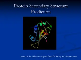
Protein Tertiary Structure and Motifs

Introduction
•
The overall three-dimensional structure of a protein
chain, including the positions of amino acid side chains,
is referred to as the tertiary structure of the protein.
Knowledge of
•
Protein tertiary structure is essential to understanding
how enzymes function and how to design, inhibit, and
activate proteins. As the tertiary structure of the
protein is specific to a particular protein sequence,
several additional terms describe the architecture of
proteins in three-dimensional space, and these terms
facilitate the comparison of different proteins on
several levels. Protein structures are often composed of
independently folding units within the same protein
chain, called domains.
Introduction
•
Every protein domain has a fold, which refers to
the arrangement of secondary structure elements
in that domain. Protein domains often contain
specific arrangements of a few contiguous
secondary structure elements.
•
Such units of secondary structure groups that are
repeatedly found in a variety of proteins are
called motifs or supersecondary structures.
Motifs are unable to fold independently and
often do not perform a specific function, thus
discriminating motifs from protein domains.
Levels of protein structure
•
Proteins can be described using four levels or
structures: the primary structure, or amino acid
sequence of the protein; the secondary structure,
or the local spatial arrangement of the
polypeptide backbone atoms, often organized
into regular elements such as alpha helices and
beta sheets; the tertiary structure of a protein, or
the overall structure of a single polypeptide
chain; and the quaternary structure, or the
arrangement of multiple polypeptide chains.
Conti…..
•
The tertiary structure describes the position of all
backbone atoms as well as side-chain atoms in a
given polypeptide chain.
•
Each different protein sequence necessarily has a
unique tertiary structure. This can be confusing
as the core arrangement of secondary structure
elements, also called the fold, can be the same
for proteins of different sequences. Therefore, in
order to more easily compare protein structures,
the folds of individual domains are often used.
Motif
•
Secondary structure elements describe local regions of polypeptide
backbone conformation and include the alpha helix and beta sheet.
•
A few of these contiguous secondary structure elements can combine in
specific three-dimensional arrangements to form super secondary
structures or structural motifs. These structural motifs may be associated
with a function, such as the DNA-binding helix -turn-helix motif (Fig. 1a),
but more often are not associated with any particular function. Other
common structural motifs include the beta-hairpin (Fig. 1b) and Greek-key
(Fig. 1c) motifs.
•
Motifs are unable to fold or evolve independently and are only found as
fragments of a protein. In some cases a series of motifs may combine to
form a stably folding domain. The TIM barrel or (beta-alpha) fold is
composed of a series of eight overlapping beta-alpha-beta motifs (Figs. 2b
and 3a).
Protein Domain
•
Within a given protein structure, there are regions that independently
form stable tertiary structures. These regions are called domains. Domains
can independently fold and evolve and are associated with a specific
function.
•
Protein domains are not always composed of contiguous stretches of
polypeptide and may have additional regions of polypeptide inserted into
the protein.
•
As domains can fold independently and have a specific function, they can
be treated as modules that can be swapped from one protein to another
or even added on to a protein.
•
Nuclear receptor proteins have a multidomain structure, including an N-
terminal hypervariable (or A/B) domain, a DNA-binding domain, and a
ligand-binding domain (Fig. 2a).
•
Studies with nuclear receptors demonstrated that swapping the DNA-
binding domains of two different receptors also swaps the DNA sequence
recognized by the protein (Giguere et al. 1987).
•
Domains may be added to the N- or C-terminus of a protein. The zinc-
finger domain is a small DNA-binding domain common in eukaryotes.
Studies have demonstrated that multiple zinc-finger domains can be
linked together to increase the region of DNA recognized by the protein,
therefore greatly increasing its specificity (Urnov et al. 2010).
Conti….
•
There are Many reasons multi domain proteins exist. Some multi
domain proteins are composed of two domains that function to
catalyze successive steps in a biosynthetic pathway.
•
The bifunctional enzyme phosphoribosylanthranilate isomerase-
indoleglycerolphosphate synthase (PRAI-IGPS) is one such enzyme
and is composed of two (beta-alpha) barrel domains fused together
(Fig. 2b).
•
In this way the product of the first reaction is shuttled directly to
the next reaction in the pathway (Wilmanns et al. 1992). Other
multidomain proteins play an important role in cell signaling and
transport. Proteins involved in signaling may have multiple PDZ and
SH3 domains in order to facilitate the formation of protein signaling
complexes (Fig. 2c).
Fold
•
The fold of a protein is closely related to the domain. Each domain
possesses a given fold, which refers to the core arrangement of secondary
structure elements, excluding side-chain position.
•
The fold of a protein may involve the insertion or deletion of regions of
polypeptide.
•
.Many proteins with different tertiary structures and a variety of functions
can utilize the same fold, and as a result most folds are not associated
with a given function.
•
The TIM barrel or (beta-alpha) fold (Fig. 3a) is the most common protein
fold in existence and can catalyze reactions in five of the six major enzyme
classification groups (Wierenga 2001).
•
. However, the Rossmann fold (beta-alpha-beta-alpha-beta unit that is
often paired with itself; Fig. 3b) is an exception to this rule, and this fold
has been demonstrated to bind nucleotides, particularly NAD+ and NADP
Protein tertiary structure, essential for enzyme function and protein design, refers to the overall three-dimensional arrangement of amino acid side chains in a protein chain. This structure is specific to each protein sequence, with domains and motifs playing key roles in protein architecture. Protein structures have four levels: primary, secondary, tertiary, and quaternary. Motifs are specific arrangements of secondary structure elements that do not fold independently but can form functional units within protein domains.

Download Presentation

Please find below an Image/Link to download the presentation.
The content on the website is provided AS IS for your information and personal use only. It may not be sold, licensed, or shared on other websites without obtaining consent from the author.If you encounter any issues during the download, it is possible that the publisher has removed the file from their server.
You are allowed to download the files provided on this website for personal or commercial use, subject to the condition that they are used lawfully. All files are the property of their respective owners.
The content on the website is provided AS IS for your information and personal use only. It may not be sold, licensed, or shared on other websites without obtaining consent from the author.
E N D
Presentation Transcript
Introduction The overall three-dimensional structure of a protein chain, including the positions of amino acid side chains, is referred to as the tertiary structure of the protein. Knowledge of Protein tertiary structure is essential to understanding how enzymes function and how to design, inhibit, and activate proteins. As the tertiary structure of the protein is specific to a particular protein sequence, several additional terms describe the architecture of proteins in three-dimensional space, and these terms facilitate the comparison of different proteins on several levels. Protein structures are often composed of independently folding units within the same protein chain, called domains.
Introduction Every protein domain has a fold, which refers to the arrangement of secondary structure elements in that domain. Protein domains often contain specific arrangements of a few contiguous secondary structure elements. Such units of secondary structure groups that are repeatedly found in a variety of proteins are called motifs or supersecondary Motifs are unable to fold independently and often do not perform a specific function, thus discriminating motifs from protein domains. structures.
Levels of protein structure Proteins can be described using four levels or structures: the primary structure, or amino acid sequence of the protein; the secondary structure, or the local spatial polypeptide backbone atoms, often organized into regular elements such as alpha helices and beta sheets; the tertiary structure of a protein, or the overall structure of a single polypeptide chain; and the quaternary structure, or the arrangement of multiple polypeptide chains. arrangement of the
Conti.. The tertiary structure describes the position of all backbone atoms as well as side-chain atoms in a given polypeptide chain. Each different protein sequence necessarily has a unique tertiary structure. This can be confusing as the core arrangement of secondary structure elements, also called the fold, can be the same for proteins of different sequences. Therefore, in order to more easily compare protein structures, the folds of individual domains are often used.
Motif Secondary structure elements describe local regions of polypeptide backbone conformation and include the alpha helix and beta sheet. A few of these contiguous secondary structure elements can combine in specific three-dimensional arrangements to form super secondary structures or structural motifs. These structural motifs may be associated with a function, such as the DNA-binding helix -turn-helix motif (Fig. 1a), but more often are not associated with any particular function. Other common structural motifs include the beta-hairpin (Fig. 1b) and Greek-key (Fig. 1c) motifs. Motifs are unable to fold or evolve independently and are only found as fragments of a protein. In some cases a series of motifs may combine to form a stably folding domain. The TIM barrel or (beta-alpha) fold is composed of a series of eight overlapping beta-alpha-beta motifs (Figs. 2b and 3a).
Protein Domain Within a given protein structure, there are regions that independently form stable tertiary structures. These regions are called domains. Domains can independently fold and evolve and are associated with a specific function. Protein domains are not always composed of contiguous stretches of polypeptide and may have additional regions of polypeptide inserted into the protein. As domains can fold independently and have a specific function, they can be treated as modules that can be swapped from one protein to another or even added on to a protein. Nuclear receptor proteins have a multidomain structure, including an N- terminal hypervariable (or A/B) domain, a DNA-binding domain, and a ligand-binding domain (Fig. 2a). Studies with nuclear receptors demonstrated that swapping the DNA- binding domains of two different receptors also swaps the DNA sequence recognized by the protein (Giguere et al. 1987). Domains may be added to the N- or C-terminus of a protein. The zinc- finger domain is a small DNA-binding domain common in eukaryotes. Studies have demonstrated that multiple zinc-finger domains can be linked together to increase the region of DNA recognized by the protein, therefore greatly increasing its specificity (Urnov et al. 2010).
Conti. There are Many reasons multi domain proteins exist. Some multi domain proteins are composed of two domains that function to catalyze successive steps in a biosynthetic pathway. The bifunctional enzyme phosphoribosylanthranilate isomerase- indoleglycerolphosphate synthase (PRAI-IGPS) is one such enzyme and is composed of two (beta-alpha) barrel domains fused together (Fig. 2b). In this way the product of the first reaction is shuttled directly to the next reaction in the pathway (Wilmanns et al. 1992). Other multidomain proteins play an important role in cell signaling and transport. Proteins involved in signaling may have multiple PDZ and SH3 domains in order to facilitate the formation of protein signaling complexes (Fig. 2c).
Fold The fold of a protein is closely related to the domain. Each domain possesses a given fold, which refers to the core arrangement of secondary structure elements, excluding side-chain position. The fold of a protein may involve the insertion or deletion of regions of polypeptide. .Many proteins with different tertiary structures and a variety of functions can utilize the same fold, and as a result most folds are not associated with a given function. The TIM barrel or (beta-alpha) fold (Fig. 3a) is the most common protein fold in existence and can catalyze reactions in five of the six major enzyme classification groups (Wierenga 2001). . However, the Rossmann fold (beta-alpha-beta-alpha-beta unit that is often paired with itself; Fig. 3b) is an exception to this rule, and this fold has been demonstrated to bind nucleotides, particularly NAD+ and NADP