Overview of Contrast Media in Radiography
P
r
o
f
.
I
b
r
a
h
i
m
A
l
r
a
s
h
i
d
A
c
o
n
t
r
a
s
t
m
e
d
i
u
m
i
s
a
s
u
b
s
t
a
n
c
e
t
h
a
t
i
s
a
d
m
i
n
i
s
t
e
r
e
d
t
o
t
h
e
p
a
t
i
e
n
t
t
h
a
t
i
s
e
i
t
h
e
r
m
o
r
e
r
a
d
i
o
p
a
q
u
e
o
r
m
o
r
e
r
a
d
i
o
l
u
c
e
n
t
t
h
a
n
t
h
e
s
u
r
r
o
u
n
d
i
n
g
t
i
s
s
u
e
.
T
h
i
s
a
l
l
o
w
s
a
s
s
e
s
s
m
e
n
t
o
f
t
h
e
p
o
s
i
t
i
o
n
,
s
i
z
e
,
s
h
a
p
e
a
n
d
i
n
t
e
r
n
a
l
a
r
c
h
i
t
e
c
t
u
r
e
o
f
t
h
e
o
r
g
a
n
t
h
a
t
w
a
s
n
o
t
a
p
p
a
r
e
n
t
o
n
t
h
e
o
r
i
g
i
n
a
l
r
a
d
i
o
g
r
a
p
h
.
:
S
e
q
u
e
n
t
i
a
l
f
i
l
m
s
o
r
t
h
e
u
s
e
o
f
i
m
a
g
e
-
i
n
t
e
n
s
i
f
i
e
d
f
l
u
o
r
o
s
c
o
p
y
m
a
y
a
l
s
o
s
h
o
w
t
h
e
f
u
n
c
t
i
o
n
o
f
a
n
o
r
g
a
n
,
e
.
g
.
,
t
h
e
r
a
t
e
o
f
s
t
o
m
a
c
h
e
m
p
t
y
i
n
g
o
r
t
h
e
p
r
e
s
e
n
c
e
o
f
p
e
r
i
s
t
a
l
s
i
s
.
P
r
o
p
e
r
t
i
e
s
r
e
q
u
i
r
e
d
i
n
a
n
i
d
e
a
l
c
o
n
t
r
a
s
t
m
e
d
i
u
m
i
n
c
l
u
d
e
D
i
f
f
e
r
e
n
t
a
b
s
o
r
p
t
i
v
e
p
o
w
e
r
f
r
o
m
t
i
s
s
u
e
,
t
h
e
r
e
b
y
p
r
o
d
u
c
i
n
g
e
f
f
e
c
t
i
v
e
r
a
d
i
o
g
r
a
p
h
i
c
c
o
n
t
r
a
s
t
;
1- No irritant or toxic side effects
2-accurate delineation of the organ
3-Persistence for sufficient time to take
radiographs
TYPES OF CONTRAST MEDIA
1-Negative contrast studies will show the
location, size and wall thickness of the
organ and will show marked wall thickening
and large luminal filling defects such as
masses or foreign bodies.
2
-
P
o
s
i
t
i
v
e
c
o
n
t
r
a
s
t
s
t
u
d
i
e
s
g
i
v
e
l
i
t
t
l
e
m
o
r
e
i
n
f
o
r
m
a
t
i
o
n
t
h
a
n
n
e
g
a
t
i
v
e
c
o
n
t
r
a
s
t
s
t
u
d
i
e
s
b
u
t
a
r
e
t
h
e
b
e
s
t
w
a
y
o
f
d
e
t
e
c
t
i
n
g
a
s
m
a
l
l
d
e
f
e
c
t
i
n
t
h
e
w
a
l
l
o
f
t
h
e
o
r
g
a
n
,
a
s
m
i
n
o
r
c
o
n
t
r
a
s
t
l
e
a
k
a
g
e
i
s
e
a
s
i
l
y
s
e
e
n
.
POSITIVE CONTRAST MEDIA
1.Barium sulphate preparations
2.Iodine preparations
a) Ionic, water-soluble iodine contrast media
b) Non-ionic, water-soluble iodine contrast
media
Radiation safety
R
a
d
i
a
t
i
o
n
s
a
f
e
t
y
p
r
i
n
c
i
p
l
e
s
a
i
m
t
o
l
i
m
i
t
e
x
p
o
s
u
r
e
t
o
i
o
n
i
s
i
n
g
r
a
d
i
a
t
i
o
n
f
o
r
r
a
d
i
a
t
i
o
n
t
h
e
r
a
p
y
p
e
r
s
o
n
n
e
l
,
p
e
o
p
l
e
a
f
f
e
c
t
e
d
b
y
c
a
n
c
e
r
a
n
d
t
h
e
g
e
n
e
r
a
l
p
u
b
l
i
c
.
W
h
e
r
e
v
e
r
t
h
e
r
e
i
s
k
n
o
w
n
r
i
s
k
o
f
e
x
p
o
s
u
r
e
t
o
i
o
n
i
s
i
n
g
r
a
d
i
a
t
i
o
n
,
h
e
a
l
t
h
p
r
o
f
e
s
s
i
o
n
a
l
s
m
u
s
t
b
e
g
u
i
d
e
d
b
y
t
h
e
A
L
A
R
A
(
a
s
l
o
w
a
s
r
e
a
s
o
n
a
b
l
y
a
c
h
i
e
v
a
b
l
e
)
p
r
i
n
c
i
p
l
e
s
o
f
r
a
d
i
a
t
i
o
n
s
a
f
e
t
y
f
o
r
t
i
m
e
,
d
i
s
t
a
n
c
e
a
n
d
s
h
i
e
l
d
i
n
g
.
T
i
m
e
T
h
e
l
e
s
s
t
i
m
e
s
p
e
n
t
n
e
a
r
a
r
a
d
i
a
t
i
o
n
s
o
u
r
c
e
,
t
h
e
l
e
s
s
r
a
d
i
a
t
i
o
n
a
b
s
o
r
b
e
d
.
T
h
i
s
i
s
e
s
p
e
c
i
a
l
l
y
i
m
p
o
r
t
a
n
t
f
o
r
p
e
r
s
o
n
n
e
l
s
u
c
h
a
s
r
a
d
i
a
t
i
o
n
t
h
e
r
a
p
i
s
t
s
D
i
s
t
a
n
c
e
T
h
e
i
n
v
e
r
s
e
-
s
q
u
a
r
e
l
a
w
s
t
a
t
e
s
t
h
a
t
r
a
d
i
a
t
i
o
n
e
x
p
o
s
u
r
e
a
n
d
d
i
s
t
a
n
c
e
a
r
e
i
n
v
e
r
s
e
l
y
r
e
l
a
t
e
d
.
S
h
i
e
l
d
i
n
g
T
h
e
t
y
p
e
o
f
s
h
i
e
l
d
i
n
g
d
e
v
i
c
e
u
s
e
d
d
e
p
e
n
d
s
o
n
t
h
e
r
a
n
g
e
o
f
e
m
i
s
s
i
o
n
o
f
t
h
e
r
a
d
i
o
a
c
t
i
v
e
s
o
u
r
c
e
.
S
t
a
n
d
a
r
d
s
h
i
e
l
d
i
n
g
d
e
v
i
c
e
s
i
n
c
l
u
d
e
l
e
a
d
a
p
r
o
n
s
,
t
h
y
r
o
i
d
s
h
i
e
l
d
s
,
a
n
d
e
y
e
s
h
i
e
l
d
s
S
p
i
l
l
m
a
n
a
g
e
m
e
n
t
I
n
t
h
e
e
v
e
n
t
o
f
a
r
a
d
i
a
t
i
o
n
i
n
c
i
d
e
n
t
,
s
u
c
h
a
s
t
h
e
l
o
s
s
o
f
a
s
o
u
r
c
e
o
r
a
s
p
i
l
l
,
a
p
p
r
o
p
r
i
a
t
e
p
r
o
c
e
d
u
r
e
s
a
n
d
n
o
t
i
f
i
c
a
t
i
o
n
s
m
u
s
t
b
e
f
o
l
l
o
w
e
d
.
T
h
e
s
e
s
h
o
u
l
d
b
e
c
l
e
a
r
l
y
o
u
t
l
i
n
e
d
i
n
t
h
e
c
l
i
n
i
c
a
l
e
n
v
i
r
o
n
m
e
n
t
a
s
p
a
r
t
o
f
r
a
d
i
a
t
i
o
n
s
a
f
e
t
y
a
n
d
h
o
s
p
i
t
a
l
p
o
l
i
c
y
.
CT (Computed Tomography) Scan
A computerized tomography scan (CT or CAT scan) uses
computers and rotating X-ray machines to create cross-
sectional images of the body. These images provide more
detailed information than normal X-ray images. They can
show the soft tissues, blood vessels, and bones in various
parts of the body. A CT scan may be used to visualize the:
head
shoulders
spine
heart
abdomen
knee
chest
M
R
I
u
s
e
s
a
s
t
r
o
n
g
m
a
g
n
e
t
i
c
f
i
e
l
d
a
n
d
r
a
d
i
o
w
a
v
e
s
t
o
c
r
e
a
t
e
d
e
t
a
i
l
e
d
i
m
a
g
e
s
o
f
t
h
e
o
r
g
a
n
s
a
n
d
t
i
s
s
u
e
s
w
i
t
h
i
n
t
h
e
b
o
d
y
.
S
i
n
c
e
i
t
s
i
n
v
e
n
t
i
o
n
,
d
o
c
t
o
r
s
a
n
d
r
e
s
e
a
r
c
h
e
r
s
c
o
n
t
i
n
u
e
t
o
r
e
f
i
n
e
M
R
I
t
e
c
h
n
i
q
u
e
s
t
o
a
s
s
i
s
t
i
n
m
e
d
i
c
a
l
p
r
o
c
e
d
u
r
e
s
a
n
d
r
e
s
e
a
r
c
h
.
T
h
e
d
e
v
e
l
o
p
m
e
n
t
o
f
M
R
I
r
e
v
o
l
u
t
i
o
n
i
z
e
d
m
e
d
i
c
i
n
e
.
F
u
n
c
t
i
o
n
a
l
m
a
g
n
e
t
i
c
r
e
s
o
n
a
n
c
e
i
m
a
g
i
n
g
o
r
f
u
n
c
t
i
o
n
a
l
M
R
I
(
f
M
R
I
)
u
s
e
s
M
R
I
t
e
c
h
n
o
l
o
g
y
t
o
m
e
a
s
u
r
e
c
o
g
n
i
t
i
v
e
a
c
t
i
v
i
t
y
b
y
m
o
n
i
t
o
r
i
n
g
b
l
o
o
d
f
l
o
w
t
o
c
e
r
t
a
i
n
a
r
e
a
s
o
f
t
h
e
b
r
a
i
n
.
T
h
e
b
l
o
o
d
f
l
o
w
i
n
c
r
e
a
s
e
s
i
n
a
r
e
a
s
w
h
e
r
e
n
e
u
r
o
n
s
a
r
e
a
c
t
i
v
e
.
T
h
i
s
g
i
v
e
s
a
n
i
n
s
i
g
h
t
i
n
t
o
t
h
e
a
c
t
i
v
i
t
y
o
f
n
e
u
r
o
n
s
i
n
t
h
e
b
r
a
i
n
.
T
h
i
s
t
e
c
h
n
i
q
u
e
h
a
s
r
e
v
o
l
u
t
i
o
n
i
z
e
d
b
r
a
i
n
m
a
p
p
i
n
g
,
b
y
a
l
l
o
w
i
n
g
r
e
s
e
a
r
c
h
e
r
s
t
o
a
s
s
e
s
s
t
h
e
b
r
a
i
n
a
n
d
s
p
i
n
a
l
c
o
r
d
w
i
t
h
o
u
t
t
h
e
n
e
e
d
f
o
r
i
n
v
a
s
i
v
e
p
r
o
c
e
d
u
r
e
s
o
r
d
r
u
g
i
n
j
e
c
t
i
o
n
s
.
Ultrasound is a type of imaging. It uses high-
frequency
sound waves to look at organs and structures
inside
the body.
Health care professionals use it to view the
heart,
blood vessels, kidneys, liver, and other organs.
During pregnancy, doctors use ultrasound to
view the
f
e
t
u
s
.
U
n
l
i
k
e
x
-
r
a
y
s
,
u
l
t
r
a
s
o
u
n
d
d
o
e
s
n
o
t
e
x
p
o
s
e
y
o
u
t
o
r
a
d
i
a
t
i
o
n
.
D
i
g
i
t
a
l
X
-
r
a
y
r
e
p
l
a
c
e
s
t
h
e
u
s
e
o
f
f
i
l
m
o
r
c
o
m
p
u
t
e
d
r
a
d
i
o
g
r
a
p
h
y
(
C
R
)
p
l
a
t
e
s
w
i
t
h
a
d
i
r
e
c
t
d
i
g
i
t
a
l
t
r
a
n
s
f
e
r
o
f
X
-
r
a
y
i
m
a
g
e
s
i
n
t
o
t
h
e
P
A
C
S
.
D
i
g
i
t
a
l
r
a
d
i
o
g
r
a
p
h
y
(
D
R
)
i
s
t
h
e
d
i
r
e
c
t
c
o
n
v
e
r
s
i
o
n
o
f
t
r
a
n
s
m
i
t
t
e
d
X
-
r
a
y
p
h
o
t
o
n
s
i
n
t
o
a
d
i
g
i
t
a
l
i
m
a
g
e
u
s
i
n
g
a
n
a
r
r
a
y
o
f
s
o
l
i
d
-
s
t
a
t
e
d
e
t
e
c
t
o
r
s
s
u
c
h
a
s
a
m
o
r
p
h
o
u
s
s
e
l
e
n
i
u
m
o
r
s
i
l
i
c
o
n
,
w
i
t
h
c
o
m
p
u
t
e
r
p
r
o
c
e
s
s
i
n
g
a
n
d
d
i
s
p
l
a
y
o
f
t
h
e
i
m
a
g
e
An electronic device that detects gamma rays
emitted by radio pharmaceautical (e.g
technetium 99m (Tc-99m) that have been
introduced into the body as tracers. The
position of the source of the
r
a
d
i
o
a
c
t
i
v
i
t
y
c
a
n
b
e
p
l
o
t
t
e
d
a
n
d
d
i
s
p
l
a
y
e
d
o
n
a
T
V
m
o
n
i
t
o
r
o
r
p
h
o
t
o
g
r
a
p
h
i
c
f
i
l
m
.
Contrast media play a crucial role in radiography by enhancing visualization of organs and structures. Types of contrast media, safety principles, and properties of ideal contrast media are discussed in detail.

Uploaded on Feb 27, 2025 | 0 Views
Download Presentation

Please find below an Image/Link to download the presentation.
The content on the website is provided AS IS for your information and personal use only. It may not be sold, licensed, or shared on other websites without obtaining consent from the author.If you encounter any issues during the download, it is possible that the publisher has removed the file from their server.
You are allowed to download the files provided on this website for personal or commercial use, subject to the condition that they are used lawfully. All files are the property of their respective owners.
The content on the website is provided AS IS for your information and personal use only. It may not be sold, licensed, or shared on other websites without obtaining consent from the author.
E N D
Presentation Transcript
A contrast medium is a substance that is administered to the patient that is either more radiopaque or more radiolucent than the surrounding tissue. This allows assessment of the position, size, shape and internal architecture of the organ that was not apparent on the original radiograph. :
Sequential films or the use of image- intensified fluoroscopy may also show the function of an organ, e.g., the rate of stomach emptying or the presence of peristalsis. Properties required in an ideal contrast medium include
Different absorptive power from tissue, thereby producing effective radiographic contrast ; 1- No irritant or toxic side effects 2-accurate delineation of the organ 3-Persistence for sufficient time to take radiographs
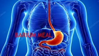
TYPES OF CONTRAST MEDIA 1-Negative contrast studies will show the location, size and wall thickness of the organ and will show marked wall thickening and large luminal filling defects such as masses or foreign bodies. 2-Positive contrast studies give little more information than negative contrast studies but are the best way of detecting a small defect in the wall of the organ, as minor contrast leakage is easily seen . TYPES OF CONTRAST MEDIA
POSITIVE CONTRAST MEDIA 1.Barium sulphate preparations 2.Iodine preparations a) Ionic, water-soluble iodine contrast media b) Non-ionic, water-soluble iodine contrast media POSITIVE CONTRAST MEDIA
Radiation safety Radiation safety principles aim to limit exposure to ionising radiation for radiation therapy personnel, people affected by cancer and the general public. Wherever there is known risk of exposure to ionising radiation, health professionals must be guided by the ALARA (as low as reasonably achievable) principles of radiation safety for time, distance and shielding. Radiation safety
Time The less time spent near a radiation source, the less radiation absorbed. This is especially important for personnel such as radiation therapists Distance The inverse-square law states that radiation exposure and distance are inversely related. Time Distance
Shielding The type of shielding device used depends on the range of emission of the radioactive source. Standard shielding devices include lead aprons, thyroid shields, and eye shields Shielding
Spill management In the event of a radiation incident, such as the loss of a source or a spill, appropriate procedures and notifications must be followed. These should be clearly outlined in the clinical environment as part of radiation safety and hospital policy. Spill management
CT (Computed Tomography) Scan A computerized tomography scan (CT or CAT scan) uses computers and rotating X-ray machines to create cross- sectional images of the body. These images provide more detailed information than normal X-ray images. They can show the soft tissues, blood vessels, and bones in various parts of the body. A CT scan may be used to visualize the: head shoulders spine heart abdomen knee chest CT (Computed Tomography) Scan
MRI uses a strong magnetic field and radio waves to create detailed images of the organs and tissues within the body. Since its invention, doctors and researchers continue to refine MRI techniques to assist in medical procedures and research. The development of MRI revolutionized medicine.
Functional magnetic resonance imaging or functional MRI (fMRI) uses MRI technology to measure cognitive activity by monitoring blood flow to certain areas of the brain. The blood flow increases in areas where neurons are active. This gives an insight into the activity of neurons in the brain. This technique has revolutionized brain mapping, by allowing researchers to assess the brain and spinal cord without the need for invasive procedures or drug injections.
Ultrasound is a type of imaging. It uses high- frequency sound waves to look at organs and structures inside the body. Health care professionals use it to view the heart, blood vessels, kidneys, liver, and other organs. During pregnancy, doctors use ultrasound to view the fetus. Unlike x-rays, ultrasound does not expose you to radiation.
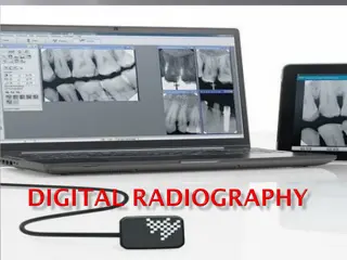
Digital X-ray replaces the use of film or computed radiography (CR) plates with a direct digital transfer of X-ray images into the PACS conversion of transmitted X-ray photons into a digital image using an array of solid-state detectors such as amorphous selenium or silicon, with computer processing and display of the image PACS. Digital radiography (DR) is the direct
An electronic device that detects gamma rays emitted by radio pharmaceautical (e.g technetium 99m (Tc-99m) that have been introduced into the body as tracers. The position of the source of the radioactivity can be plotted and displayed on a TV monitor or photographic film.


 Scan")
 Scan")