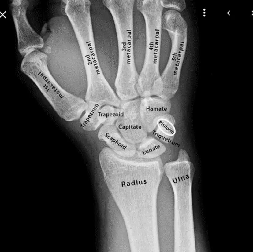
Overview of Carpal Bones and Carpal Tunnel Anatomy
Carpal bones are arranged in two rows with distinct features such as the flexor retinaculum and extensor retinaculum that form the carpal tunnel. Radiological features and nutrient arteries of the scaphoid bone are crucial in understanding carpal anatomy. Ossification of the carpal bones occurs in a specific order. Dr. Zaid Saad Al-Nasrawi, a specialist in trauma and orthopedic surgery, provides insights into the structure and function of the carpal bones.
Download Presentation

Please find below an Image/Link to download the presentation.
The content on the website is provided AS IS for your information and personal use only. It may not be sold, licensed, or shared on other websites without obtaining consent from the author. Download presentation by click this link. If you encounter any issues during the download, it is possible that the publisher has removed the file from their server.
E N D
Presentation Transcript
The carpal bones Dr. Zaid Saad Al-Nasrawi Specialist trauma and Orthopedics surgery
The carpal bones are arranged in two rows of four each In the proximal row, from lateral to medial, are the scaphoid, lunate and triquetral bones, with the pisiform on the anterior surface of the triquetral The trapezium, trapezoid, capitate and hamate make up the distal row
The Carpal tunnel Together the carpal bones form an arch, with its concavity situated anteriorly
The flexor retinaculum is attached laterally to the scaphoid and the ridge of the trapezium, and medially to the pisiform and the hook of the hamate. It converts the arch of bones into a tunnel, the carpal tunnel, which conveys the superficial and deep flexor tendons of the fingers and the thumb (except flexor carpi ulnaris and palmaris longus tendons) and the median nerve
The extensor retinaculum on the dorsum of the wrist attaches to the pisiform and triquetrum medially and the radius laterally Six separate synovial sheaths run beneath it
Radiological features of the carpal bones These are radiographed in the anteroposterior, lateral and oblique positions. Carpal tunnel views are obtained by extending the wrist and taking an inferosuperior view that is centred over the anterior part of the wrist
Nutrient arteries of the scaphoid In 13% of subjects these enter the scaphoid exclusively in its distal half If such a bone fractures across its midportion, the blood supply to the proximal portion is cut off and ischaemic necrosis is inevitable This occurs in 50% of patients with dis placed scaphoid fractures
Ossification of the carpal bones These ossify from a single centre each. The capitate ossifies first and the pisiform last, but the order and timing of the ossification of the other bones is variable Excluding the pisi- form. they ossify in a clockwise direction from capitate to trapezoid as follows: the capitate at 4 months; the hamate at 4 months; the triquetral at 3 years; the lunate bone at 5 years; and the scaphoid, trapezium and trapezoid at 6 years The pisiform ossifies at 11 years of age
The metacarpals and phalanges The five metacarpals are numbered from the lateral to the medial side. Each has a base proximally that articulates with that of the other metacarpals, except in the case of the first metacarpal, which is as a result more mobile and less likely to fracture The phalanges are 14 in number, three for each finger and two for the thumb Like the metacarpals, each has a head, a shaft and a base
The metacarpal sign A line tangential to the heads of the fourth and fifth metacarpals does not cross the head of the third metacarpal in 90% of normal hands this is called the metacarpal sign
The carpal angle This is formed by lines tangential to the proximal ends of the scaphoid and lunate bones In normal hands the average angle is 138
Ossification of the metacarpals and phalanges These ossify between the ninth and twelfth fetal weeks Secondary ossification centres appear in the distal end of the metacarpals of the fingers at 2 years and fuse at 20 years of age Secondary centres for the thumb metacarpal and for the phalanges are at their proximal end and appear between 2 and 3 years, and fuse between 18 and 20 years of age


