Overview of Bacterial and Fungal Corneal Ulcer
Bacterial and fungal corneal ulcers, known as suppurative keratitis, are a significant cause of blindness, included in national blindness control programs. They result from infections by exogenous organisms like S. aureus, S. pneumoniae, Aspergillus, and Candida. Predisposing factors include trauma, topical steroids, and neurotrophic keratitis. Pathophysiology involves progressive infiltration, necrosis, and eventual healing with scar formation. Understanding the stages of corneal ulcers is crucial for effective management.

Download Presentation

Please find below an Image/Link to download the presentation.
The content on the website is provided AS IS for your information and personal use only. It may not be sold, licensed, or shared on other websites without obtaining consent from the author.If you encounter any issues during the download, it is possible that the publisher has removed the file from their server.
You are allowed to download the files provided on this website for personal or commercial use, subject to the condition that they are used lawfully. All files are the property of their respective owners.
The content on the website is provided AS IS for your information and personal use only. It may not be sold, licensed, or shared on other websites without obtaining consent from the author.
E N D
Presentation Transcript
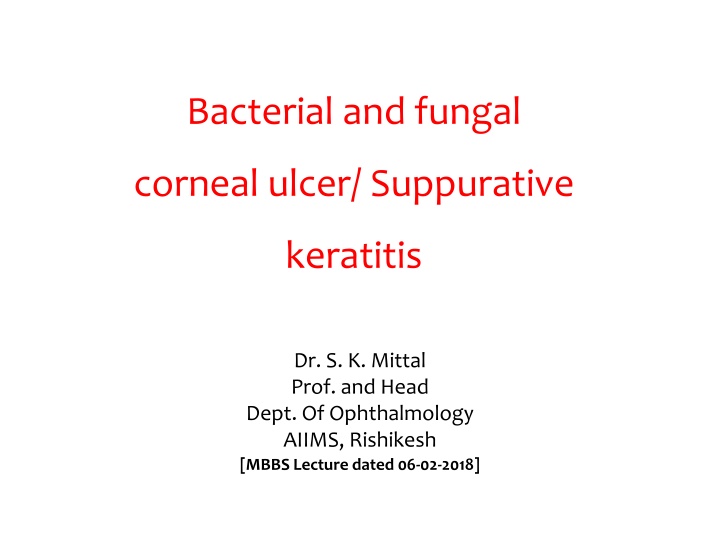
Bacterial and fungal corneal ulcer/ Suppurative keratitis Dr. S. K. Mittal Prof. and Head Dept. Of Ophthalmology AIIMS, Rishikesh [MBBS Lecture dated 06-02-2018]
Keratitis-Inflammation of cornea Corneal ulcer- Loss of corneal epithelium with inflammation of surrounding tissue and stroma and suppuration, with or without hypopyon
CORNEAL ULCER One the common cause of blindness Included in National Programme for Control of Blindness
Classification of Keratitis Bacterial A] Superficial l Infective Fungal Non-infective Viral Central Acanthamoebal Peripheral B] Deep Keratitis
Causative Organisms Infections are almost always exogenous Causative organism (BACTERIAL): S. aureus, S. epidermidis, S. pneumoniae, Pseudomonas aeruginosa. Uncommon: Neisseria gonorrhoeae, E. Coli FUNGAL : Aspergillus and Fusarium sp.(most common), Candida sp
Predisposing factors Trauma: e.g. Contact lenses, trichiasis, surgery (in fungal typical history of trauma with vegetable matter, mostly in harvesting season) Topical steroids Lagophthalmos : e.g. Facial nerve palsy
Predisposing factors Neurotrophic keratitis resulting from viral infections and lesions of ophthalmic division of Trigeminal nerve Dry eye syndrome Deficiency states ( Vit. A ) and metabolic diseases ( DM) Poor local hygiene and local infection ( chronic dacryocystitis)
Pathophysiology of ulcer Progressive infiltration Lymphocytes infiltrates in epithelium Necrosis Active ulceration Greyish infiltration with circumcorneal hyperaemia Hypopyon and descemetocele Phagocytosis Ulcers begin to heal Regression Epithelium covers the ulcers Scars and opacities formation Cicatrization
Assessment of Corneal ulcer History, general, and systemic examination Visual acuity: may be low Eye and Ocular adnexa: Eye lid , lacrimal sac Conjunctiva: circumcorneal congestion, chemosis Corneal ulcer: size, site ,surface, margin, slough, corneal sensation, thinning , satellite lesions Anterior chamber: Cells, flare, hypopyon Pupil
Clinical Features of Corneal ulcer SYMPTOMS: 1. Pain/Foreign body sensation 2. photophobia 3. DV/Blurred vision 4. Discharge/Watering 5. Redness 6. White spot on Cornea
Clinical Features of Corneal ulcer Signs: 1. Bleparospasm 2. Lid edema 3. Ciliary congestion of conjunctiva 4. Ulcer with greyish- white necrotic slough 5. Hypopyon+-
Symptoms in Mycotic Corneal Ulcer are less prominent than an equal size Bacterial ulcer
signs SIGNS BACTERIAL FUNGAL Lids Swelling of lids Might be present present Blepharospasm present Conjunctival chemosis and hyperemia Present+++ Present++ Ciliary congestion +++ +++ Elevated rolled out margins Satellite lesion Dense suppuration Endothelial palque Ulcer Greyish-white circumscribed infiltrate, Yellowish-white oval/ irregular area of ulcer. Stromal edema Hypopyon Hypopyon (sterile, whitish, mobile Hypopyon(infected,imm obile,yellowish), common Complications Corneal perforation ,endophthalmitis Endophthalmitis
Symptoms are less as compared to signs FUNGAL CORNEAL ULCER
Differential Diagnosis of ulcer Acute conjunctivitis Acute iridocyclitis Acute congestive glaucoma Corneal Opacity
Complications of Corneal Ulcer I. Descematocele
II. Perforation and its complications : Anterior synechia , Iris prolapse, expulsion of lens and vitreous, Intraocular hemorrhage, iii. Endophthalmitis / panophthalmitis iv. Secondary glaucoma v. Anterior capsular cataract vi. Staphyloma formation
Microbiological Investigations The majority are managed without smears or cultures. Scraping done: from ulcer margins and base of ulcer Examination of Smear stained with Gram stain, Giemsa stain, KOH mount for fungi. Culture on blood agar, chocolate agar, thioglycollate broth, and Sabouraud s dextrose agar
Management Principles: Control of infection Symptomatic relief Prevention of complications
Control of Infection(for bacterial ulcer) Topical antibiotics Fluoroquinolone eye drop: Cipro/ ofloxacin moxifloxacin(0.5%) drop. Gatifloxacin 0.3 % drop. Alternatives Fortified cephazolin eye drop Fortified tobramycin eye drop Fortified vancomycin eye drop
Antimicrobials for Fungal corneal ulcer Topical antifungal drops: - Natamycin 5 % 1 hourly by day and 2 hourly by night for 6 weeks to 6 months - Amphotericin B 0.15/ 0.3 % frequent instillation Oral antifungal agents; Ketoconazole 200-600 mg/ day .Fluconazole 200- 400mg/ day Scrapping done to help removal of slough and penetration of the drug Along with antibiotics support to prevent secondary bacterial infections
Treatment Cycloplegics : Atropine 1 % eye drop t.i.d. Hot fomentation Systemic analgesics : Anti-inflammatory drugs such as paracetamol & ibuprofen Removal of local predisposing factor Vitamins ( A, B-complex & C)
Treatment ( Non Healing Corneal Ulcer) Debridement of ulcer Chemical Cuterization Cyano acrylate glue Therapeutic penetrating keratoplasty Treatment of complications: perforation, secondary glaucoma
Outcome of corneal ulcer Healing with out opacity Healing with opacity Staphyloma Secondary glaucoma Cataract Phthisis bulbi
Source Text-Kanski, Parson s, Samar Basak, Pradeep Sharma Photographs- above , Archives & Website


")


")