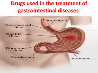
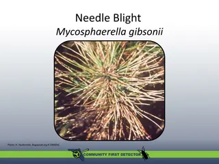
Peptic Ulcer Disease: Causes, Symptoms, and Treatment

PEPTIC ULCER. PERFORATED ULCER.
GASTROINTESTINAL BLEEDING
ODESSA NATIONAL MEDICAL UNIVERSITY
UNIVERSITY CLINIC DEPARTMENT of SURGERY #3
 
 
Peptic ulcer disease
Peptic ulcer disease (PUD) 
refers to the full-thickness ulcerations of duodenal or
gastric mucosa. The ulcerations form when exposure to acid and digestive
enzymes overcomes 
mucosal defense mechanisms
.
The most common etiologies include 
Helicobacter pylori
 (
H. pylori
) 
infection and
prolonged use of 
non-steroidal anti-inflammatory drugs (NSAIDs). 
Patients may
be asymptomatic or may present with abdominal pain, nausea, and early satiety.
Peptic ulcer disease typically 
responds 
well to medical treatment consisting of 
H.
pylori eradication, eliminating risk factors, and proton pump inhibitors (PPIs).
If left untreated, it can lead to 
bleeding, perforation, gastric outlet obstruction,
and gastric cancer
.
 
 
Introduction
Definition
A peptic ulcer is a mucosal defect in the wall of the stomach or duodenum that
penetrates the muscularis mucosa.
Epidemiology
Incidence:
Uncomplicated peptic ulcer disease (PUD): 1 case per 1,000 person-years in the
general population
PUD complications: 0.7 cases per 1,000 person-years in the general population
Duodenal ulcers > gastric ulcers (3:1)
Affects men and women equally
Duodenal ulcers occur, on average, 2 decades earlier than gastric ulcers.
PUD rates have been falling over the past few decades.
Etiology
Infection:
Helicobacter pylori 
or
 H.
pylori
 
(most common):
80%–90% duodenal ulcers
70%–80% gastric ulcers
Viruses:
Herpes simplex virus (HSV)
Cytomegalovirus (CMV)
Rare infections:
Tuberculosis (TB)
Syphilis
Mucormycosis
Medications:
Non-steroidal anti-inflammatory drugs
(NSAIDs; most common)
Medications linked to PUD or risk
exacerbated by combination with NSAIDs:
Bisphosphonates
Clopidogrel
Corticosteroids
Spironolactone
Chemotherapy
Sirolimus
Mycophenolate mofetil
Potassium chloride
Etiology
Hormonal:
Gastrinoma (Zollinger-Ellison
syndrome)
Antral G-cell
hyperplasia/hyperfunction
(gastritis)
Post-surgical:
Antral exclusion
Gastric bypass
Ischemic: cocaine use
Decompensated chronic diseases:
Cirrhosis
Renal failure
Chronic obstructive pulmonary disease
(COPD)
Prolonged intensive care unit (ICU) stay
for any reason
Idiopathic
Other rare causes:
Annular pancreas
Sarcoidosis
Crohn’s disease
Radiation
Risk factors
In many cases, the listed etiologies are not enough to produce PUD.
Contributing risk factors to PUD development:
Smoking
Alcohol
Stress (severe illness–related, psychologic)
Diet
Genetic predisposition
Blood types A and O
Pathophysiology
Anatomic location of ulcers
Duodenal ulcer:
 an ulcer located in the duodenum,
typically in the duodenal bulb (1st part)
Gastric ulcer:
 an ulcer in the stomach lining, commonly
in the lesser curvature of the stomach near the
junction of the fundus and antral mucosa (or type I)


Pathophysiology
Stomach and duodenum:
Normally exposed to a toxic environment (acid + pepsin)
Imbalance between offending agents and defense mechanisms
leads to PUD.
Defense preventing mucosal injury:
Mucus-bicarbonate-phospholipid layer
Epithelial layer (repair of which is regulated by prostaglandins)
Subepithelial defense (mucosal blood flow for supply of nutrients
and oxygen, disposal of noxious agents)
Pathophysiology
Mechanisms by offending agents:
Increased gastric acid secretion:
H. pylori
 gastritis or inflammation: ↑ gastric acid, inhibits somatostatin, ↓ mucus
NSAID inhibition of COX 1 → ↓ prostaglandin (↓ mucus, ↓ mucosal blood flow, ↓
epithelial proliferation)
NSAID inhibition of COX 2 → delays healing
Impaired duodenal bicarbonate secretion (in patients with duodenal ulcers)
Effects of other etiologies or risk factors:
Smoking → ↑ acid secretion, ↓ prostaglandin
Zollinger-Ellison syndrome (gastrinoma) → ↑ gastrin
Brain injury increases intracranial pressure, overstimulates vagus nerve → ↑ gastric acid
(
Cushing’s ulcer
)
Burn injury (stress) → reduced volume, cell necrosis → gastric mucosal sloughing →
impaired barrier (
Curling’s ulcer
)
 
Clinical Presentation
Asymptomatic
70% of PUD patients
Older adults
Patients taking NSAIDs
Complication(s) (bleeding or perforation) may be the 1st
clinical presentation without antecedent symptoms.
Clinical Presentation
Symptomatic
Abdominal pain:
Epigastric with radiation to left or right upper quadrants
Sometimes radiates to the back
Classic duodenal ulcer pain: 2–5 hours after eating and at night (when acid is secreted
without food)
Sometimes exacerbated by eating (pyloric channel ulcers)
Other symptoms:
Nausea/vomiting
Early satiety
Postprandial fullness/bloating
Belching
Diagnosis
History
Review of common risk factors:
NSAID/aspirin use
Concomitant use of high-risk medications
Underlying chronic disease
Prior gastric surgery or radiation
Laboratory studies
Anemia (occult bleeding and/or iron malabsorption)
High fasting gastrin levels (if Zollinger-Ellison syndrome is suspected)
Diagnosis
Esophagogastroduodenoscopy (EGD)
Most accurate diagnostic test
Findings:
Gastric ulcer: 
Usually solitary discrete mucosal lesions, with punched-out smooth base
Benign lesions have smooth, rounded edges (as opposed to irregular edges noted in
malignant lesions).
Typically in lesser curvature
Duodenal ulcer:
Small breaks in the mucosa, often < 1 cm
Noted usually in the 1st part of the duodenum
 
 
Diagnosis
Indications for
 biopsy
:
Suspected malignancy (ulcerated mass, irregular margins, abnormal mucosal
folds)
Any gastric ulcer in areas with a high incidence of gastric cancer
If unusual etiology is suspected (e.g., sarcoidosis)
Gastric mucosal biopsy of antrum and body (in addition to
 
the ulcer itself)
for 
H. pylori 
detection
Tests for 
H. pylori
Non-invasive:
Stool antigen assay:
For initial diagnosis
To confirm eradication
Urea breath test:
Patient is given radioactively labeled urea orally.
Urease produced by 
H. pylori 
splits urea and liberates CO
2
.
Radioactive CO
2
 is detected in the breath.
Serology:
Detection of serum IgG against
 H. pylori
Low accuracy
IgG remains positive after eradication.
Tests for 
H. pylori
Invasive 
(require sampling of gastric mucosa):
Biopsy urease test:
Urease produced by 
H. pylori 
liberates ammonia from urea.
Alkaline pH changes color of a pH reagent.
Histology:
Accuracy improved by stains (immunohistochemical, Giemsa stains)
Curved, flagellated gram-negative rods are seen.
Bacterial culture and sensitivity
 
 
Management
Risk factor modification
Discontinue NSAIDs.
Stop smoking, and discontinue intake of alcohol and drugs.
Bland diet
Medications
H. pylori 
eradication:
A combination of an antibiotic regimen and proton pump inhibitors (PPIs):
Triple therapy: PPI + clarithromycin + amoxicillin or metronidazole
Bismuth-containing quadruple therapy: PPI + bismuth + tetracycline + metronidazole
Confirm 
H. pylori
 eradication after treatment.
Management
PPIs:
Uncomplicated ulcers:
Given for 2 weeks (plus antibiotic treatment for 
H. pylori
 if positive)
Longer if the patient has indications for maintenance therapy
Complicated ulcers (bleeding, penetration, obstruction, or perforation):
Duodenal ulcer: 4–8 weeks
Gastric ulcer: 8–12 weeks (confirmation of ulcer healing needed)
Longer if the patient has indications for maintenance therapy
Maintenance therapy:
If NSAIDs or aspirin need to be continued
Giant ulcer (> 2 cm)
H. pylori 
negative, NSAID-negative ulcers
Recurrent PUD (> 2/year)
Management
Repeat endoscopy
Duodenal ulcers:
Usually not necessary
If symptoms persist with treatment > 4 weeks
If evidence of ongoing bleeding
Gastric ulcers:
Persistent symptoms despite 8–12 weeks of medical therapy
Unclear etiology
Giant ulcer (> 2 cm)
Ulcer was suspicious for malignancy on the 1st endoscopy (even with negative biopsy).
Evidence of ongoing bleeding
Failure to eradicate 
H. pylori
 infection
Risk factors for gastric cancer:
Age > 50
H. pylori
Family history
Immigrants from endemic areas (Japan, Korea)
Gastric atrophy, metaplasia/dysplasia, adenoma
Surgical treatment
Uncommon because medical therapy is very effective
Indications 
for surgery:
Management of complications:
Bleeding
Perforation
Gastric outlet obstruction
PUD refractory to medical management
Non-compliance or intolerable side effects of medications
Surgical treatment
Surgical principles:
Prevent ulcer complications: ulcer resection
Reduce acid secretion: vagotomy, antrectomy
Minimize postoperative physiologic disturbances: pyloroplasty to facilitate gastric
emptying after vagotomy
Procedures:
Highly selective vagotomy: Only the vagal branches stimulating acid secretion are
transected.
Vagotomy and drainage (pyloroplasty or gastrojejunostomy)
Procedures involving partial stomach resection:
Vagotomy and antrectomy: removes the acid-producing portion of the stomach
Partial gastrectomy: removes the portion of the stomach containing the gastric ulcer
Reconstruction needed for a partial gastrectomy: by gastroduodenal (Billroth I) or gastrojejunal
(Billroth II) anastomosis
Complications
Gastrointestinal bleeding
Patients present with hematemesis and/or melena.
May be chronic/low grade and present as anemia
Acute bleeding is usually approached with endoscopic intervention.
Dieulafoy lesion: 
vascular malformation in the stomach (submucosa) that ulcerates and causes massive bleeding
Surgery may be required if unable to achieve control endoscopically.
Perforation
Sudden onset of severe diffuse abdominal pain, peritonitis, tachycardia
Emergent surgery is required.
Penetration
Walled-off perforation or perforation into the retroperitoneal space
Symptoms not as pronounced as with a free perforation; subacute onset
Often results in an abscess formation or fistulas to the surrounding organs (colon, biliary tree, blood vessels)
 
 
Complications
Gastric outlet obstruction
Mechanical obstruction from peri-pyloric inflammation and scarring
1st-line treatment is 
H. pylori 
eradication and PPIs.
Endoscopic dilation for failure of medical management
Surgery as a last resort
Gastric cancer
Adenocarcinoma
MALToma (mucosa-associated lymphoid tissue)
Both associated with chronic 
H. pylori
 infection and atrophic gastritis
Complications
Upper GI bleeding is the most common complication of peptic ulcer disease. The next most
common complication is perforation.
The annual incidence of upper GI bleeding secondary to a peptic ulcer is estimated to be
between 19 to 57 cases per 100,000 individuals. In comparison, ulcer perforation is expected to
be 4 to 14 cases per 100,000 individuals.
Advanced age is a risk factor as 60% of patients with PUD are older than 60. Infections with
Helicobacter pylori and the use of nonsteroidal anti-inflammatory drugs (NSAIDs) are each
identified as risk factors for the development of bleeding ulcers and peptic ulcer perforation.
Perforating Ulcer
Clinical Presentation
Most patients with a perforated peptic ulcer will present with symptoms. The most common symptom in patients
with peptic ulcer disease is dyspepsia or upper abdominal pain. This pain may be vague upper abdominal
discomfort or it may be localized to either the right upper quadrant, left upper quadrant, or epigastrium.
Gastric ulcers may be worsened by food whereas pain from a duodenal ulcer may be delayed 2-5 hours after
eating. Patients who are experiencing bleeding from a peptic ulcer may complain of nausea, hematemesis, or
melanotic stools. Some patients may report bright red blood per rectum or maroon-colored stool if the upper GI
bleeding is brisk.
A thorough physical examination should be done on all patients complaining of abdominal pain. Those with a
perforated peptic ulcer are likely to have diffuse abdominal tenderness that progresses to guarding and rigidity. 
Perforating Ulcer
E
valuation
The evaluation of a patient in whom perforated peptic ulcer is suspected should be done
quickly as the morbidity and mortality increase significantly with time. Even if a perforated
peptic ulcer is suspected due to history and physical examination, diagnostic studies should be
obtained to confirm the diagnosis and to rule out other possible etiologies.
Typical workup includes labs and imaging studies. Standard labs should include complete blood
count (CBC), chemistry panel, liver function tests, coagulation profile, and lipase levels (to rule
out pancreatitis). Blood type and screening should be done as well.
Perforating Ulcer
E
valuation
Duodenal perforation can cause acute pain associated with free perforation, or less acute symptoms
associated with abscess or fistula formation.
Perforation of the duodenum with spillage of intraluminal contents into the peritoneal cavity causes
acute chemical peritonitis. This is followed by a systemic inflammatory response syndrome (SIRS),
which can progress to secondary bacterial peritonitis and sepsis. Patients with retroperitoneal
perforation may lack peritoneal signs and present more indolently.
Double-contrast computed tomography (CT) scan is the most valuable method for diagnosing
duodenal perforation. It should be performed whenever there is a clinical suspicion and the patient
does not need immediate surgery. CT features of perforation include discontinuity of the duodenal
wall and the presence of extraluminal air or extravasated oral contrast. Other CT findings include
duodenal wall thickening, fat stranding and periduodenal fluid collection.


Management
The main goals of treatment are
resuscitation, control of infection,
nutritional support and restoration of
gastrointestinal tract continuity.
Management
The main goals of treatment are
resuscitation, control of infection, nutritional
support and restoration of gastrointestinal
tract continuity.
Management
Conservative treatment
Initial conservative management consists of nil per os, intravenous fluid therapy, broad-
spectrum antibiotics, intravenous PPIs, nasogastric tube insertion and 
H.
pylori
 eradication. The added value of somatostatin remains controversial.
Non-operative management of perforated duodenal ulcers is feasible in selected
patients. Perforated ulcers may seal spontaneously with fibrin, omentum or by fusion of
the duodenum to the underside of the liver between the gallbladder and the falciform
ligament.
Operative management is usually recommended if there is free leakage of contrast
medium into the peritoneal cavity. Progressive abdominal signs or intra-abdominal
sepsis should warrant surgery.
In high-risk patients, who cannot tolerate surgical treatment, conservative management
may also include percutaneous drainage of fluid collections.
Management
Endoscopic management
Endoscopic treatment is an attractive treatment modality due to its minimally invasive nature. Early endoscopic
closure (<24 h) is considered to be technically easier because the inflammatory changes are less pronounced.
TTSC
Through-the-scope clips (TTSC) can be used for endoscopic closure of small duodenal perforations. Linear
perforations <1 cm are most suitable for the use of TTSC.
OTSC
In contrast to common endoscopic clips, the over-the-scope clips (OTSC) are able to compress larger
quantities of tissue. The OTSC technique can be used for perforations ranging from 1 to 3 cm.
Endoloop with clips
A combined technique using TTSC and an endoloop can be used if the OTSC technique is unavailable.
SEMS
Self-expandable metal stents (SEMS) are alternative endoscopic treatment options for duodenal
perforations.
Management
Surgical treatment
The choice of surgical treatment depends on the size and localization of the perforation, the viability
of the duodenal walls, the degree of local contamination and underlying etiology.
Simple surgical repair
The main surgical treatment is simple repair of the perforation site. This can be performed as a
primary closure with or without the addition of an omental patch.
Alternatively, a pedicled omental flap (Cellan–Jones repair) or free omental plug (Graham patch) can
be sutured into the perforation.
Sutureless techniques have also been developed using a gelatin sponge and fibrin glue to seal off
the perforation. There seem to be no significant differences in terms of postoperative morbidity and
mortality rates when comparing primary closure, omentopexy or tegmentation (without closure).
Surgical repair can be performed either with conventional open surgery or with laparoscopy.
 
Management
Surgical treatment
Abdominal drains
The routine placement of abdominal drains after surgical repair is controversial. The
literature suggests no benefit in preventing postoperative fluid collections or abscesses.
Furthermore, drains may be associated with increased morbidity such as drain wound
site infection.
Pyloric exclusion
Pyloric exclusion involves surgical repair of the duodenum, gastrotomy and closure of
the pylorus from within and finally the formation of a gastrojejunostomy. The rationale
behind this procedure is to divert all gastric and biliary secretions from the duodenum.
The added benefit of using a gastric diversion procedure such as pyloric exclusion for
duodenal perforations has been questioned in recent years. Importantly, the procedure
has been associated with more postoperative complications and longer hospital stay
compared to simple repair without pyloric exclusion.
Management
Surgical treatment
Reconstructive surgery
For large duodenal perforations, a duodeno-duodenostomy may be necessary. If this is
not possible, a Roux-en-Y duodenojejunostomy may be performed over the
perforation. A Billroth II operation may be necessary if the perforation is to the first or
proximal second portion of the duodenum. If the duodeno-pancreatic head complex is
destroyed, a pancreaticoduodenectomy may be necessary.
Tube duodenostomy
Tube duodenostomy is a damage control procedure for large duodenal perforations
when other repair techniques are not possible due to the magnitude of duodenal
damage, hemodynamic instability of the patient or the lack of surgical expertise for
complex reconstruction. The perforation is sutured around a catheter inserted into the
perforation to enhance directed fistulation of the perforation. The catheter is removed
after a minimum of 6 weeks. A feeding jejunostomy may be placed for enteral
nutritional support.
Prognostic factors
The main prognostic factor remains the time interval between the perforation
and treatment. Mortality increases when the delay is greater than 24 h.
Other prognostic factors have been reported but are mainly related to clinical
signs of sepsis, such as increased Acute Physiology and Chronic Health
Evaluation II (APACHE II) score.
Old age and co-morbidity are also strong adverse prognostic factors.
Gastrointestinal Bleeding
Gastrointestinal bleeding (GIB) 
is a symptom of multiple diseases within the gastrointestinal
(GI) tract.
Gastrointestinal bleeding is designated as 
upper or lower 
based on the etiology’s location to
the 
ligament of Treitz
.
It is more common to have bleeding in the upper GI tract, with 
peptic ulcer disease 
being
the most frequent cause.
Depending on the location of the bleeding, the patient may present with 
hematemesis
(
vomiting blood
), 
melena
 (
black, tarry stool
), or 
hematochezia
 (
fresh blood in stools
).
Some patients presenting with GIB can be 
hemodynamically unstable 
and require emergent
stabilization and evaluation. The source of the bleed can often be 
located and treated with
endoscopy.
 
 
Epidemiology
Upper gastrointestinal bleeding (UGIB):
Incidence of about 100 per 100,000 adults per year
Twice as common in men
Increased risk with age (> 60 years)
Lower gastrointestinal bleeding (LGIB):
Incidence of about 20.5 per 100,000 adults per year
Increased risk with age (200-fold increase in the 3rd to 9th decades)
Somewhat more common in men
Risk factors:
Helicobacter pylori
 infection
Nonsteroidal anti-inflammatory drugs (NSAIDs)
Alcohol use
Cirrhosis
Vascular disease
Etiology
Upper gastrointestinal bleed (proximal to the ligament of Treitz): 
Esophagus
Esophageal varices
Esophagitis: infection or inflammation in the esophagus
Esophageal cancer
Mallory-Weiss tear: a tear in the esophageal lining due to forceful vomiting
Stomach
Gastric ulcer
Erosive gastritis
Gastric antral vascular ectasia (GAVE): dilated small blood vessels in the pyloric antrum (uncommon)
Portal hypertensive gastropathy
Dieulafoy lesion: large, tortuous vessel that can erode and bleed
Angiodysplasia: abnormal collection of blood vessels
Duodenum
Duodenal ulcer
Angiodysplasia
Aortoenteric fistula (rare)
Gastric and duodenal ulcers are the most common causes.
Etiology
Lower GIB (distal to the ligament of Treitz):
Diverticular disease
Diverticulosis: sac-like protrusion of the colonic wall (common)
Diverticulitis: less commonly associated with GI bleed
Meckel’s diverticulum: congenital outpouching in the ileum
Vascular disease
Angiodysplasia
Ischemia (e.g., mesenteric ischemia, ischemic colitis)
Hemorrhoids: venous structures of the anorectum that engorge, prolapse, and bleed
Trauma
Anal fissure: a small tear in the anal mucosa
Neoplasm
Colon polyp
Colorectal cancer
Inflammatory disease
Infectious colitis
Ulcerative colitis and Crohn’s disease: autoimmune inflammatory bowel diseases that cause inflammation and ulcers
Iatrogenic
After biopsy or polypectomy
Radiation colitis: radiation-induced inflammation in the colon
Aortoenteric fistula (rare, but serious)
Clinical Presentation
Clinical manifestations
Patients with occult bleeding
may be asymptomatic.
Symptoms of anemia:
Fatigue, weakness
Dyspnea
Pallor
Lightheadedness
Syncope
Abdominal pain or heartburn → ulcers,
gastritis, ischemia
Coffee-ground emesis → UGIB
Hematemesis → UGIB
Melena (black, tarry stool)
Hematochezia (bright-red blood in stools)
Usually seen in LGIB
Can be from brisk, large-volume UGIB
Weight loss → malignancy
Clinical Presentation
Physical exam
Patients with mild or occult bleeding
may not have significant findings.
Evidence of hemodynamic instability:
Tachycardia
Hypotension
Altered mental status
Orthostatic hypotension: seen with acute blood loss
of ≥ 2 units
Pale skin color and conjunctiva
Some may have abdominal tenderness (e.g.,
ischemia).
Rectal exam:
o
Black or bloody stools
o
Normal, brown stool may be seen in occult bleeding
o
Hemorrhoids
o
Anal fissure
o
Rectal mass
Evaluate for signs of chronic liver disease:
o
Spider angiomata
o
Splenomegaly
o
Abdominal distension and ascites
o
Asterixis
Diagnosis
Diagnosis and management of GIB tend to go hand-in-hand and will vary
depending on the hemodynamic stability of the patient.
Laboratory evaluations
Complete blood count → anemia from blood loss
Hemoglobin may initially be normal in acute bleeds.
Potential thrombocytopenia may be seen in cirrhosis.
Fecal occult blood testing → detect occult bleeding
Coagulation factors → coagulopathy, which may need reversal
Liver function tests → underlying liver disease
Basic metabolic panel → ↑ BUN (blood urea nitrogen) may signal upper GIB
Iron, ferritin → iron deficiency
Diagnosis
Procedures
Esophagoduodenoscopy (EGD)
Modality of choice in UGIB
Visualize the site of hemorrhage within the esophagus, stomach, or duodenum.
Collect pathology specimens.
Colonoscopy
Modality of choice in LGIB
Visualize the site of hemorrhage within the large intestine and terminal ileum.
Collect pathology specimens.
Colon preparation is required
 
 
Diagnosis
Procedures
Capsule endoscopy
Provides imaging of the small intestine
Patient swallows a wireless camera, which takes pictures along the digestive tract.
Most often used for continued or intermittent bleeding when EGD and colonoscopy are
unremarkable
Angiography
Bleeding rate of at least 0.5
1 mL/min is required for detection.
Reserved for patients who cannot undergo endoscopy due to hemodynamic instability
Management
Initial steps
Assess the patient’s hemodynamics and stabilize:
Protect the patient’s airway:
Patient may need intubation for severe hematemesis
Prevent aspiration
Obtain adequate IV access: 2 large-gauge peripheral IVs and/or central line
IV fluid resuscitation
Send a blood type and screen evaluation.
Blood transfusion
Management
Medications
Proton pump inhibitors (pantoprazole)
Octreotide
Somatostatin analog, which causes splanchnic vasoconstriction
Used for esophageal bleeding
Management
Interventions
Around 80% will stop bleeding without intervention.
EGD and colonoscopy
Injection of epinephrine around bleeding point
Thermal hemostasis (electrocoagulation)
Endoclips
Angiography
Vasoconstriction via vasopressin
Embolization
Procedure runs the risk of bowel ischemia or infarction.
Management
Interventions
Balloon tamponade
Used for esophageal varices
Tube is inserted into the esophagus, and balloon is inflated.
Provides short-term hemostasis until definitive treatment can be arranged.
Surgery
Considered when bleeding cannot be contained through the
above interventions (rare)
Localization of the source is important before pursuing surgery.
Sengstaken–Blakemore Tube
Upper Gastrointestinal Bleeding
Balloon Tamponade
 
 
Management
Special considerations
Patients with cirrhosis and variceal bleeding should have antibiotic
prophylaxis to prevent spontaneous bacterial peritonitis.
Reverse any anticoagulation.
Hold antihypertensive medications.
THANK YOU FOR
ATTENTION
ODESSA NATIONAL MEDICAL UNIVERSITY
UNIVERSITY CLINIC DEPARTMENT of SURGERY #3
Slide Note
Embed
Share

Peptic ulcer disease (PUD) involves ulcerations in the duodenal or gastric mucosa caused by factors like Helicobacter pylori infection and NSAID use. Symptoms include abdominal pain and nausea, and treatment often involves H. pylori eradication and proton pump inhibitors to prevent complications like bleeding and gastric cancer.

  • Peptic Ulcer
  • Helicobacter pylori
  • Gastric Mucosa
  • NSAIDs
  • Gastrointestinal Bleeding

Uploaded on Mar 26, 2024 | 1 Views


Download Presentation

Please find below an Image/Link to download the presentation.

The content on the website is provided AS IS for your information and personal use only. It may not be sold, licensed, or shared on other websites without obtaining consent from the author. Download presentation by click this link. If you encounter any issues during the download, it is possible that the publisher has removed the file from their server.

E N D

Presentation Transcript


  1. ODESSA NATIONAL MEDICAL UNIVERSITY UNIVERSITY CLINIC DEPARTMENT of SURGERY #3 PEPTIC ULCER. PERFORATED ULCER. GASTROINTESTINAL BLEEDING

  2. Peptic ulcer disease Peptic ulcer disease (PUD) refers to the full-thickness ulcerations of duodenal or gastric mucosa. The ulcerations form when exposure to acid and digestive enzymes overcomes mucosal defense mechanisms. The most common etiologies include Helicobacter pylori (H. pylori) infection and prolonged use of non-steroidal anti-inflammatory drugs (NSAIDs). Patients may be asymptomatic or may present with abdominal pain, nausea, and early satiety. Peptic ulcer disease typically responds well to medical treatment consisting of H. pylori eradication, eliminating risk factors, and proton pump inhibitors (PPIs). If left untreated, it can lead to bleeding, perforation, gastric outlet obstruction, and gastric cancer.

  3. Introduction Definition A peptic ulcer is a mucosal defect in the wall of the stomach or duodenum that penetrates the muscularis mucosa. Epidemiology Incidence: Uncomplicated peptic ulcer disease (PUD): 1 case per 1,000 person-years in the general population PUD complications: 0.7 cases per 1,000 person-years in the general population Duodenal ulcers > gastric ulcers (3:1) Affects men and women equally Duodenal ulcers occur, on average, 2 decades earlier than gastric ulcers. PUD rates have been falling over the past few decades.

  4. Etiology Infection: Helicobacter pylori or H. pylori (most common): 80% 90% duodenal ulcers 70% 80% gastric ulcers Viruses: Herpes simplex virus (HSV) Cytomegalovirus (CMV) Rare infections: Tuberculosis (TB) Syphilis Mucormycosis Medications: Non-steroidal anti-inflammatory drugs (NSAIDs; most common) Medications linked to PUD or risk exacerbated by combination with NSAIDs: Bisphosphonates Clopidogrel Corticosteroids Spironolactone Chemotherapy Sirolimus Mycophenolate mofetil Potassium chloride

  5. Etiology Hormonal: Gastrinoma (Zollinger-Ellison syndrome) Antral G-cell hyperplasia/hyperfunction (gastritis) Decompensated chronic diseases: Cirrhosis Renal failure Chronic obstructive pulmonary disease (COPD) Prolonged intensive care unit (ICU) stay for any reason Post-surgical: Antral exclusion Gastric bypass Idiopathic Other rare causes: Annular pancreas Sarcoidosis Crohn s disease Radiation Ischemic: cocaine use

  6. Risk factors In many cases, the listed etiologies are not enough to produce PUD. Contributing risk factors to PUD development: Smoking Alcohol Stress (severe illness related, psychologic) Diet Genetic predisposition Blood types A and O

  7. Pathophysiology Anatomic location of ulcers Duodenal ulcer: an ulcer located in the duodenum, typically in the duodenal bulb (1st part) Gastric ulcer: an ulcer in the stomach lining, commonly in the lesser curvature of the stomach near the junction of the fundus and antral mucosa (or type I)

  8. Table: Classification of gastric ulcers by location (modified Johnson classification) Type Location Acid level Lesser curve of the stomach at the incisura angularis I Low to normal Gastric body; coexists with duodenal ulcer II Increased In the pyloric channel (within 3 cm of pylorus) III Increased Proximal gastroesophageal ulcer IV Normal Anywhere in the stomach (aspirin/NSAID induced) V Normal

  9. Pathophysiology Stomach and duodenum: Normally exposed to a toxic environment (acid + pepsin) Imbalance between offending agents and defense mechanisms leads to PUD. Defense preventing mucosal injury: Mucus-bicarbonate-phospholipid layer Epithelial layer (repair of which is regulated by prostaglandins) Subepithelial defense (mucosal blood flow for supply of nutrients and oxygen, disposal of noxious agents)

  10. Pathophysiology Mechanisms by offending agents: Increased gastric acid secretion: H. pylori gastritis or inflammation: gastric acid, inhibits somatostatin, mucus NSAID inhibition of COX 1 prostaglandin ( mucus, mucosal blood flow, epithelial proliferation) NSAID inhibition of COX 2 delays healing Impaired duodenal bicarbonate secretion (in patients with duodenal ulcers) Effects of other etiologies or risk factors: Smoking acid secretion, prostaglandin Zollinger-Ellison syndrome (gastrinoma) gastrin Brain injury increases intracranial pressure, overstimulates vagus nerve gastric acid (Cushing s ulcer) Burn injury (stress) reduced volume, cell necrosis gastric mucosal sloughing impaired barrier (Curling s ulcer)

  11. Clinical Presentation Asymptomatic 70% of PUD patients Older adults Patients taking NSAIDs Complication(s) (bleeding or perforation) may be the 1st clinical presentation without antecedent symptoms.

  12. Clinical Presentation Symptomatic Abdominal pain: Epigastric with radiation to left or right upper quadrants Sometimes radiates to the back Classic duodenal ulcer pain: 2 5 hours after eating and at night (when acid is secreted without food) Sometimes exacerbated by eating (pyloric channel ulcers) Other symptoms: Nausea/vomiting Early satiety Postprandial fullness/bloating Belching

  13. Diagnosis History Review of common risk factors: NSAID/aspirin use Concomitant use of high-risk medications Underlying chronic disease Prior gastric surgery or radiation Laboratory studies Anemia (occult bleeding and/or iron malabsorption) High fasting gastrin levels (if Zollinger-Ellison syndrome is suspected)

  14. Diagnosis Esophagogastroduodenoscopy (EGD) Most accurate diagnostic test Findings: Gastric ulcer: Usually solitary discrete mucosal lesions, with punched-out smooth base Benign lesions have smooth, rounded edges (as opposed to irregular edges noted in malignant lesions). Typically in lesser curvature Duodenal ulcer: Small breaks in the mucosa, often < 1 cm Noted usually in the 1st part of the duodenum

  15. Diagnosis Indications for biopsy: Suspected malignancy (ulcerated mass, irregular margins, abnormal mucosal folds) Any gastric ulcer in areas with a high incidence of gastric cancer If unusual etiology is suspected (e.g., sarcoidosis) Gastric mucosal biopsy of antrum and body (in addition to the ulcer itself) for H. pylori detection

  16. Tests for H. pylori Non-invasive: Stool antigen assay: For initial diagnosis To confirm eradication Urea breath test: Patient is given radioactively labeled urea orally. Urease produced by H. pylori splits urea and liberates CO2. Radioactive CO2is detected in the breath. Serology: Detection of serum IgG against H. pylori Low accuracy IgG remains positive after eradication.

  17. Tests for H. pylori Invasive (require sampling of gastric mucosa): Biopsy urease test: Urease produced by H. pylori liberates ammonia from urea. Alkaline pH changes color of a pH reagent. Histology: Accuracy improved by stains (immunohistochemical, Giemsa stains) Curved, flagellated gram-negative rods are seen. Bacterial culture and sensitivity

  18. Management Risk factor modification Discontinue NSAIDs. Stop smoking, and discontinue intake of alcohol and drugs. Bland diet Medications H. pylori eradication: A combination of an antibiotic regimen and proton pump inhibitors (PPIs): Triple therapy: PPI + clarithromycin + amoxicillin or metronidazole Bismuth-containing quadruple therapy: PPI + bismuth + tetracycline + metronidazole Confirm H. pylori eradication after treatment.

  19. Management PPIs: Uncomplicated ulcers: Given for 2 weeks (plus antibiotic treatment for H. pylori if positive) Longer if the patient has indications for maintenance therapy Complicated ulcers (bleeding, penetration, obstruction, or perforation): Duodenal ulcer: 4 8 weeks Gastric ulcer: 8 12 weeks (confirmation of ulcer healing needed) Longer if the patient has indications for maintenance therapy Maintenance therapy: If NSAIDs or aspirin need to be continued Giant ulcer (> 2 cm) H. pylori negative, NSAID-negative ulcers Recurrent PUD (> 2/year)

  20. Management Repeat endoscopy Duodenal ulcers: Usually not necessary If symptoms persist with treatment > 4 weeks If evidence of ongoing bleeding Gastric ulcers: Persistent symptoms despite 8 12 weeks of medical therapy Unclear etiology Giant ulcer (> 2 cm) Ulcer was suspicious for malignancy on the 1st endoscopy (even with negative biopsy). Evidence of ongoing bleeding Failure to eradicate H. pylori infection Risk factors for gastric cancer: Age > 50 H. pylori Family history Immigrants from endemic areas (Japan, Korea) Gastric atrophy, metaplasia/dysplasia, adenoma

  21. Surgical treatment Uncommon because medical therapy is very effective Indications for surgery: Management of complications: Bleeding Perforation Gastric outlet obstruction PUD refractory to medical management Non-compliance or intolerable side effects of medications

  22. Surgical treatment Surgical principles: Prevent ulcer complications: ulcer resection Reduce acid secretion: vagotomy, antrectomy Minimize postoperative physiologic disturbances: pyloroplasty to facilitate gastric emptying after vagotomy Procedures: Highly selective vagotomy: Only the vagal branches stimulating acid secretion are transected. Vagotomy and drainage (pyloroplasty or gastrojejunostomy) Procedures involving partial stomach resection: Vagotomyand antrectomy: removes the acid-producing portion of the stomach Partial gastrectomy: removes the portion of the stomach containing the gastric ulcer Reconstruction needed for a partial gastrectomy: by gastroduodenal (Billroth I) or gastrojejunal (Billroth II) anastomosis

  23. Complications Gastrointestinal bleeding Patients present with hematemesis and/or melena. May be chronic/low grade and present as anemia Acute bleeding is usually approached with endoscopic intervention. Dieulafoy lesion: vascular malformation in the stomach (submucosa) that ulcerates and causes massive bleeding Surgery may be required if unable to achieve control endoscopically. Perforation Sudden onset of severe diffuse abdominal pain, peritonitis, tachycardia Emergent surgery is required. Penetration Walled-off perforation or perforation into the retroperitoneal space Symptoms not as pronounced as with a free perforation; subacute onset Often results in an abscess formation or fistulas to the surrounding organs (colon, biliary tree, blood vessels)

  24. Complications Gastric outlet obstruction Mechanical obstruction from peri-pyloric inflammation and scarring 1st-line treatment is H. pylori eradication and PPIs. Endoscopic dilation for failure of medical management Surgery as a last resort Gastric cancer Adenocarcinoma MALToma (mucosa-associated lymphoid tissue) Both associated with chronic H. pylori infection and atrophic gastritis

  25. Complications Upper GI bleeding is the most common complication of peptic ulcer disease. The next most common complication is perforation. The annual incidence of upper GI bleeding secondary to a peptic ulcer is estimated to be between 19 to 57 cases per 100,000 individuals. In comparison, ulcer perforation is expected to be 4 to 14 cases per 100,000 individuals. Advanced age is a risk factor as 60% of patients with PUD are older than 60. Infections with Helicobacter pylori and the use of nonsteroidal anti-inflammatory drugs (NSAIDs) are each identified as risk factors for the development of bleeding ulcers and peptic ulcer perforation.

  26. Perforating Ulcer Clinical Presentation Most patients with a perforated peptic ulcer will present with symptoms. The most common symptom in patients with peptic ulcer disease is dyspepsia or upper abdominal pain. This pain may be vague upper abdominal discomfort or it may be localized to either the right upper quadrant, left upper quadrant, or epigastrium. Gastric ulcers may be worsened by food whereas pain from a duodenal ulcer may be delayed 2-5 hours after eating. Patients who are experiencing bleeding from a peptic ulcer may complain of nausea, hematemesis, or melanotic stools. Some patients may report bright red blood per rectum or maroon-colored stool if the upper GI bleeding is brisk. A thorough physical examination should be done on all patients complaining of abdominal pain. Those with a perforated peptic ulcer are likely to have diffuse abdominal tenderness that progresses to guarding and rigidity.

  27. Perforating Ulcer Evaluation The evaluation of a patient in whom perforated peptic ulcer is suspected should be done quickly as the morbidity and mortality increase significantly with time. Even if a perforated peptic ulcer is suspected due to history and physical examination, diagnostic studies should be obtained to confirm the diagnosis and to rule out other possible etiologies. Typical workup includes labs and imaging studies. Standard labs should include complete blood count (CBC), chemistry panel, liver function tests, coagulation profile, and lipase levels (to rule out pancreatitis). Blood type and screening should be done as well.

  28. Perforating Ulcer Evaluation Duodenal perforation can cause acute pain associated with free perforation, or less acute symptoms associated with abscess or fistula formation. Perforation of the duodenum with spillage of intraluminal contents into the peritoneal cavity causes acute chemical peritonitis. This is followed by a systemic inflammatory response syndrome (SIRS), which can progress to secondary bacterial peritonitis and sepsis. Patients with retroperitoneal perforation may lack peritoneal signs and present more indolently. Double-contrast computed tomography (CT) scan is the most valuable method for diagnosing duodenal perforation. It should be performed whenever there is a clinical suspicion and the patient does not need immediate surgery. CT features of perforation include discontinuity of the duodenal wall and the presence of extraluminal air or extravasated oral contrast. Other CT findings include duodenal wall thickening, fat stranding and periduodenal fluid collection.

  29. Management The main goals of treatment are resuscitation, control of infection, nutritional support and restoration of gastrointestinal tract continuity.

  30. Management The main goals of treatment are resuscitation, control of infection, nutritional support and restoration of gastrointestinal tract continuity.

  31. Management Conservative treatment Initial conservative management consists of nil per os, intravenous fluid therapy, broad- spectrum antibiotics, intravenous PPIs, pylori eradication. The added value of somatostatin remains controversial. nasogastric tube insertion and H. Non-operative management of perforated duodenal ulcers is feasible in selected patients. Perforated ulcers may seal spontaneously with fibrin, omentum or by fusion of the duodenum to the underside of the liver between the gallbladder and the falciform ligament. Operative management is usually recommended if there is free leakage of contrast medium into the peritoneal cavity. Progressive abdominal signs or intra-abdominal sepsis should warrant surgery. In high-risk patients, who cannot tolerate surgical treatment, conservative management may also include percutaneous drainage of fluid collections.

  32. Management Endoscopic management Endoscopic treatment is an attractive treatment modality due to its minimally invasive nature. Early endoscopic closure (<24 h) is considered to be technically easier because the inflammatory changes are less pronounced. TTSC Through-the-scope clips (TTSC) can be used for endoscopic closure of small duodenal perforations. Linear perforations <1 cm are most suitable for the use of TTSC. OTSC In contrast to common endoscopic clips, the over-the-scope clips (OTSC) are able to compress larger quantities of tissue. The OTSC technique can be used for perforations ranging from 1 to 3 cm. Endoloop with clips A combined technique using TTSC and an endoloop can be used if the OTSC technique is unavailable. SEMS Self-expandable metal stents (SEMS) are alternative endoscopic treatment options for duodenal perforations.

  33. Management Surgical treatment The choice of surgical treatment depends on the size and localization of the perforation, the viability of the duodenal walls, the degree of local contamination and underlying etiology. Simple surgical repair The main surgical treatment is simple repair of the perforation site. This can be performed as a primary closure with or without the addition of an omental patch. Alternatively, a pedicled omental flap (Cellan Jones repair) or free omental plug (Graham patch) can be sutured into the perforation. Sutureless techniques have also been developed using a gelatin sponge and fibrin glue to seal off the perforation. There seem to be no significant differences in terms of postoperative morbidity and mortality rates when comparing primary closure, omentopexy or tegmentation (without closure). Surgical repair can be performed either with conventional open surgery or with laparoscopy.

  34. Management Surgical treatment Abdominal drains The routine placement of abdominal drains after surgical repair is controversial. The literature suggests no benefit in preventing postoperative fluid collections or abscesses. Furthermore, drains may be associated with increased morbidity such as drain wound site infection. Pyloric exclusion Pyloric exclusion involves surgical repair of the duodenum, gastrotomy and closure of the pylorus from within and finally the formation of a gastrojejunostomy. The rationale behind this procedure is to divert all gastric and biliary secretions from the duodenum. The added benefit of using a gastric diversion procedure such as pyloric exclusion for duodenal perforations has been questioned in recent years. Importantly, the procedure has been associated with more postoperative complications and longer hospital stay compared to simple repair without pyloric exclusion.

Related


More Related Content

giItT1WQy@!-/#giItT1WQy@!-/#giItT1WQy@!-/#giItT1WQy@!-/#giItT1WQy@!-/#giItT1WQy@!-/#giItT1WQy@!-/#giItT1WQy@!-/#giItT1WQy@!-/#giItT1WQy@!-/#giItT1WQy@!-/#