Spasticity and Increased Muscle Tone in Neurological Disorders

Spasticity and Increased
Muscle Tone
Muscle Tone
Dr. Salah Elmalik
Dr. Salah Elmalik
OBJECTIVES


At the end of this lecture you should be able to:
 
Define spasticity and rigidity .
Describe the neurophysiology of spasticity
Describe the causes of spasticity
MUSCLE TONE
 
Resistance of a muscle to stretch is often
referred to as its 
referred to as its 
tone or tonus
tone or tonus
.
.
Muscle tone is static component of stretch
reflex 
reflex 
.It is a continuous  mild muscle
contraction that acts as background to
actual movement.
actual movement.
A 
A 
hypertonic 
hypertonic 
muscle is one in which the
resistance to stretch is high  because of
hyperactive stretch reflexes
 
 
Hypertonia is of two types:
 
1.
Spasticity
Spasticity
 
2. Rigidity
2. Rigidity
 
Spasticity:
Spasticity:
As described by Lance (1980): “it is a motor
disorder, characterised by increase in tonic stretch
reflexes (muscle tone) with exaggerated tendon
jerks, resulting from hyper-excitability of the
dynamic stretch reflex as one component of the
upper motor neurone (UMN) syndrome
 
 
Clinically it can be defined as increased resistance
to passive stretch.
to passive stretch.
-Spasticity is velocity dependent increased
resistance to passive movement of the muscle due to
abnormally high muscle tone (hypertonia) which
varies with the speed of displacement of a joint.
- 
- 
The faster you stretch the muscle the greater the
resistance.
resistance.
-- Spasticity is clearly neural in nature and is a associated
with the – UMNL
with the – UMNL
-Involvement of the corticospinal tract is often
associated with UMNL and spasticity. There are a
number of clinical features that are associated with
spasticity :-
spasticity :-
-hypereflexia, flexor spasticity in the upper limb &
extensor spasticity in the lower limb.
I
I
-UMN lesions Spasticity is of Clasp Knife Type
Spasticity
Spasticity
 
 
- 
Spasticity is characterized by hyper-
excitability of both types of stretch reflex:-
 1- increase in tonic static stretch reflexes
 1- increase in tonic static stretch reflexes
(muscle tone) as one component of the upper
motor neurone (UMN) syndrome
 2- Exaggerated tendon jerks, resulting from
 2- Exaggerated tendon jerks, resulting from
hyper-excitability of the dynamic stretch
reflex as one component of the upper motor
neurone (UMN) syndrome
Rigidity
Rigidity
 
Rigidity is increased neural
activity throughout the range of
muscle movement and is not
velocity dependent.
velocity dependent.
Rigidity is present in both agonist
and antagonist muscles. It is
often  associated with basal
ganglia disease such as Parkinson’s
disease
disease
 
R
R
i
i
g
g
i
i
d
d
i
i
t
t
y
y
 
 
i
i
n
n
 
 
P
P
a
a
r
r
k
k
i
i
n
n
s
s
o
o
n
n
i
i
s
s
m
m
:
:
1
1
.
.
 
 
L
L
e
e
a
a
d
d
-
-
p
p
i
i
p
p
e
e
 
 
r
r
i
i
g
g
i
i
d
d
i
i
t
t
y
y
 
 
:
:
     Passive movement of an extremity
     Passive movement of an extremity
meets with a constant dead feeling
resistance like a lead pipe
throughout the range of movement
.
.
Rigidity-2
Rigidity-2
 
2. Cog-wheel rigidity :
In cogwheel rigidity one feels
that resistance varies
rhythmically when applying a
passive movement.
passive movement.
 It is because of an
 It is because of an
underlying resting tremor
associated with rigidity.
Other types of Rigidity
 
 
- 
Decerebrate rigidity :
  (extension of head & 4 limbs extensors )
  (extension of head & 4 limbs extensors )
- 
- 
Decorticate rigidity:
(extensor rigidity in legs & moderate
flexion of arms if head unturned )
 
Features of UMN Syndrome
(1
) 
Weakness and decreased muscle control .
(2) No remarkable muscle wasting, but  disuse atrophy
(3) Spasticity ( hypertonia ) , frequently called
“ clasp-knife spasticity ”,increased extensor muscle tone
then a sudden collapse in resistance due to inhibition of
extensor motor neurons by GTOs (golgi tendon organs)
(4) Clonus
 
Repetitive jerky motions (clonus), especially
when  limb moved & stretched suddenly
(5)  exaggerated  tendon jerks
(6) Extensor plantar reflex = Babinski sign ( dorsiflexion
of the big toe and fanning out of the other toes )
(7) Absent abdominal reflexes
 
 
Mechanism of spasticity in UMN lesions:
In UMN syndrome the motor neurones are free from
the descending inhibitory influence of the Higher
Motor-Controlling centers ( medullary RF, red nucleus ,
basal ganglia)resulting in un antagonized excitatory
input ( pontine RF, vestibulo-spinal) to gamma motor
neurones causing hypertonia &spasticity
- This results in
 ( 1) State of ongoing ( unremitting )contraction of
muscles .( due to hyperactive gamma activity )
 (2) decreased ability to control movement
 (3) increased resistance felt on passive stretch.
 
Causes of spasticity:-
A-(UMNS) syndrome include 
:
(1) Cerebral palsy
(2) Stroke
(3) Spinal cord injury
(4) Multiple Sclerosis
(5) Acquired brain injury (
trauma , etc )
B-Parkinsonism
C-Decerebrate & decorticate
rigidity
 
 
(1) 
Cerebral palsy
Caused by brain damage
due to  lack of oxygen,
that cause damage to the
motor control centers of
the developing brain ,it
can occur during pregnancy
, during childbirth  ( or
after birth up to about
age three by meningitis)
(2) Multiple Sclerosis
 
Is an auto-immune demyelinating disease , in which the body's
own immune system attacks and damages the myelin sheath of
myelinated nerves mainly of brain ,SC ,and optic nerve
Loss of myelin sheath (demyelination) prevents axons from
saltatory conduction of action potentials  causing muscle
weakness& wasting.
weakness& wasting.
 Disease onset usually occurs in young adults, and it is more
 Disease onset usually occurs in young adults, and it is more
common in females .
common in females .
 The disease can attack any part of the CNS , and when it
 The disease can attack any part of the CNS , and when it
causes demyelination of  descending motor tracts in the
brainstem & spinal cord , the subject develops spasticity and
other signs of UMNS .
The disease frequently remits and relapses  because of
remylination & restore of function and during acute attacks
intravenous corticosteroids can improve symptoms
3-STROKE:-
 
 a-Haemorrhagic stroke as in cerebral haemorrhage
 b- Ischaemic stroke as in thrombosis or embolism
-all cause death of brain tissues, results in paralysis
in the opposite half of the body .
 A lesion in Corona Radiata on one 
side can cause
Monoplegia in a contralateral limb (UL or LL ,
according to site).
A lesion in the Internal Capsule on one side may cause
Hemiplegia or Hemiparesis on the contralateral
side ( with the picture of upper motor
neuron syndrome ).
Complete transection of spinal cord-1
 
 
The higher the level of the section, the more serious are the
consequences.
consequences.
 
If the transection is in the upper cervical region  immediate
death follows, due to paralysis of all respiratory muscles.
 
In the lower cervical region below the 5
th
th
 cervical segment
 cervical segment
diaphragmatic respiration is still possible, but the patient
suffers complete paralysis of all four limbs 
(quadriplegia
(quadriplegia
).
).
 
Transection lower down in the thoracic region allows normal
respiration but the patient ends up with paralysis of both lower
limbs 
limbs 
(paraplegia)
(paraplegia)
Complete transection of spinal cord-2
 
Stages :-
A/ Spinal shock ( 2-6 weeks )
B/ Recovery of reflex activity
C/ Paraplegia in extension
A/ Spinal shock
A/ Spinal shock
In the immediate period following transection there is :
(1) complete loss of spinal reflex activity 
below 
below 
t
t
he level of the lesion .
(2) Loss of all sensations 
(anesthesia
(anesthesia
) and voluntary movement
( paralysis) 
( paralysis) 
below
below
 the level of the lesion , due to interruption of all
 the level of the lesion , due to interruption of all
sensory and motor tracts
(3) Loss of tendon reflexes and superficial reflexes (abdominal ,
plantar & withdrawal reflexes ) .
(4) The loss of muscle tone (flaccidity) and absence of any muscle
activity (muscle pump ) lead to decreased venous return causing the
lower limbs to become cold and blue in cold weather
Complete transection of spinal cord-3
 
(5)The wall of the urinary bladder becomes paralysed and
urine is retained until the pressure in the bladder overcomes
the resistance offered by the tone of the sphincters and
dribbling occurs. This is known as
 
 
retention with overflow.
(7)Loss of vasomotor tone occurs, due to interruption of
fibers that connect the vasomotor centres in the medulla
oblongata with the lateral horn cells of the spinal cord,
which project sympathetic vasoconstrictor impulses to blood
vessels.
vessels.
 
 
vasodilatation
vasodilatation
 
 
causes a fall in blood pressure; the
higher the level of the section, the lower the blood pressure.
(8) Bedsores due to pressure of body-weight against
underlining support
underlining support
-
-
This stage varies in duration but usually lasts a maximum of 2-6
weeks, after which some reflex activity recovers.
B/ Stage of return of reflex activity
 
As the spinal shock ends , spinal reflex activity appears
again this partial recovery may be due to , increase in the
natural degree of excitability of the spinal cord neurons
below the level of the section ,  to make up for  the loss of
supraspinal facilitatory influences. It may also be due to
sprouting of fibrers from remaining other  inputs.
 
Features of the stage of recovery of reflex activity:
 
(1) Gradual rise of arterial blood pressure 
due to return of
spinal vasomotor activity in the lateral horn cells. But, since
vasomotor control from the medulla is absent, the blood
pressure is not stable
 
(2
(2
) Return of spinal reflexes
Flexor reflexes return earlier than extensor ones.
Babiniski sign ( extensor plantar reflex) is one of the earliest signs of this
stage.
    -Tendon reflexes also recover earlier in flexors.
     - As a result, 
flexor spastic  tone 
causes the lower limbs to
take a position of slight flexion, a state referred to 
as paraplegia in flexion.
-The return of the stretch reflex ( & consequently 
muscle tone
) , and
vasoconstrictor tone in arterioles and venules , improve the circulation
through the limbs
 
(
3) Recovery of visceral reflexes: 
return of micturition, defecation &
erection reflexes.
-
However , voluntary control over micturition and defecation , and the sensation
of bladder and rectal fullness are permanently lost.
 
(4) Sexual reflexes
(4) Sexual reflexes
, consisting of erection or ejaculation on genital manipulation
, recover.
 
(5) 
(5) 
Mass reflex 
appears in this stage
   - A minor painful stimulus to the skin of the lower limbs will not
only cause withdrawal of that limb but will evoke  many other
reflexes through spread of excitation (by irradiation) to many
autonomic centers. So the bladder and rectum will also empty,
the skin will sweat, the blood pressure will rise
 
 Since effective regeneration never occurs in the human central
nervous system, patients with complete transection never
recover fully.
 Voluntary movements and sensations are permanently lost.
however, patients who are rehabilitated and properly managed
may enter into a more advanced stage of rec
overy.
C/ 
Stage of  extensor paraplegia
 
(1) During this stage the tone in extensor muscles
returns gradually to exceed that in the flexors. The
lower limbs become spastically extended.
 
Extensor reflexes become exaggerated, as shown by
tendon jerks and by the appearance of clonus.
The positive supportive reaction becomes well
developed and the patient can stand on his feet with
appropriate support.
 (2) The flexor withdrawal reflex which appeared in
the earlier stage is associated during this stage with
the crossed extensor reflex.
Hemisection of the Spinal Cord
( Brown-Sequard syndrome)
H
e
m
i
s
e
c
t
i
o
n
 
o
f
 
t
h
e
 
S
p
i
n
a
l
 
C
o
r
d
 
(
B
r
o
w
n
-
S
e
q
u
a
r
d
 
s
y
n
d
r
o
m
e
)
 
A/ At the level of the lesion
:
 
all manifestations occur on the same side:
    1. Paralysis of the lower motor neuron type
    1. Paralysis of the lower motor neuron type
     2. Loss of all sensations in the areas supplied by the afferent fibers that
     2. Loss of all sensations in the areas supplied by the afferent fibers that
enter the spinal cord in the damaged segments
     3. Vasodilatation of the blood vessels that receive vasoconstrictor fibers
     3. Vasodilatation of the blood vessels that receive vasoconstrictor fibers
from the damaged segment
 
B/ Ipsilaterally below the level of the lesion :
   1. spastic lower limb .
   1. spastic lower limb .
    2.Fine touch, position and vibration sense are lost
    2.Fine touch, position and vibration sense are lost
    3.Vasodilatation
    3.Vasodilatation
C/ Contralaterally below the level of the lesion :
Pain and temperature sensations are lost, Why ?
Slide Note
Embed
Share

Spasticity and increased muscle tone are common features in neurological disorders, characterized by hyperactive stretch reflexes and muscle contractions. Spasticity is velocity-dependent and associated with upper motor neuron lesions, leading to increased resistance to passive movement. Rigidity, on the other hand, involves increased neural activity throughout the muscle range. This lecture discusses the definitions, neurophysiology, causes, and clinical features of spasticity and hypertonia, offering insights into these complex motor abnormalities.

  • Spasticity
  • Muscle Tone
  • Neurological Disorders
  • Motor Abnormalities
  • Neurophysiology

Uploaded on Sep 16, 2024 | 0 Views


Download Presentation

Please find below an Image/Link to download the presentation.

The content on the website is provided AS IS for your information and personal use only. It may not be sold, licensed, or shared on other websites without obtaining consent from the author. Download presentation by click this link. If you encounter any issues during the download, it is possible that the publisher has removed the file from their server.

E N D

Presentation Transcript


  1. Spasticity and Increased Muscle Tone Dr. Salah Elmalik

  2. OBJECTIVES At the end of this lecture you should be able to: Define spasticity and rigidity . Describe the neurophysiology of spasticity Describe the causes of spasticity

  3. MUSCLE TONE Resistance of a muscle to stretch is often referred to as its tone or tonus. Muscle tone is static component of stretch reflex .It is a continuous mild muscle contraction that acts as background to actual movement. A hypertonic muscle is one in which the resistance to stretch is high because of hyperactive stretch reflexes

  4. Hypertonia is of two types: 1. Spasticity 2. Rigidity

  5. Spasticity: As described by Lance (1980): it is a motor disorder, characterised by increase in tonic stretch reflexes (muscle tone) with exaggerated tendon jerks, resulting from hyper-excitability of the dynamic stretch reflex as one component of the upper motor neurone (UMN) syndrome

  6. Clinically it can be defined as increased resistance to passive stretch. -Spasticity is velocity dependent increased resistance to passive movement of the muscle due to abnormally high muscle tone (hypertonia) which varies with the speed of displacement of a joint. - The faster you stretch the muscle the greater the resistance. -- Spasticity is clearly neural in nature and is a associated with the UMNL -Involvement of the corticospinal tract is often associated with UMNL and spasticity. There are a number of clinical features that are associated with spasticity :- -hypereflexia, flexor spasticity in the upper limb & extensor spasticity in the lower limb. I-UMN lesions Spasticity is of Clasp Knife Type

  7. Spasticity - Spasticity is characterized by hyper- excitability of both types of stretch reflex:- 1- increase in tonic static stretch reflexes (muscle tone) as one component of the upper motor neurone (UMN) syndrome 2- Exaggerated tendon jerks, resulting from hyper-excitability of the dynamic stretch reflex as one component of the upper motor neurone (UMN) syndrome

  8. Rigidity Rigidity is increased neural activity throughout the range of muscle movement and is not velocity dependent. Rigidity is present in both agonist and antagonist muscles. It is often associated with basal ganglia disease such as Parkinson s disease Rigidity in Parkinsonism: 1. Lead-pipe rigidity : Passive movement of an extremity meets with a constant dead feeling resistance like a lead pipe throughout the range of movement.

  9. Rigidity-2 2. Cog-wheel rigidity : In cogwheel rigidity one feels that resistance varies rhythmically when applying a passive movement. It is because of an underlying resting tremor associated with rigidity.

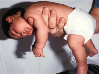
  10. Other types of Rigidity - Decerebrate rigidity : (extension of head & 4 limbs extensors ) - Decorticate rigidity: (extensor rigidity in legs & moderate flexion of arms if head unturned )

  11. Features of UMN Syndrome (1) Weakness and decreased muscle control . (2) No remarkable muscle wasting, but disuse atrophy (3) Spasticity ( hypertonia ) , frequently called clasp-knife spasticity ,increased extensor muscle tone then a sudden collapse in resistance due to inhibition of extensor motor neurons by GTOs (golgi tendon organs) (4) Clonus Repetitive jerky motions (clonus), especially when limb moved & stretched suddenly (5) exaggerated tendon jerks (6) Extensor plantar reflex = Babinski sign ( dorsiflexion of the big toe and fanning out of the other toes ) (7) Absent abdominal reflexes

  12. Mechanism of spasticity in UMN lesions: In UMN syndrome the motor neurones are free from the descending inhibitory influence of the Higher Motor-Controlling centers ( medullary RF, red nucleus , basal ganglia)resulting in un antagonized excitatory input ( pontine RF, vestibulo-spinal) to gamma motor neurones causing hypertonia &spasticity - This results in ( 1) State of ongoing ( unremitting )contraction of muscles .( due to hyperactive gamma activity ) (2) decreased ability to control movement (3) increased resistance felt on passive stretch.

  13. Causes of spasticity:- A-(UMNS) syndrome include : (1) Cerebral palsy (2) Stroke (3) Spinal cord injury (4) Multiple Sclerosis (5) Acquired brain injury ( trauma , etc ) B-Parkinsonism C-Decerebrate & decorticate rigidity

  14. (1) Cerebral palsy Caused by brain damage due to lack of oxygen, that cause damage to the motor control centers of the developing brain ,it can occur during pregnancy , during childbirth ( or after birth up to about age three by meningitis)

  15. (2) Multiple Sclerosis Is an auto-immune demyelinating disease , in which the body's own immune system attacks and damages the myelin sheath of myelinated nerves mainly of brain ,SC ,and optic nerve Loss of myelin sheath (demyelination) prevents axons from saltatory conduction of action potentials causing muscle weakness& wasting. Disease onset usually occurs in young adults, and it is more common in females . The disease can attack any part of the CNS , and when it causes demyelination of descending motor tracts in the brainstem & spinal cord , the subject develops spasticity and other signs of UMNS . The disease frequently remits and relapses because of remylination & restore of function and during acute attacks intravenous corticosteroids can improve symptoms

  16. 3-STROKE:- a-Haemorrhagic stroke as in cerebral haemorrhage b- Ischaemic stroke as in thrombosis or embolism -all cause death of brain tissues, results in paralysis in the opposite half of the body . A lesion in Corona Radiata on one side can cause Monoplegia in a contralateral limb (UL or LL , according to site). A lesion in the Internal Capsule on one side may cause Hemiplegia or Hemiparesis on the contralateral side ( with the picture of upper motor neuron syndrome ).

  17. Complete transection of spinal cord-1 The higher the level of the section, the more serious are the consequences. If the transection is in the upper cervical region immediate death follows, due to paralysis of all respiratory muscles. In the lower cervical region below the 5thcervical segment diaphragmatic respiration is still possible, but the patient suffers complete paralysis of all four limbs (quadriplegia). Transection lower down in the thoracic region allows normal respiration but the patient ends up with paralysis of both lower limbs (paraplegia)

  18. Complete transection of spinal cord-2 Stages :- A/ Spinal shock ( 2-6 weeks ) B/ Recovery of reflex activity C/ Paraplegia in extension A/ Spinal shock In the immediate period following transection there is : (1) complete loss of spinal reflex activity below the level of the lesion . (2) Loss of all sensations (anesthesia) and voluntary movement ( paralysis) below the level of the lesion , due to interruption of all sensory and motor tracts (3) Loss of tendon reflexes and superficial reflexes (abdominal , plantar & withdrawal reflexes ) . (4) The loss of muscle tone (flaccidity) and absence of any muscle activity (muscle pump ) lead to decreased venous return causing the lower limbs to become cold and blue in cold weather

  19. Complete transection of spinal cord-3 (5)The wall of the urinary bladder becomes paralysed and urine is retained until the pressure in the bladder overcomes the resistance offered by the tone of the sphincters and dribbling occurs. This is known as retention with overflow. (7)Loss of vasomotor tone occurs, due to interruption of fibers that connect the vasomotor centres in the medulla oblongata with the lateral horn cells of the spinal cord, which project sympathetic vasoconstrictor impulses to blood vessels. vasodilatation causes a fall in blood pressure; the higher the level of the section, the lower the blood pressure. (8) Bedsores due to pressure of body-weight against underlining support -This stage varies in duration but usually lasts a maximum of 2-6 weeks, after which some reflex activity recovers.

  20. B/ Stage of return of reflex activity As the spinal shock ends , spinal reflex activity appears again this partial recovery may be due to , increase in the natural degree of excitability of the spinal cord neurons below the level of the section , to make up for the loss of supraspinal facilitatory influences. It may also be due to sprouting of fibrers from remaining other inputs. Features of the stage of recovery of reflex activity: (1) Gradual rise of arterial blood pressure due to return of spinal vasomotor activity in the lateral horn cells. But, since vasomotor control from the medulla is absent, the blood pressure is not stable

  21. (2) Return of spinal reflexes Flexor reflexes return earlier than extensor ones. Babiniski sign ( extensor plantar reflex) is one of the earliest signs of this stage. -Tendon reflexes also recover earlier in flexors. - As a result, flexor spastic tone causes the lower limbs to take a position of slight flexion, a state referred to as paraplegia in flexion. -The return of the stretch reflex ( & consequently muscle tone) , and vasoconstrictor tone in arterioles and venules , improve the circulation through the limbs (3) Recovery of visceral reflexes: return of micturition, defecation & erection reflexes. -However , voluntary control over micturition and defecation , and the sensation of bladder and rectal fullness are permanently lost. (4) Sexual reflexes, consisting of erection or ejaculation on genital manipulation , recover.

  22. (5) Mass reflex appears in this stage - A minor painful stimulus to the skin of the lower limbs will not only cause withdrawal of that limb but will evoke many other reflexes through spread of excitation (by irradiation) to many autonomic centers. So the bladder and rectum will also empty, the skin will sweat, the blood pressure will rise Since effective regeneration never occurs in the human central nervous system, patients with complete transection never recover fully. Voluntary movements and sensations are permanently lost. however, patients who are rehabilitated and properly managed may enter into a more advanced stage of recovery.

  23. C/ Stage of extensor paraplegia (1) During this stage the tone in extensor muscles returns gradually to exceed that in the flexors. The lower limbs become spastically extended. Extensor reflexes become exaggerated, as shown by tendon jerks and by the appearance of clonus. The positive supportive reaction becomes well developed and the patient can stand on his feet with appropriate support. (2) The flexor withdrawal reflex which appeared in the earlier stage is associated during this stage with the crossed extensor reflex.

  24. Hemisection of the Spinal Cord ( Brown-Sequard syndrome)

  25. Hemisection of the Spinal Cord ( Brown-Sequard syndrome) A/ At the level of the lesion: all manifestations occur on the same side: 1. Paralysis of the lower motor neuron type 2. Loss of all sensations in the areas supplied by the afferent fibers that enter the spinal cord in the damaged segments 3. Vasodilatation of the blood vessels that receive vasoconstrictor fibers from the damaged segment B/ Ipsilaterally below the level of the lesion : 1. spastic lower limb . 2.Fine touch, position and vibration sense are lost 3.Vasodilatation C/ Contralaterally below the level of the lesion : Pain and temperature sensations are lost, Why ?

More Related Content

giItT1WQy@!-/#giItT1WQy@!-/#giItT1WQy@!-/#giItT1WQy@!-/#giItT1WQy@!-/#giItT1WQy@!-/#giItT1WQy@!-/#giItT1WQy@!-/#giItT1WQy@!-/#giItT1WQy@!-/#