Management of Fluid Control and Soft Tissue in Prosthodontics

F
L
U
I
D
 
C
O
N
T
R
O
L
 
A
N
D
S
O
F
T
 
T
I
S
S
U
E
M
A
N
A
G
E
M
E
N
T
R
R
U
U
N
N
G
G
T
T
A
A
 
 
C
C
O
O
L
L
L
L
E
E
G
G
E
E
 
 
O
O
F
F
 
 
D
D
E
E
N
N
T
T
A
A
L
L
S
S
C
C
I
I
E
E
N
N
C
C
E
E
S
S
 
 
A
A
N
N
D
D
 
 
R
R
E
E
S
S
E
E
A
A
R
R
C
C
H
H
DEPT OF PROSTHODONTICS
PRESENTATED BY DKASHYAP
PRESENTATED  BY DR. RUCHA KASHYAP
CONENTS
INTRODUCTION
CLASSIFICATION
ADVANTAGE/ DIS ADVANTAGE
INDIACATION/ CONTRAINDICATION
SELECTION OF ABUTMENT TEETH
TYPES OF ATTCHMENTS
SUMMARY
TAKE HOME MESSAGE
REFERENCES
2
SPECIFIC LEARNING OBJECTIVES
3
INTRODUCTION
INTRODUCTION
    
    
Restorative procedures in the mouth cannot be executed
efficiently unless the moisture is controlled . moisture
control includes the exclusion of sulcular fluid ,saliva and
gingival bleeding from the operating field. It also refers to
preventing the aspiration or swallowing of fluid sprayed
from the handpiece and restorative debris . primary
objectives of moisture control are isolation ,retraction and
accessibility.
accessibility.
   The gingiva must be displaced to make a complete
impression and some time even to permit completion of the
preparation and cementation of the restoration.
FLUID CONTROL (ISOLATION)
      The need for removal of fluid varies
depending upon the task being performed .
Isolation
 of the operating site is done for the
 of the operating site is done for the
following reasons:
following reasons:
      To obtain a dry, clean ,operating field
      To obtain a dry, clean ,operating field
      For easy access and visibility
      For easy access and visibility
      To improve properties of dental materials
      To improve properties of dental materials
      To protect the patient and operator
      To protect the patient and operator
      To improve the operating efficiency
      To improve the operating efficiency
METHOD OF FLUID CONTROL
MECHANICAL 
CHEMICAL
OTHER
1.
Rubber dam
2.
High volume vacuum
3.
Saliva  ejector
4.
svedopter
1.
Anti-sialogogues
2.
Local anesthetics
1.
Cotton rolls
2.
Cellulose wafers
3.
Throat shields
RUBBER DAM
RUBBER DAM
Introduced by S.C BARNUM  in 1864 .
It is the most effective of all isolation devices utilized in restorative dentistry .
valuable in the removal of old restorations or excavation of caries when
exposure of the pulp is a possibility .
It is used to define the operating field by isolating one or more teeth from the
oral environment.
It eliminates the saliva from the operating side and retracts the soft tissue.
I
t
 
s
h
o
u
l
d
 
n
o
t
 
b
e
 
u
s
e
d
 
w
h
i
l
e
 
m
a
k
i
n
g
 
a
 
p
o
l
y
v
i
n
y
l
 
s
i
l
o
x
a
n
e
 
i
m
p
r
e
s
s
i
o
n
,
b
e
c
a
u
s
e
 
i
t
 
w
i
l
l
 
i
n
h
i
b
i
t
 
i
t
s
 
p
o
l
y
m
e
r
i
z
a
t
i
o
n
 
.
It also provides excellent isolation and access when a pin retained amalgam
or composite resin core is required before a cast restoration can be
fabricated .
   parts of rubber dam
         Rubber dam sheet
         Rubber dam holder
         Rubber dam retainer
         Rubber dam punch
         Rubber dam retainer forceps
         Rubber dam napkin
     Certain condition preclude the use of the rubber dam
(1)
    Teeth that have not erupted sufficiently to receive a retainer
(2)
    some third molars
(3)
    extremely malpositioned teeth
(4)
    Patient suffering from asthma may not tolerate the rubber dam if breathing through
the nose  is difficult .
High volume vacuum
High volume vacuum
A high –volume suction tip is
extremely useful during the
preparation phase and is most
effectively utilized with an assistant
It  makes an excellent lip retractor
while the operator uses a mirror to
retract and protect the tongue .
Mcwherter (1957) showed that one
type of evacuator would remove 0.5
L water in 2 seconds ,had a 75% to
95 % pickup of water in air ,and
would remove 100 % of solid during
cutting procedure .
cutting procedure .
SALIVA EJECTOR
SALIVA EJECTOR
  It is most useful as an adjunct to
high volume evacuation .
  It can be used for evacuation
when the maxillary arch is being
treated .
  It is not very  effective   during
impression and cementation .
  It is placed at the corner of the
mouth, opposite to the quadrant
being operated and the patient ‘s
head is turned toward it.
S
S
V
V
E
E
D
D
O
O
P
P
T
T
E
E
R
R
It is used for isolating the
mandibular teeth .
It is the metal saliva ejector
attached with a tongue deflector .
D
i
s
a
d
v
a
n
t
a
g
e
s
 
o
f
 
s
v
e
d
o
p
t
e
r
Access to the lingual surface of
mandibular teeth is limited
Since it is a metallic device , care
must be taken to avoid any injury to
the floor of the mouth .
Presences of mandibular tori
precludes  its  use .
PRECAUTION
    
Selection of an oversized
reflector should be avoided. Since it
could cut into the palate above or
trigger the gag reflex .
   For better positioning ,the
anterior part of the svedopter should
be placed in the incisor region with
the tubing under the patient 
,
arm .
    The svedopter is most effective when it is
used with the patient in a nearly upright
position .In this position ,water and other fluid
collect on the floor of the mouth ,where they
are pulled off by the vacuum.
       These are helpful for short period of
Isolation .Especially in   conjunction with
profound anesthesia ,absorbents provide
acceptable dryness for procedures such as
impression –taking and cementation .
     Now a days cotton roll holder are used for
holding cotton roll in position .But this is
inconvenient and time-consuming .
  Advantage of cotton roll holders is that the
cheeks and tongue are slightly retracted from the
teeth, which enhances access and visibility .
      when removing cotton rolls or cellulose
wafers, it may be necessary to moisten them
using the air/water syringe to prevent inadvertent
removal of the epithelium from cheek ,floor of the
mouth ,or lips .
.
Cotton roll and cellulose wafers
      Throat shield are used during try-in and
removal of indirect restorations.
     This is particularly important when treating teeth
in the maxillary arch .
     A gauze sponge (2x2 inch ) ,unfolded and
spread over the tongue and the posterior part of the
mouth , is helpful in recovering a restoration should it
be dropped .
THROAT SHIELD
CHEMICAL METHOD
CHEMICAL METHOD
A
N
T
I
-
S
I
A
L
O
G
O
G
U
E
S
They are gastrointestinal anti-cholinergic that act on the smooth
muscles of gastrointestinal ,urinary and biliary tracts , producing
a dry mouth as a side effect .
Commonly used anti-sialogogues
Methantheline bromide (banthine) :50 mg 1 hr before procedure
Propantheline bromide : 15 mg 1 hr before procedure
Clonidine hydrochloride (antihypertensive) : 0.2 mg 1 hr before
procedure .
 
 
C
o
n
t
r
a
i
n
d
i
c
a
t
i
o
n
          History of hypersensitivity to the drugs
          History of hypersensitivity to the drugs
         Obstructive conditions of the GIT or urinary tract
         Obstructive conditions of the GIT or urinary tract
         Patient with glaucoma
         Patient with glaucoma
         Asthma
         Asthma
         Congestive heart failure .
         Congestive heart failure .
 
 
s
i
d
e
 
e
f
f
e
c
t
          Drowsiness
          Blurred vision
          Bitter taste
      
Propantheline can be made tasteless by injecting 2 to 6 mg in
solution intraorally .Onset of action occurs in 5 to 10 minutes , and the
duration of a dry working environment is approximately 2  hours
       Importance of finish line exposure
       
The gingival tissue must be healthy and free of inflammation before restoration are
fabricated
      The finish line must be reproduced in the impression . The marginal fit is very important in
preventing recurrent caries and gingival inflammation .(marginal integrity)
      Obtaining a complete impression is complicated when some or all of the preparation finish
line lies at or apical to the crest of the free gingiva .
     Control of fluid in the sulcus , particularly when a hydrophobic impression material is used
,is also necessary , because liquids can cause an incomplete impression of the critical finish line
area .
Need for gingival retraction
        To widen the gingival sulcus to provide access for the
impression materials to reach the sub-gingival margins and to
record accurately the finished margins
        Recording the contour beyond the finish line helps to
correctly contour the restoration and smoothly blend the margins
of the restorations with the unprepared tooth structure.
        While cementing a restoration ,it helps in removal of excess
cement without injuring the gingival tissue.
         In case of sub gingival preparation ,it prevents injury to the
crest of the gingiva.
(
A
)


M
e
c
h
a
n
i
c
a
l
 
 
m
e
t
h
o
d


(
a
)


c
o
p
p
e
r
 
b
a
n
d
s
                                                              (b)   retraction cord
                                                              (c)   Rubber dam
(
B
)


C
h
e
m
i
c
o
-
m
e
c
h
a
n
i
c
a
l


g
i
n
g
i
v
a
l
 
r
e
t
r
a
c
t
i
o
n
 
c
o
r
d
(
C
)


S
u
r
g
i
c
a
l
 
m
e
t
h
o
d


(
a
)


r
o
t
a
r
y
 
c
u
r
e
t
t
a
g
e
                                                               (b)    electrosurgery


M
E
T
H
O
D
S
 
 
F
O
R
 
G
I
N
G
I
V
A
L
 
R
E
T
R
A
C
T
I
O
N
(A) 
(A) 
MECHANICAL METHOD OF RETRACTION
(
a
)


C
O
P
P
E
R
 
B
A
N
D
 
/
T
U
B
E
 
I
M
P
R
E
S
S
I
O
N
       I
       I
st
st
 describe by john j .Lucca (1959)
 describe by john j .Lucca (1959)
It is used to carry the impression materials as well as
to displace the gingiva to expose the finish line .
impression compound  or elastomeric impression
materials  can be used  along with this band .
T
E
C
H
N
I
Q
U
E
    A copper band is welded to form a    tube
    A copper band is welded to form a    tube
corresponding to the size of the prepared tooth . one
end of the tube is festooned ,or trimmed to follow the
profile of  the finish line. After positioning and contouring
the tube over the prepared tooth, it is filled with
modelling compound and then it is  seated carefully in
place along the path of insertion of the tooth preparation
and the impression is made .
Disadvantage
                   It can cause injury to the gingival tissues.
(
b
)
 
R
E
T
R
A
C
T
I
O
N
 
C
O
R
D
     Plain cotton cord was used for sulcus enlargement physically pushing away the
gingiva from the finish line .
    Its effectiveness is limited because the use of pressure alone often will not control
sulcular hemorrhage .
(c)


R
U
B
B
E
R
 
D
A
M
   Generally it is used when a limited number of teeth in one quadrant are being
restored and in situations in which preparations do not have to be extended very far
subgingivally .It can be used with modified tray if the bow and wings of the clamp are
blocked out .
Chemico mechanical method
          
It is a method of combining a
chemical with pressure packing ,which
leads to enlargement of the gingival sulcus
as well as control  of fluids seeping from
the sulcus .
  gingival retraction cord soaked in a
chemical (which promote gingival
contraction ) will provide better gingival
retraction compared to a plain retraction
cord .
C
H
E
M
I
C
A
L
 
U
S
E
D
 
These are generally local
vasoconstrictors which produce
transient gingival shrinkage.
(A)     8% racemic epinephrine
(B)     Aluminium chloride
(C) Alum (aluminium potassium
sulphate )
(d)     Aluminium sulphate
(E)      Ferric sulphate
Ideal requirement for chemical used with gingival retraction
cords
cords
            
It should produce effective  gingival
displacement and haemostasis.
            It should not produce any irreversible
damage to the gingiva  .
            it should not have any systemic side
effect .


c
o
n
t
r
a
i
n
d
i
c
a
t
i
o
n
s
 
f
o
r
 
e
p
i
n
e
p
h
r
i
n
e
     CVS  disease
     Diabetes
     Hyperthyroidism
     Known hypersensitivity to epinephrine
     patient taking ganglionic blocker ,or
epinephrine potentiating drug
Newer gingival retraction
agents are
  Phenylephrine
hydrochloride  0.25 %
  Oxymetazoline
hydrochloride  0.05 %
  Tetrahydrozoline
hydrochloride  0.05 %
        The amount of epinephrine absorbed is highly variable
,depending on the degree of exposure of the vascular bed as well as the
time of contact and the amount of medication in the cord .
        The amount of epinephrine absorbed  from 2.5 cm of typical
retraction cord during 5 to 15 minutes in the gingival sulcus is 71 µg .It is
approximately 1/3 
rd 
the maximum dose of 0.2 mg (200 µg ) for a healthy
adult and nearly twice the recommended amount of 0.04 mg (40 µg ) for
a cardiac patient .
         If cord is placed around more than one tooth ,if more than one
impression is made of a single tooth , and /or if the epinephrine-
containing anesthetic is used , a patient could easily exceed the
recommended maximum dose of epinephrine .
          WEIR and WILLIAMS (1984) ,in an in vivo study of 120 human
teeth ,found no significant difference between the hemorrhage control
offered by cords impregnated with aluminum sulphate ,and those
impregnated with epinephrine
S
o
m
e
 
s
i
l
e
n
t
 
p
o
i
n
t
 
 
a
b
o
u
t
 
e
p
i
n
e
p
h
r
i
n
e
        TECHNIQUE
        TECHNIQUE
      CORD PLACEMENT IS A FINESSE MOVE , NOT A POWER PLAY
 .
         The operating area should be dry. Fluid control  should be done with an
evacuating device and the quadrant containing the prepared tooth is isolated
with cotton rolls.
         Hemorrhage can be controlled by using haemostatic agent like
hemodent liquid (aluminium chloride)
Retraction cord is looped around the
tooth and held tightly with the thumb
and forefinger and apply slight
tension in an apical direction
Cord is twisted to make it
tight and small
Placement of cord is begun by
pushing it into the gingival sulcus
on the mesial surface of the tooth
.It should be tacked lightly into the
distal crevice .
As the cord is being placed subgingivally
,the instrument must be pushed slightly
towards the area already tucked into place .If
the force of  the instrument is directed away
from the area previously packed, the cord
already packed will be pulled out .
Occasionally it is necessary
to hold the cord with one
instrument while packing
with the second .
The instrument used for
packing should be angled
slightly towards the root to
facilitate the sub-gingival
placement of the cord
If it is held parallel  to the long axis of the
tooth , the cord will be pushed against the
wall of the gingival crevice ,and will
rebounce .
Excess cord is cut off near the inter
proximal area such that a slight overlap of
the cord occur in this region . if the overlap
occur on the facial and lingual surfaces ,
the gingival finish line in that area may not
be replicated properly in the impression .
At least 2-3 mm of cord is left protruding
out-side the sulcus so that it can be
grasped for easy removal .
After cutting off the excess at the
mesial end ,the distal end of the
cord is a tucked in until it overlaps
the tucked mesial end .
TECHNIQUE (contd)
TECHNIQUE (contd)
The cord can be packed with special
instrument like Fischer packing  instrument .It
is a double ended, serrated or smooth edges
stainless steel instrument facilitates placing
of retraction cord around the tooth.
Both ends of the retraction cord packers are
shaped at an angle which allows the cord to
be packed swiftly right around the tooth.
.


R
e
t
r
a
c
t
i
o
n
 
c
o
r
d
 
s
c
i
s
s
o
r
s
B
l
u
n
t
-
t
i
p
p
e
d
 
r
e
t
r
a
c
t
i
o
n
 
c
o
r
d
 
s
c
i
s
s
o
r
s
w
i
t
h
 
l
e
s
s
 
r
i
s
k
 
t
o
 
t
i
s
s
u
e
.
 
1
/
2
"
 
l
o
n
g
 
s
p
r
i
n
g
s
t
e
e
l
 
b
l
a
d
e
s
 
f
l
e
x
 
f
o
r
 
a
 
c
o
n
s
i
s
t
e
n
t
 
c
u
t
 
t
o
t
h
e
 
t
i
p
 
a
n
d
 
a
 
l
o
n
g
e
r
 
s
e
r
v
i
c
e
 
l
i
f
e
.
U
n
i
b
a
n
d
 
s
p
r
i
n
g
 
h
a
n
d
l
e
 
p
r
o
v
i
d
e
s
 
f
o
r
s
m
o
o
t
h
 
c
o
n
t
r
o
l
.
                  After 4-6 minutes , the cord should be removed slowly in
order to avoid bleeding .If active bleeding persists , a cord soaked in
ferric sulphate should be placed in the sulcus and removed after 3
minutes
                  The impression should be made only after cessation of
bleeding .
                  The retraction cord must be slightly moist before removal .
removing dry cord from the crevice can injure the delicate epithelial lining
of the gingiva .


M
a
g
i
c
 
F
o
a
m
 
C
o
r
d
i
s
 
t
h
e
 
f
i
r
s
t
 
e
x
p
a
n
d
i
n
g
V
P
S
 
m
a
t
e
r
i
a
l
 
d
e
s
i
g
n
e
d
f
o
r
 
e
a
s
y
 
a
n
d
 
f
a
s
t
r
e
t
r
a
c
t
i
o
n
 
o
f
 
t
h
e
 
s
u
l
c
u
s
w
i
t
h
o
u
t
 
p
o
t
e
n
t
i
a
l
l
y
t
r
a
u
m
a
t
i
c
 
p
a
c
k
i
n
g
 
o
r
p
r
e
s
s
u
r
e
.
Magic Foam Cord
 
 
P
e
r
f
e
c
t
 
r
e
t
r
a
c
t
i
o
n
 
o
f
 
t
h
e
 
S
u
l
c
u
s
,
 
s
t
o
p
s
 
b
l
e
e
d
i
n
g
w
i
t
h
o
u
t
 
i
n
v
a
s
i
v
e
 
m
a
t
e
r
i
a
l
s
 
o
r
 
t
e
c
h
n
i
q
u
e
s
 
 
E
a
s
i
e
r
 
t
o
 
u
s
e
 
(
s
a
m
e
 
a
s
 
i
m
p
r
e
s
s
i
o
n
 
t
a
k
i
n
g
)
.
 
F
l
o
w
s
d
i
r
e
c
t
l
y
 
i
n
t
o
 
t
h
e
 
S
u
l
c
u
s
.
 
N
o
 
n
e
e
d
 
f
o
r
 
t
e
c
h
n
i
q
u
e
s
e
n
s
i
t
i
v
e
 
a
p
p
l
i
c
a
t
i
o
n
 
t
e
c
h
n
i
q
u
e
.
 
 
N
o
 
t
r
a
u
m
a
 
(
n
o
 
p
a
c
k
i
n
g
 
o
r
 
p
r
e
s
s
u
r
e
,
 
n
o
 
b
l
e
e
d
i
n
g
c
a
u
s
e
d
 
b
y
 
t
h
e
 
p
r
o
c
e
d
u
r
e
)
 
 
A
s
t
r
i
n
g
e
n
t
 
i
s
 
n
o
t
 
r
e
q
u
i
r
e
d
 
 
n
o
 
n
e
e
d
 
t
o
 
r
i
n
s
e
 
 
M
o
r
e
 
e
f
f
i
c
i
e
n
t
 
 
w
h
e
n
 
d
o
i
n
g
 
m
u
l
t
i
p
l
e
 
p
r
e
p
a
r
a
t
i
o
n
s
M
M
a
a
g
g
i
i
c
c
 
 
F
F
o
o
a
a
m
m
C
C
o
o
r
r
d
d
H
H
o
o
w
w
 
 
D
D
o
o
e
e
s
s
 
 
I
I
t
t
 
 
W
W
o
o
r
r
k
k
S
y
r
i
n
g
e
 
F
o
a
m
C
o
r
d
a
r
o
u
n
d
 
t
h
e
 
p
r
e
p
a
r
a
t
i
o
n
P
l
a
c
e
 
p
r
e
-
f
i
t
t
e
d
C
o
m
p
r
e
c
a
p
 
o
v
e
r
t
o
o
t
h
 
a
n
d
 
a
s
k
p
a
t
i
e
n
t
 
t
o
 
b
i
t
e
d
o
w
n
W
a
i
t
 
5
 
m
i
n
.
 
t
o
a
l
l
o
w
 
F
o
a
m
C
o
r
d
m
a
t
e
r
i
a
l
 
t
o
 
f
u
l
l
y
s
e
t
 
a
n
d
 
s
u
l
c
u
s
 
t
o
e
x
p
a
n
d
P
r
e
p
a
r
a
t
i
o
n
 
r
e
a
d
y
f
o
r
 
f
i
n
a
l
i
m
p
r
e
s
s
i
o
n
1. Initial Situation 
2. Pre-fit the Comprecap
3
.
 
A
p
p
l
y
 
M
a
g
i
c
 
F
o
a
m
C
o
r
d
 
a
r
o
u
n
d
 
t
h
e
 
p
r
e
p
a
r
a
t
i
o
n
s
6. Comprecap After
6. Comprecap After
Removal
Removal
4
.
 
P
l
a
c
e
 
C
o
m
p
r
e
c
a
p
 
5
.
 
L
e
t
 
t
h
e
 
p
a
t
i
e
n
t
 
b
i
t
e
 
o
n
 
t
h
e
 
C
o
m
p
r
e
c
a
p
SURGICAL METHODS


R
O
T
A
R
Y
 
C
U
R
E
T
T
A
G
E
 
(
G
I
N
G
E
T
T
A
G
E
)
         It has been compared with periodontal curettage but periodontal curettage is used
to debride diseased tissue from the sulcus to allow reepithelization and healing .
         kamansky et al (1984 ) reported that less change in gingival height with rotary
curettage than with lateral gingival displacement using retraction cord .With curettage
there was an apparent disruption of the apical sulcular and attachment epithelium
,resulting in apical repositioning and an increase in sulcus depth .
R
o
t
a
r
y
 
c
u
r
e
t
t
a
g
e
 
(
g
i
n
g
e
t
t
a
g
e
)
     The concept of rotary curettage was described by Amsterdam in 1954 . The
technique describe by Hansing.
      It is a troughing technique , the purpose of which is to produce limited removal of
the epithelial tissue in the sulcus while a chamfer finish line is being created in tooth
structure.
       It must be done only on healthy , inflammation free tissue to avoid the tissue
shrinkage that occur when disease tissue heals .


T
h
e
 
f
o
l
l
o
w
i
n
g
 
c
r
i
t
e
r
i
a
 
s
h
o
u
l
d
 
b
e
 
f
u
l
f
i
l
l
e
d
 
f
o
r
 
g
i
n
g
e
t
t
a
g
e
               Absence of bleeding upon probing from the gingiva
               The depth of the sulcus is less than 3 mm
               Presence of adequate keratinized gingiva .


T
T
E
E
C
C
H
H
N
N
I
I
Q
Q
U
U
E
E
                                        
Disadvantages
            Technique sensitive as the instrument offers poor tactile
sensation.
            It can potentially damage the periodontium if used incorrectly.
Prior to rotary curettage ,a
shoulder finish line is
formed at the level of the
gingival crest .
A torpedo diamond point
simultaneously forms a
chamfer finish line and
removes the epithelial
lining of the sulcus.
ELECTROSURGICAL RETRACTION
Often “electrocautery” is used to describe electrosurgery. This is
incorrect. Electrocautery refers to direct current (electrons flowing in one
direction) whereas electrosurgery uses alternating current. During
electrocautery, current does not enter the patient’s body. Only the heated
wire comes in contact with tissue. In electrosurgery, the patient is
included in the circuit and current enters the patient’s body. In
electrosurgery cutting electrode remains cold whereas in electrocautery
a hot electrode is applied to the tissue .
ELECTROCAUTERY Vs ELECTROSURGERY
        Electrosurgery  denotes surgical reduction of sulcular
epithelium using an electrode to produce gingival retraction .It has
been recommended for enlargement of the gingival sulcus and
removal of irritated tissue that has proliferated over preparation
finish line .
        Electrosurgery unit is  a high frequency oscillator or radio
transmitter that uses either a vacuum tube or a transistor to deliver
a high frequency electrical current of at least 1.0 MHZ (one million
cycles per second )  . the procedure is also called as surgical
diathermy .
         Electrosurgery cannot stop bleeding once it starts , if
hemorrhage occurs ,it first must be controlled with pressure and /
or chemicals , and then vessels can be sealed with a coagulating
ball electrode .
         Credit for being the direct progenitor of electrosurgery is
generally given to D
,
 ARSONVAL (1981)
 
 
C
o
n
t
r
a
i
n
d
i
c
a
t
i
o
n
             
Patient with cardiac pacemakers because external electromagnetic
interference hinders the pacemakers function .
             The use of topical anesthetics such as ethyl chloride and other
inflammable aerosols should be avoided when electrosurgery is to be used .


A
d
v
a
n
t
a
g
e
s
       Sophisticated technique
       Can be done in case with gingival inflammation
       Produce little to no bleeding .
       Quick procedure


D
i
s
a
d
v
a
n
t
a
g
e
      Very technique sensitive
      Application of excessive pressure may produce severe tissue
damage
     Difficult to control lateral dissipation of heat
     The operatory area should be very moist during the procedure .
this leads to compromised access and visibility .
  
Mechanism
A.
Current flows from a small cutting electrode that produces a high current
density and a rapid temperature rise at its point of contact with the tissue
B.
The cells directly adjacent to the electrode are destroyed by this
temperature increases .
C.
The circuit is completed by contact between the patient and a ground
electrode that will not generate heat in the tissue because its large
surface area produces a low current density , even though the same
amount of current passes through it .
       
Type of current used for electrosurgery
U
N
R
E
C
T
I
F
I
E
D
 
,
D
A
M
P
E
D
 
C
U
R
R
E
N
T
 
(
O
U
D
I
N
 
O
R
T
E
L
S
A
 
C
U
R
R
E
N
T
)
It  is characterized by recurring peaks of power that
rapidly diminishes. It produces intense lack of moisture
(dehydration) ,necrosis and coagulation of the cells .It
produces slow and painful healing , hence it is avoided .
P
A
R
T
I
A
L
L
Y
 
R
E
C
T
I
F
I
E
D
 
,
 
D
A
M
P
E
D
 
C
U
R
R
E
N
T
 
(
H
A
L
F
W
A
V
E
 
M
O
D
U
L
A
T
E
D
 
)
 Here the current during the second half of each cycle is
damped so that only the peak waves act on the
electrode .
It produces good coagulation and haemostasis . but it
also produces slow and painful  healing with
considerable tissue destruction because the electrical
flow is intermittent .
F
U
L
L
Y
 
R
E
C
T
I
F
I
E
D
 
 
C
U
R
R
E
N
T
 
(
F
U
L
L
 
W
A
V
E
M
O
D
U
L
A
T
E
D
 
)
Here the frequency is similar to a partially rectified
current but it is continuous .It produces adequate
sulcus enlargement with good cutting characteristics
along with good haemostasis .
F
U
L
L
Y
 
R
E
C
T
I
F
I
E
D
 
,
F
I
L
T
E
R
E
D
 
C
U
R
R
E
N
T
Here the peak waves are repeated so that there is
continuous flow without  any dip . lower frequency
waves are filtered in this current . it produces excellent
cutting . hence it is most preferred .


E
L
E
C
T
R
O
S
U
R
G
I
C
A
L
 
U
N
I
T
 
 
(
A
)


G
R
O
U
N
D
 
E
L
E
C
T
R
O
D
E
    Also known as ground plate, indifferent
plate, indifferent electrode, neutral electrode,
dispersive electrode, passive electrode or
patient return. Proper grounding of a patient is
considered to be the single most important
safety factor when electrosurgery is used .
      ORINGER recommends that the ground
be placed under the thigh rather than behind
the back , as is often done . contact with a
small bony protuberance , such as a vertebra
or bony tubercles could produce a high current
density to cause a burn .The only precaution to
be observed in placing the ground under the
legs is that the patient does not have keys in a
pants pocket or is not wearing metal garters .
(
B
)


S
U
R
G
I
C
A
L
 
E
L
E
C
T
R
O
D
E
 
O
R
 
C
U
T
T
I
N
G
 
E
L
E
C
T
R
O
D
E
It is designed to fit on to the hand piece of the electrosurgical unit. This heat helps to
vaporize the target tissue. An electrosurgical probe comprises of a shank and a
cutting edge. The shank is designed to fit into the piece of the electrosurgical unit. It
may be either straight or j- shaped . numerous cutting edge designs are available but
the most commonly used ones are
A.
     Coagulating probe
B.
     Diamond loop
C.
     Round loop
D.
     Small straight probe
E.
     Small loop
    Local anesthesia should be given.
           During electro-surgery, considerable
unpleasant odor will be liberated due to tissue necrosis
.Aromatic oils such as peppermint oil can be applied at
the vermilion border of lip so that it masks the
unpleasant odor that will arise from the electrode
during the procedure.
      Grounding should be done before the usage of
the electrode in order to protect the patient from
electrical accidents.
B
a
s
i
c
 
p
r
i
n
c
i
p
a
l
s
 
d
u
r
i
n
g
 
e
l
e
c
t
r
o
s
u
r
g
i
c
a
l
 
p
r
o
c
e
d
u
r
e
s
Electrodes must be completely seated in the
hand piece . if any uninsulated portion of it
other than the cutting tip is exposed outside
the handpiece chuck , it could produce an
accidental burn on the patient 
,
s lip .
During its use, the electrode should be applied with
very light pressure and swift strikes. Tactile control for
the operator is vital for this procedures because it is
sufficient to just run the probe along the sulcus
without any pressure. Pressure should  not be
applied, because it may produce excessive tissue
damage .
      The electrode should never be placed stagnant at any one
point as it may lead to lateral dissipation of heat producing gingival
injury.
     In order to prevent lateral heat dissipation, the probe should be
moved at a minimal speed of 7 mm per second.
     Moist tissues can be cut best. If it is necessary to redo the
cutting in a particular  region, a rest period  of 8-10 seconds should
be allowed to elapse before beginning the second stroke.
     The electrode should pass through the tissue in a very smooth
motion without dragging or charring the tissue.
A wooden tongue depressor , plastic handle
mirror and a plastic vacuum tip should be
kept close to the surgical site .
Effectively all the basic principles to be followed during electrosurgery can be
condensed in to the following triad.
1.
Proper power setting
2.
Quick  passage of the electrode
3.
Adequate time intervals between strokes
The operator should stop frequently to clean any
fragments of tissue from the electrode . the
electrode  can be cleaned by wiping it with an
alcohol –soaked sponge.
     Before any tissue is removed , it is important to
assess the width of the band of attached gingiva .
     To enlarge the gingival sulcus for impression
making , a small (straight or J shaped ) electrode is
selected .
     With the electrosurgery unit off , the electrode is
held over the tooth to be operated and the cutting
strokes are traced over the tissue .
      A whole tooth can be covered in four separate
motion namely :facial ,mesial ,lingual and distal .
Gingival sulcus enlargement
          
Crown  lengthening is done when the clinical crown is short in
comparison to the anatomical crown .
         If there is a wide band of attached gingiva surrounding the tooth,
clinical crown lengthening  can be done to increase  the accessibility. Crown
lengthening is nothing  but removal of the hyperplastic  gingiva
(gingivectomy) in order to expose the anatomical crown.
          It provides better and more easily maintainable tissue contour.
          The diamond electrode is run over the tissues such that one of its
surfaces follows the incline of the tooth where the procedure is carried out.
          The bevel should be done on attached gingiva to prevent re-growth.
         When there is an extensive wound, a periodontal dressing is given (it
should be changed weekly).
Technique for surgical crown lengthening


T
e
c
h
n
i
q
u
e
 
f
o
r
 
r
e
m
o
v
a
l
 
o
f
 
e
d
e
n
t
u
l
o
u
s
 
 
c
u
f
f
      An edentulous  cuff is  nothing but the remnant of the interdental
papilla, which forms a roll of tissue , adjacent to the proximal surface
of the teeth  , adjacent to the edentulous spaces .
      Roll or cuff will make it difficult to fabricate a pontic with
cleanable embrasures and strong connectors .So before a pontic is
fabricated ,an edentulous ridge should be examined carefully , if there
are cuffs they should be removed .
      A large loop electrode is used for planing away the large roll of
tissue . when this larger electrode is used , it requires a higher power
setting of the unit .
summary
Precision attachments serve the function of retention, stress
distribution and aesthetics successfully. Appropriate selection of
attachments, plays a major role in the success of treatment. 
Use of precision attachment strengthens the aspects of retention
and particularly, esthetics when compared to conventional
removable partial dentures.
55
TAKE HOME MESSAGE
The clinicians who familiarize himself with precision
attachments will add a new dimensions to his treatment
options
Proper maintenance and care by the patient and regular
follow up decides on the long term success of the
attachment and prosthesis.
56
references
Kanathila H. et al.,  An insight into various attachments used in
prosthodontics: A review, International Journal of Applied Dental
Sciences,  2018; 4(4): 157-160
Mensor MC. Classification and selection of attachments. J Prosthet
Dent. 1973; 29:494-97
Preiskel H.W. Precision attachments in Prosthodontics: The
applications of intracoronal and extracoronal attachments, volume 1,
Quintessence books
Preiskel H.W.  Precision attachments for partially dentate mouth,
Annals of the Royal College of Surgeons of England (I974),  vol 55,
294-298
57
QUESTIONS
1.DESCRIBE THE METHOD TO CONTROL SALIVA  AND SOFT TISSUE
MANAGEMENT FOR FPD
2.DEFINE GINGIVAL RETRACTION. WHAT ARE THE VARIOUS
METHODS OF GINGIVAL RETRACTION FOLLOWED IN FPD WORK.
SHORT ANSWER-
1.TISSUE MANAGEMENT IN FPD
2.RETRACTION CORD
3.FLUID CONTROL
THANK
 
YOU
Slide Note
Embed
Share

Restorative procedures in dentistry require efficient control of moisture to achieve successful outcomes. This involves isolating the operating field to create a dry, clean environment for better access and visibility. Various methods such as rubber dam usage, high-volume vacuum, and saliva ejectors are employed for effective fluid control. Rubber dam, introduced in 1864, is a highly effective device for isolation in restorative dentistry, providing excellent moisture control and tissue retraction during procedures.

  • Prosthodontics
  • Fluid Control
  • Soft Tissue Management
  • Dental Procedures
  • Moisture Control

Uploaded on Sep 17, 2024 | 0 Views


Download Presentation

Please find below an Image/Link to download the presentation.

The content on the website is provided AS IS for your information and personal use only. It may not be sold, licensed, or shared on other websites without obtaining consent from the author. Download presentation by click this link. If you encounter any issues during the download, it is possible that the publisher has removed the file from their server.

E N D

Presentation Transcript


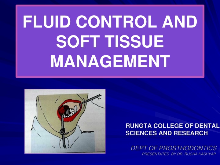
  1. FLUID CONTROL AND SOFT TISSUE MANAGEMENT RUNGTA COLLEGE OF DENTAL SCIENCES AND RESEARCH DEPT OF PROSTHODONTICS PRESENTATED BY DR. RUCHA KASHYAP PRESENTATED BY DKASHYAP

  2. CONENTS INTRODUCTION CLASSIFICATION ADVANTAGE/ DIS ADVANTAGE INDIACATION/ CONTRAINDICATION SELECTION OF ABUTMENT TEETH TYPES OF ATTCHMENTS SUMMARY TAKE HOME MESSAGE REFERENCES 2

  3. SPECIFIC LEARNING OBJECTIVES SR.N CORE AREA DOMAIN CATEGORY O 1. INTRODUCTION Cognitive Must Know CLASSIFICATION 2. ADVANTAGE/ DIS Cognitive Must Know ADVANTAGE INDIACATION/ CONTRAINDICATION SELECTION OF ABUTMENT TEETH 3. TYPES OF ATTCHMENTS Affirmative Must Know 3

  4. INTRODUCTION Restorative procedures in the mouth cannot be executed efficiently unless the moisture is controlled . moisture control includes the exclusion of sulcular fluid ,saliva and gingival bleeding from the operating field. It also refers to preventing the aspiration or swallowing of fluid sprayed from the handpiece and restorative debris . primary objectives of moisture control are isolation ,retraction and accessibility. The gingiva must be displaced to make a complete impression and some time even to permit completion of the preparation and cementation of the restoration.

  5. FLUID CONTROL (ISOLATION) The need for removal of fluid varies depending upon the task being performed . Isolation of the operating site is done for the following reasons: To obtain a dry, clean ,operating field For easy access and visibility To improve properties of dental materials To protect the patient and operator To improve the operating efficiency

  6. METHOD OF FLUID CONTROL MECHANICAL CHEMICAL OTHER 1. Rubber dam 1. Anti-sialogogues 1. Cotton rolls 2. High volume vacuum 2. Local anesthetics 2. Cellulose wafers 3. Saliva ejector 3. Throat shields 4. svedopter

  7. RUBBER DAM Introduced by S.C BARNUM in 1864 . It is the most effective of all isolation devices utilized in restorative dentistry . valuable in the removal of old restorations or excavation of caries when exposure of the pulp is a possibility . It is used to define the operating field by isolating one or more teeth from the oral environment. It eliminates the saliva from the operating side and retracts the soft tissue. It should not be used while making a polyvinyl siloxane impression, because it will inhibit its polymerization . It also provides excellent isolation and access when a pin retained amalgam or composite resin core is required before a cast restoration can be fabricated .

  8. parts of rubber dam Rubber dam sheet Rubber dam holder Rubber dam retainer Rubber dam punch Rubber dam retainer forceps Rubber dam napkin Certain condition preclude the use of the rubber dam (1) Teeth that have not erupted sufficiently to receive a retainer (2) some third molars (3) extremely malpositioned teeth (4) Patient suffering from asthma may not tolerate the rubber dam if breathing through the nose is difficult .

  9. High volume vacuum A high volume suction tip is extremely useful preparation phase and is most effectively utilized with an assistant during the It makes an excellent lip retractor while the operator uses a mirror to retract and protect the tongue . Mcwherter (1957) showed that one type of evacuator would remove 0.5 L water in 2 seconds ,had a 75% to 95 % pickup of water in air ,and would remove 100 % of solid during cutting procedure .

  10. SALIVA EJECTOR It is most useful as an adjunct to high volume evacuation . It can be used for evacuation when the maxillary arch is being treated . It is not very effective during impression and cementation . It is placed at the corner of the mouth, opposite to the quadrant being operated and the patient s head is turned toward it.

  11. SVEDOPTER It mandibular teeth . It is the metal saliva ejector attached with a tongue deflector . is used for isolating the Disadvantages of svedopter Access to the lingual surface of mandibular teeth is limited Since it is a metallic device , care must be taken to avoid any injury to the floor of the mouth . Presences of precludes its use . mandibular tori

  12. The svedopter is most effective when it is used with the patient in a nearly upright position .In this position ,water and other fluid collect on the floor of the mouth ,where they are pulled off by the vacuum. PRECAUTION Selection reflector should be avoided. Since it could cut into the palate above or trigger the gag reflex . of an oversized For anterior part of the svedopter should be placed in the incisor region with the tubing under the patient ,arm . better positioning ,the

  13. Cotton roll and cellulose wafers These are helpful for short period of Isolation .Especially in conjunction with profound anesthesia acceptable dryness for procedures such as impression taking and cementation . ,absorbents provide Now a days cotton roll holder are used for holding cotton roll in position .But this is inconvenient and time-consuming . Advantage of cotton roll holders is that the cheeks and tongue are slightly retracted from the teeth, which enhances access and visibility . when removing cotton rolls or cellulose wafers, it may be necessary to moisten them using the air/water syringe to prevent inadvertent removal of the epithelium from cheek ,floor of the mouth ,or lips . .

  14. THROAT SHIELD Throat shield are used during try-in and removal of indirect restorations. This is particularly important when treating teeth in the maxillary arch . A gauze sponge (2x2 inch ) ,unfolded and spread over the tongue and the posterior part of the mouth , is helpful in recovering a restoration should it be dropped .

  15. CHEMICAL METHOD ANTI-SIALOGOGUES They are gastrointestinal anti-cholinergic that act on the smooth muscles of gastrointestinal ,urinary and biliary tracts , producing a dry mouth as a side effect . Commonly used anti-sialogogues Methantheline bromide (banthine) :50 mg 1 hr before procedure Propantheline bromide : 15 mg 1 hr before procedure Clonidine hydrochloride (antihypertensive) : 0.2 mg 1 hr before procedure .

  16. Contraindication History of hypersensitivity to the drugs Obstructive conditions of the GIT or urinary tract Patient with glaucoma Asthma Congestive heart failure . side effect Drowsiness Blurred vision Bitter taste Propantheline can be made tasteless by injecting 2 to 6 mg in solution intraorally .Onset of action occurs in 5 to 10 minutes , and the duration of a dry working environment is approximately 2 hours

  17. Importance of finish line exposure The gingival tissue must be healthy and free of inflammation before restoration are fabricated The finish line must be reproduced in the impression . The marginal fit is very important in preventing recurrent caries and gingival inflammation .(marginal integrity) Obtaining a complete impression is complicated when some or all of the preparation finish line lies at or apical to the crest of the free gingiva . Control of fluid in the sulcus , particularly when a hydrophobic impression material is used ,is also necessary , because liquids can cause an incomplete impression of the critical finish line area .

  18. Need for gingival retraction To widen the gingival sulcus to provide access for the impression materials to reach the sub-gingival margins and to record accurately the finished margins Recording the contour beyond the finish line helps to correctly contour the restoration and smoothly blend the margins of the restorations with the unprepared tooth structure. While cementing a restoration ,it helps in removal of excess cement without injuring the gingival tissue. In case of sub gingival preparation ,it prevents injury to the crest of the gingiva.

  19. METHODS FOR GINGIVAL RETRACTION (A) Mechanical method (a) copper bands (b) retraction cord (c) Rubber dam (B) Chemico-mechanical gingival retraction cord (C) Surgical method (a) rotary curettage (b) electrosurgery

  20. (A) MECHANICAL METHOD OF RETRACTION (a) COPPER BAND /TUBE IMPRESSION Ist describe by john j .Lucca (1959) It is used to carry the impression materials as well as to displace the gingiva to expose the finish line . impression compound or elastomeric impression materials can be used along with this band .

  21. TECHNIQUE A copper band is welded to form a tube corresponding to the size of the prepared tooth . one end of the tube is festooned ,or trimmed to follow the profile of the finish line. After positioning and contouring the tube over the prepared tooth, it is filled with modelling compound and then it is seated carefully in place along the path of insertion of the tooth preparation and the impression is made . Disadvantage It can cause injury to the gingival tissues.

  22. (b) RETRACTION CORD Plain cotton cord was used for sulcus enlargement physically pushing away the gingiva from the finish line . Its effectiveness is limited because the use of pressure alone often will not control sulcular hemorrhage . (c) RUBBER DAM Generally it is used when a limited number of teeth in one quadrant are being restored and in situations in which preparations do not have to be extended very far subgingivally .It can be used with modified tray if the bow and wings of the clamp are blocked out .

  23. Chemico mechanical method CHEMICAL USED It is a method of combining a chemical with pressure packing ,which leads to enlargement of the gingival sulcus as well as control of fluids seeping from the sulcus . gingival retraction cord soaked in a chemical (which contraction ) will provide better gingival retraction compared to a plain retraction cord . These are generally local vasoconstrictors which produce transient gingival shrinkage. (A) 8% racemic epinephrine promote gingival (B) Aluminium chloride (C) Alum (aluminium potassium sulphate ) (d) Aluminium sulphate (E) Ferric sulphate

  24. Ideal requirement for chemical used with gingival retraction cords It should produce effective gingival displacement and haemostasis. It should not produce any irreversible damage to the gingiva . it should not have any systemic side effect . Newer gingival retraction agents are Phenylephrine hydrochloride 0.25 % Oxymetazoline hydrochloride 0.05 % Tetrahydrozoline hydrochloride 0.05 % contraindications for epinephrine CVS disease Diabetes Hyperthyroidism Known hypersensitivity to epinephrine patient taking ganglionic blocker ,or epinephrine potentiating drug

  25. Some silent point about epinephrine The amount of epinephrine absorbed is highly variable ,depending on the degree of exposure of the vascular bed as well as the time of contact and the amount of medication in the cord . The amount of epinephrine absorbed from 2.5 cm of typical retraction cord during 5 to 15 minutes in the gingival sulcus is 71 g .It is approximately 1/3 rd the maximum dose of 0.2 mg (200 g ) for a healthy adult and nearly twice the recommended amount of 0.04 mg (40 g ) for a cardiac patient . If cord is placed around more than one tooth ,if more than one impression is made of a single tooth , and /or if the epinephrine- containing anesthetic is used , a patient could easily exceed the recommended maximum dose of epinephrine . WEIR and WILLIAMS (1984) ,in an in vivo study of 120 human teeth ,found no significant difference between the hemorrhage control offered by cords impregnated with aluminum sulphate ,and those impregnated with epinephrine

  26. TECHNIQUE CORD PLACEMENT IS A FINESSE MOVE , NOT A POWER PLAY . The operating area should be dry. Fluid control should be done with an evacuating device and the quadrant containing the prepared tooth is isolated with cotton rolls. Hemorrhage can be controlled by using haemostatic agent like hemodent liquid (aluminium chloride) Retraction cord is looped around the tooth and held tightly with the thumb and forefinger and apply slight tension in an apical direction Cord is twisted to make it tight and small

  27. As the cord is being placed subgingivally ,the instrument must be pushed slightly towards the area already tucked into place .If the force of the instrument is directed away from the area previously packed, the cord already packed will be pulled out . Placement of cord is begun by pushing it into the gingival sulcus on the mesial surface of the tooth .It should be tacked lightly into the distal crevice .

  28. Occasionally it is necessary to hold the cord with one instrument while packing with the second . The instrument used for packing should be angled slightly towards the root to facilitate the sub-gingival placement of the cord If it is held parallel to the long axis of the tooth , the cord will be pushed against the wall of the gingival crevice ,and will rebounce .

  29. Excess cord is cut off near the inter proximal area such that a slight overlap of the cord occur in this region . if the overlap occur on the facial and lingual surfaces , the gingival finish line in that area may not be replicated properly in the impression . At least 2-3 mm of cord is left protruding out-side the sulcus so that it can be grasped for easy removal . After cutting off the excess at the mesial end ,the distal end of the cord is a tucked in until it overlaps the tucked mesial end .

  30. TECHNIQUE (contd) The cord can be packed with special instrument like Fischer packing instrument .It is a double ended, serrated or smooth edges stainless steel instrument facilitates placing of retraction cord around the tooth. Both ends of the retraction cord packers are shaped at an angle which allows the cord to be packed swiftly right around the tooth. Retraction cord scissors . Blunt-tipped retraction cord scissors with less risk to tissue. 1/2" long spring steel blades flex for a consistent cut to the tip and a longer service life. Uniband spring handle provides for smooth control.

  31. After 4-6 minutes , the cord should be removed slowly in order to avoid bleeding .If active bleeding persists , a cord soaked in ferric sulphate should be placed in the sulcus and removed after 3 minutes The impression should be made only after cessation of bleeding . The retraction cord must be slightly moist before removal . removing dry cord from the crevice can injure the delicate epithelial lining of the gingiva .

  32. Magic Foam Cord MagicFoam Cord is the first expanding VPS material designed for easy and fast retraction of the sulcus without potentially traumatic packing or pressure.

  33. Magic FoamCord Perfect retraction of the Sulcus, stops bleeding without invasive materials or techniques Easier to use (same as impression taking). Flows directly into the Sulcus. No need for technique sensitive application technique. No trauma (no packing or pressure, no bleeding caused by the procedure) Astringent is not required no need to rinse More efficient when doing multiple preparations

  34. Magic FoamCord How Does It Work Syringe FoamCord around the preparation Place pre-fitted Comprecap over tooth and ask patient to bite down Wait 5 min. to allow FoamCord material to fully set and sulcus to expand Preparation ready for final impression

  35. 1. Initial Situation 2. Pre-fit the Comprecap 3. Apply MagicFoamCord around the preparations

  36. 5. Let the patient bite on the Comprecap 4. Place Comprecap 6. Comprecap After Removal

  37. SURGICAL METHODS ROTARY CURETTAGE (GINGETTAGE) It has been compared with periodontal curettage but periodontal curettage is used to debride diseased tissue from the sulcus to allow reepithelization and healing . kamansky et al (1984 ) reported that less change in gingival height with rotary curettage than with lateral gingival displacement using retraction cord .With curettage there was an apparent disruption of the apical sulcular and attachment epithelium ,resulting in apical repositioning and an increase in sulcus depth .

  38. Rotary curettage (gingettage) The concept of rotary curettage was described by Amsterdam in 1954 . The technique describe by Hansing. It is a troughing technique , the purpose of which is to produce limited removal of the epithelial tissue in the sulcus while a chamfer finish line is being created in tooth structure. It must be done only on healthy , inflammation free tissue to avoid the tissue shrinkage that occur when disease tissue heals . The following criteria should be fulfilled for gingettage Absence of bleeding upon probing from the gingiva The depth of the sulcus is less than 3 mm Presence of adequate keratinized gingiva .

  39. TECHNIQUE Prior to rotary curettage ,a shoulder finish formed at the level of the gingival crest . A torpedo diamond point simultaneously forms a chamfer finish line and removes the lining of the sulcus. line is epithelial Disadvantages Technique sensitive as the instrument offers poor tactile sensation. It can potentially damage the periodontium if used incorrectly.

  40. ELECTROSURGICAL RETRACTION ELECTROCAUTERY Vs ELECTROSURGERY Often electrocautery is used to describe electrosurgery. This is incorrect. Electrocautery refers to direct current (electrons flowing in one direction) whereas electrosurgery uses alternating current. During electrocautery, current does not enter the patient s body. Only the heated wire comes in contact with tissue. In electrosurgery, the patient is included in the circuit and current enters the patient s body. In electrosurgery cutting electrode remains cold whereas in electrocautery a hot electrode is applied to the tissue .

  41. Electrosurgery denotes surgical reduction of sulcular epithelium using an electrode to produce gingival retraction .It has been recommended for enlargement of the gingival sulcus and removal of irritated tissue that has proliferated over preparation finish line . Electrosurgery unit is a high frequency oscillator or radio transmitter that uses either a vacuum tube or a transistor to deliver a high frequency electrical current of at least 1.0 MHZ (one million cycles per second ) . the procedure is also called as surgical diathermy . Electrosurgery cannot stop bleeding once it starts , if hemorrhage occurs ,it first must be controlled with pressure and / or chemicals , and then vessels can be sealed with a coagulating ball electrode . Credit for being the direct progenitor of electrosurgery is generally given to D , ARSONVAL (1981)

  42. Contraindication Patient with cardiac pacemakers because external electromagnetic interference hinders the pacemakers function . The use of topical anesthetics such as ethyl chloride and other inflammable aerosols should be avoided when electrosurgery is to be used . Advantages Sophisticated technique Can be done in case with gingival inflammation Produce little to no bleeding . Quick procedure

  43. Disadvantage Very technique sensitive Application of excessive pressure may produce severe tissue damage Difficult to control lateral dissipation of heat The operatory area should be very moist during the procedure . this leads to compromised access and visibility . Mechanism A. Current flows from a small cutting electrode that produces a high current density and a rapid temperature rise at its point of contact with the tissue B. The cells directly adjacent to the electrode are destroyed by this temperature increases . C. The circuit is completed by contact between the patient and a ground electrode that will not generate heat in the tissue because its large surface area produces a low current density , even though the same amount of current passes through it .

  44. Type of current used for electrosurgery UNRECTIFIED ,DAMPED CURRENT (OUDIN OR TELSA CURRENT) It is characterized by recurring peaks of power that rapidly diminishes. It produces intense lack of moisture (dehydration) ,necrosis and coagulation of the cells .It produces slow and painful healing , hence it is avoided . PARTIALLY RECTIFIED , DAMPED CURRENT (HALF WAVE MODULATED ) Here the current during the second half of each cycle is damped so that only the peak waves act on the electrode . It produces good coagulation and haemostasis . but it also produces slow and painful healing with considerable tissue destruction because the electrical flow is intermittent .

  45. FULLY RECTIFIED CURRENT (FULL WAVE MODULATED ) Here the frequency is similar to a partially rectified current but it is continuous .It produces adequate sulcus enlargement with good cutting characteristics along with good haemostasis . FULLY RECTIFIED ,FILTERED CURRENT Here the peak waves are repeated so that there is continuous flow without any dip . lower frequency waves are filtered in this current . it produces excellent cutting . hence it is most preferred .

  46. ELECTROSURGICAL UNIT (A) GROUND ELECTRODE Also known as ground plate, indifferent plate, indifferent electrode, neutral electrode, dispersive electrode, passive electrode or patient return. Proper grounding of a patient is considered to be the single most important safety factor when electrosurgery is used . ORINGER recommends that the ground be placed under the thigh rather than behind the back , as is often done . contact with a small bony protuberance , such as a vertebra or bony tubercles could produce a high current density to cause a burn .The only precaution to be observed in placing the ground under the legs is that the patient does not have keys in a pants pocket or is not wearing metal garters .

  47. (B) SURGICAL ELECTRODE OR CUTTING ELECTRODE It is designed to fit on to the hand piece of the electrosurgical unit. This heat helps to vaporize the target tissue. An electrosurgical probe comprises of a shank and a cutting edge. The shank is designed to fit into the piece of the electrosurgical unit. It may be either straight or j- shaped . numerous cutting edge designs are available but the most commonly used ones are A. Coagulating probe B. Diamond loop C. Round loop D. Small straight probe E. Small loop

  48. Basic principals during electrosurgical procedures Local anesthesia should be given. During unpleasant odor will be liberated due to tissue necrosis .Aromatic oils such as peppermint oil can be applied at the vermilion border of lip so that it masks the unpleasant odor that will arise from the electrode during the procedure. electro-surgery, considerable Grounding should be done before the usage of the electrode in order to protect the patient from electrical accidents.

  49. Electrodes must be completely seated in the hand piece . if any uninsulated portion of it other than the cutting tip is exposed outside the handpiece chuck , it could produce an accidental burn on the patient ,s lip . During its use, the electrode should be applied with very light pressure and swift strikes. Tactile control for the operator is vital for this procedures because it is sufficient to just run the probe along the sulcus without any pressure. Pressure should not be applied, because it may produce excessive tissue damage .

  50. The electrode should never be placed stagnant at any one point as it may lead to lateral dissipation of heat producing gingival injury. In order to prevent lateral heat dissipation, the probe should be moved at a minimal speed of 7 mm per second. Moist tissues can be cut best. If it is necessary to redo the cutting in a particular region, a rest period of 8-10 seconds should be allowed to elapse before beginning the second stroke. The electrode should pass through the tissue in a very smooth motion without dragging or charring the tissue. A wooden tongue depressor , plastic handle mirror and a plastic vacuum tip should be kept close to the surgical site .

Related


More Related Content

giItT1WQy@!-/#giItT1WQy@!-/#giItT1WQy@!-/#giItT1WQy@!-/#giItT1WQy@!-/#giItT1WQy@!-/#giItT1WQy@!-/#giItT1WQy@!-/#