Insights into Liver Anatomy, Function, and Imaging Studies


l
i
v
e
r
A
n
a
t
o
m
y
—
l
i
g
a
m
e
n
t
s
a
n
d
p
e
r
i
t
o
n
e
a
l
r
e
f
l
e
c
t
i
o
n
1
-
l
f
t
r
i
a
n
g
u
l
a
r
l
i
g
.
f
i
x
e
d
l
f
l
o
b
e
o
f
l
i
v
e
r
t
o
t
h
e
d
i
a
p
h
r
a
g
m
-
-
-
d
i
v
i
s
i
o
n
a
n
t
.
a
n
d
p
o
s
t
.
L
e
a
f
a
l
l
o
w
t
o
m
o
b
i
l
i
z
e
l
f
l
o
b
e
o
f
l
i
v
e
r
f
r
o
m
l
f
l
a
t
.
w
a
l
l
o
f
i
v
c
2
-
R
T
t
r
i
a
n
g
u
l
a
r
l
i
g
a
m
e
n
t
.
f
i
x
e
d
R
T
l
o
b
e
o
f
l
i
v
e
r
t
o
t
h
e
R
T
H
e
m
i
d
i
a
g
h
r
a
g
m
3
-
f
a
l
c
i
f
o
r
m
l
i
g
a
m
e
n
t
w
h
i
c
h
i
t
’
s
a
r
e
m
n
a
n
t
o
f
u
m
b
i
l
i
c
a
l
v
e
i
n
r
u
n
s
f
r
o
m
u
m
b
i
l
i
c
u
s
t
o
t
h
e
l
i
v
e
r
4
-
l
e
s
s
e
r
o
m
e
n
t
u
m
c
o
n
n
e
c
t
l
i
v
e
r
t
o
t
h
e
s
t
o
m
a
c
h
B
l
o
o
d
s
u
p
p
l
y
o
f
l
i
v
e
r
8
0
%
f
r
o
m
p
o
r
t
a
l
v
e
i
n
,
2
0
%
f
r
o
m
h
e
p
a
t
i
c
a
r
t
e
r
y
w
h
i
c
h
b
r
a
n
c
h
f
r
o
m
c
a
e
l
i
c
t
r
u
n
k
w
h
i
c
h
c
o
m
e
f
r
o
m
a
o
r
t
a
.
H
e
p
a
t
i
c
a
r
t
e
r
y
d
i
v
i
d
i
n
g
t
o
R
T
a
n
d
L
F
h
e
p
a
t
i
c
a
r
t
e
r
y
t
o
s
u
p
p
l
y
t
h
e
R
T
a
n
d
l
L
F
l
o
b
e
o
f
S
t
r
u
c
t
u
r
e
i
n
h
i
l
u
m
o
f
l
i
v
e
r
H
e
p
a
t
i
c
a
r
t
e
r
y
(
a
b
o
v
e
a
n
d
m
e
d
i
a
l
)
,
p
o
r
t
a
l
v
e
i
n
(
p
o
s
t
e
r
i
o
r
)
a
n
d
c
b
d
(
a
b
o
v
e
a
n
d
l
a
t
.
)
,
p
r
e
s
e
n
t
s
w
i
t
h
f
r
e
e
a
g
e
d
o
f
l
e
s
s
e
r
o
m
i
n
t
u
m
,
V
e
n
o
u
s
d
r
a
i
n
a
g
e
o
f
t
h
e
l
i
v
e
r
V
i
a
h
e
p
a
t
i
c
v
e
i
n
s
i
n
t
o
t
h
e
i
n
f
e
r
i
o
r
v
e
n
a
c
a
v
a
A
n
a
t
o
m
y
o
f
l
i
v
e
r
(
i
n
t
e
r
n
l
)
;
l
i
v
e
r
d
i
v
i
d
e
d
t
o
r
t
a
n
d
l
f
l
o
b
e
.
r
t
l
o
b
e
h
a
s
s
e
g
m
e
n
t
f
r
o
m
V
t
o
V
I
I
I
S
U
P
P
L
Y
b
y
r
t
h
e
p
a
t
i
c
a
r
t
e
r
y
a
n
d
r
t
b
r
a
n
c
h
o
f
p
o
r
t
a
l
v
e
i
n
a
n
d
d
r
a
i
n
v
i
a
r
t
h
e
p
a
t
i
c
d
u
c
t
.
L
f
l
o
b
e
o
f
l
i
v
e
r
c
o
n
t
a
i
n
f
r
o
m
1
t
o
1
v
s
e
g
m
e
n
t
s
s
u
p
p
l
y
b
y
l
f
h
e
p
a
t
i
c
a
r
t
e
r
y
a
n
d
l
f
b
r
a
n
c
h
o
f
p
o
r
t
a
l
v
e
i
n
a
n
d
d
r
a
i
n
v
i
a
l
f
h
e
p
a
t
i
c
d
u
c
t
.
M
a
i
n
f
u
n
c
t
i
o
n
o
f
l
i
v
e
r
1
-
m
a
i
n
t
a
i
n
c
o
r
e
b
o
d
y
t
e
m
p
e
r
a
t
u
r
e
2
-
P
H
b
a
l
a
n
c
e
a
n
d
c
o
r
r
e
c
t
i
o
n
o
f
l
a
c
t
i
c
a
c
i
d
o
s
i
s
3
-
S
Y
N
T
H
E
S
I
S
O
F
C
L
O
T
T
I
N
G
F
A
C
T
O
R
4
-
g
l
u
c
o
s
e
m
e
t
a
b
o
l
i
s
m
g
l
y
c
o
l
y
s
i
s
a
n
d
g
l
u
c
o
n
e
o
g
e
n
s
i
s
5
-
u
r
e
a
f
o
r
m
a
t
i
o
n
f
r
o
m
p
r
o
t
e
i
n
c
a
t
a
b
o
l
i
s
m
6
-
b
i
l
i
r
o
b
i
n
f
o
r
m
a
t
i
o
n
f
r
o
m
h
g
s
e
g
r
e
g
a
t
i
o
n
7
-
d
r
u
g
a
n
d
h
o
r
m
o
n
e
m
e
t
a
b
o
l
i
s
m
8
-
r
e
m
o
v
a
l
o
f
g
u
t
e
n
d
o
t
o
x
i
n
a
n
d
f
o
r
e
i
g
n
a
n
t
i
g
e
n
.
L
i
v
e
r
f
u
n
c
t
i
o
n
t
e
s
t
1
-
T
S
B
D
I
R
E
C
T
A
N
D
I
N
D
I
R
E
C
T
2
-
A
L
P
(
a
l
k
a
l
i
n
p
h
o
s
p
h
a
t
a
s
e
)
3
5
-
1
3
0
I
U
3
-
A
S
T
(
A
S
P
E
R
T
A
T
E
T
R
A
N
S
A
M
I
N
A
S
E
)
4
-
4
0
I
U
4
-
A
L
T
(
A
L
A
N
I
N
T
R
A
N
S
A
M
I
N
A
S
E
)
5
-
4
0
5
-
G
A
M
M
A
G
L
U
T
A
M
Y
L
T
R
A
N
S
P
E
P
T
U
D
A
S
E
(
G
G
T
)
1
0
-
4
8
I
U
6
-
S
E
R
I
E
M
A
L
B
U
M
I
N
3
5
-
5
0
G
M
7
-
P
R
O
T
H
R
O
M
P
I
N
T
I
M
I
M
E
1
2
-
1
6
S
E
C
O
N
D
L
i
v
e
r
f
a
i
l
u
r
e
E
i
t
h
e
r
a
c
u
t
e
o
r
c
h
r
o
n
i
c
M
a
i
n
f
e
a
t
u
r
e
o
f
c
h
r
o
n
i
c
l
i
v
e
r
f
a
i
l
u
r
e
a
r
e
L
e
t
h
a
r
g
y
,
f
e
v
e
r
,
j
a
u
n
d
i
c
e
,
w
a
s
t
i
n
g
(
p
r
o
t
e
i
n
c
a
t
a
b
o
l
i
s
m
)
,
c
o
a
g
u
l
o
p
a
t
h
y
,
h
y
p
e
r
d
y
n
a
m
i
c
c
i
r
c
u
l
a
t
i
o
n
,
h
e
p
a
t
i
c
e
n
c
e
p
h
a
l
o
p
a
t
h
y
,
p
o
r
t
a
l
h
y
p
e
r
t
e
n
s
i
o
n
,
a
s
c
i
t
i
s
,
e
s
o
p
h
a
g
e
a
l
v
a
r
e
c
i
e
s
,
s
p
l
e
n
o
m
e
g
a
l
y
,
c
o
e
t
a
n
e
o
u
s
-
-
(
s
p
i
d
e
r
n
a
i
v
e
,
p
a
l
m
e
r
e
r
y
t
h
e
m
a
)
I
m
a
g
i
n
g
s
t
u
d
y
o
f
t
h
e
l
i
v
e
r
U
/
S
,
S
P
I
R
A
L
C
T
S
C
A
N
(
g
i
v
e
i
n
f
o
r
m
a
t
i
o
n
f
o
r
l
e
s
i
o
n
d
o
w
n
t
o
l
e
s
s
t
h
a
n
1
c
m
i
n
d
i
a
m
e
t
e
r
a
n
d
g
i
v
e
i
n
f
o
r
m
a
t
i
o
n
o
n
t
h
e
r
e
n
a
t
u
r
e
)
,
M
R
I
,
E
R
C
P
(
v
e
r
y
u
s
e
f
u
l
i
n
o
b
s
t
r
u
c
t
i
v
e
c
a
u
s
e
o
f
l
i
v
e
r
f
a
i
l
u
r
e
b
u
t
m
o
s
t
c
h
e
c
k
c
o
a
g
u
l
a
t
i
o
n
)
,
P
T
C
,
A
N
G
I
O
G
R
A
P
H
Y
,
N
U
C
L
E
A
R
M
E
D
I
C
I
N
S
C
A
N
I
N
G
r
a
d
i
o
i
s
o
t
o
p
e
s
c
a
n
n
i
n
g
,
L
A
P
A
R
A
S
C
O
P
Y
A
N
D
L
A
P
A
R
A
S
C
O
P
I
C
U
/
S
(
i
s
u
s
e
f
u
l
i
n
s
t
a
g
i
n
g
o
f
h
e
p
a
t
o
p
a
n
c
r
e
a
t
o
b
i
l
i
a
r
y
c
a
n
c
e
r
L
i
v
e
r
t
r
a
u
m
a
L
i
v
e
r
i
n
j
u
r
y
s
e
r
i
o
u
s
a
n
d
a
s
s
o
c
i
a
t
e
d
w
i
t
h
s
i
g
n
i
f
i
c
a
n
t
m
o
r
b
i
d
i
t
y
a
n
d
m
o
r
t
a
l
i
t
y
e
v
e
n
w
i
t
h
p
r
o
m
p
t
a
n
d
a
p
p
r
o
p
r
i
a
t
e
m
a
n
a
g
e
m
e
n
t
.
B
e
c
a
u
s
e
i
t
s
h
i
g
h
l
y
v
a
s
c
u
l
a
r
.
L
i
v
e
r
t
r
a
u
m
a
d
i
v
i
d
e
d
t
o
b
l
u
n
t
(
a
v
u
l
s
i
o
n
,
l
a
c
e
r
a
t
i
o
n
,
c
o
n
t
u
s
i
o
n
)
a
n
d
p
e
n
e
t
r
a
t
i
n
g
t
r
a
u
m
a
9
s
t
a
b
w
o
u
n
d
o
r
g
u
n
s
h
o
o
t
)
.
D
i
a
g
n
o
s
i
s
o
f
l
i
v
e
r
i
n
j
u
r
y
;
A
l
l
l
o
w
e
r
r
t
c
h
e
s
t
a
n
d
u
p
p
e
r
a
b
d
o
m
i
n
a
l
s
t
a
b
w
o
u
n
d
s
h
o
u
l
d
b
e
s
u
s
p
e
c
t
l
i
v
e
r
i
n
j
u
r
y
a
n
d
m
a
y
b
e
a
s
s
o
c
i
a
t
e
d
w
i
t
h
o
t
h
e
r
i
n
t
r
a
a
b
d
o
m
i
n
a
l
o
r
g
a
n
i
n
j
u
r
y
e
s
p
e
c
i
a
l
l
y
i
f
t
h
e
r
e
i
s
s
i
g
n
a
n
d
s
y
m
p
t
o
m
s
o
f
b
l
o
o
d
l
o
s
s
.
I
f
p
t
h
a
e
m
o
d
i
a
n
a
m
i
c
l
y
u
n
s
t
a
b
l
e
b
e
c
a
u
s
e
o
f
b
l
o
o
d
l
o
s
s
o
r
p
t
w
i
t
h
p
e
n
e
t
r
a
t
i
n
g
t
r
a
u
m
a
n
e
e
d
s
u
r
g
e
n
t
l
a
p
a
r
o
t
o
m
y
,
i
f
p
t
s
t
a
b
l
e
c
a
n
d
o
c
t
s
c
a
n
w
i
t
h
c
o
n
t
r
a
s
t
e
n
h
a
n
c
e
d
.
-
-
-
-
p
e
r
i
t
o
n
e
a
l
l
a
v
a
g
e
i
f
h
a
e
m
o
p
e
r
t
o
n
e
a
l
-
-
-
-
l
a
p
a
r
o
s
c
o
p
y
b
y
w
h
i
c
h
c
a
n
s
e
e
i
f
t
h
e
r
e
i
s
d
i
a
p
h
r
a
g
m
a
t
i
c
r
a
p
t
u
r
e
.
M
a
n
a
g
e
m
e
n
t
s
;
F
i
r
s
t
m
a
n
a
g
e
a
n
y
p
t
w
i
t
h
t
r
a
u
m
a
a
s
a
b
c
d
e
.
i
n
c
a
s
e
o
f
p
t
w
i
t
h
a
b
n
o
r
m
a
l
c
i
r
c
u
l
a
t
i
o
n
;
2
l
a
r
g
e
b
o
r
e
c
a
n
u
l
a
e
s
e
n
d
b
l
o
o
d
f
o
r
b
l
o
o
d
g
r
o
u
p
a
n
d
c
r
o
s
s
m
a
t
c
h
1
0
p
i
n
t
s
.
s
a
m
p
l
e
f
o
r
f
u
l
l
b
l
o
o
d
c
o
u
n
t
,
u
r
e
a
a
n
d
e
l
e
c
t
r
o
l
y
t
e
l
i
v
e
r
f
u
n
c
t
i
o
n
t
e
s
t
c
l
o
t
t
i
n
g
s
c
r
e
e
n
i
n
g
,
g
l
u
c
o
s
e
a
n
d
a
m
y
l
a
s
e
.
T
h
e
n
s
t
a
r
t
w
i
t
h
f
l
u
i
d
r
e
p
l
a
c
e
m
e
n
t
s
t
a
r
t
w
i
t
h
c
o
l
l
o
i
d
o
r
o
n
e
g
a
t
i
v
e
b
l
o
o
d
(
w
h
a
t
t
h
e
d
i
f
f
e
r
e
n
c
e
b
e
t
w
e
e
n
c
o
l
l
o
i
d
a
n
d
c
r
y
s
t
a
l
l
o
i
d
?
?
)
A
r
t
e
r
i
a
l
b
l
o
o
d
g
a
s
s
h
o
u
l
d
o
b
t
a
i
n
a
n
d
t
h
e
p
t
i
n
t
u
b
a
t
e
a
n
d
v
e
n
t
i
l
a
t
e
i
f
g
a
s
e
x
c
h
a
n
g
e
i
n
a
d
e
q
u
a
t
e
.
C
h
e
s
t
t
u
b
e
i
f
t
h
e
r
e
i
s
h
a
e
m
o
o
r
p
n
e
m
o
t
h
o
r
a
x
.
S
u
r
g
i
c
a
l
a
p
p
r
o
a
c
h
t
o
t
h
e
l
i
v
e
r
;
R
o
o
f
t
o
p
i
n
c
i
s
i
o
n
p
r
o
v
i
d
e
e
x
c
e
l
l
e
n
t
v
i
s
u
a
l
i
z
a
t
i
o
n
o
f
t
h
e
l
i
v
e
r
a
n
d
s
p
l
e
e
n
.
(
W
h
a
t
i
s
P
r
i
n
g
l
e
m
a
n
e
u
v
e
r
)
a
t
r
a
u
m
a
t
i
c
c
l
u
m
p
a
c
r
o
s
s
f
o
r
a
m
e
n
o
f
W
i
n
s
l
o
w
t
o
c
o
n
t
r
o
l
o
n
b
l
e
e
d
i
n
g
f
r
o
m
l
i
v
e
r
.
P
a
c
k
i
n
g
o
f
l
i
v
e
r
m
a
y
b
e
e
s
s
e
n
t
i
a
l
i
f
t
h
e
r
e
i
s
d
i
f
f
u
s
e
b
l
e
e
d
i
n
g
f
r
o
m
p
a
r
e
n
c
h
y
m
a
o
f
l
i
v
e
r
.
p
r
e
s
s
u
r
e
f
r
o
m
b
e
l
o
w
u
s
u
a
l
l
y
p
a
c
k
s
f
o
r
4
8
h
o
u
r
s
i
n
m
o
s
t
o
f
p
t
s
n
o
f
u
r
t
h
e
r
t
r
e
a
t
m
e
n
t
n
e
e
d
.
C
o
m
p
l
i
c
a
t
i
o
n
o
f
l
i
v
e
r
t
r
a
u
m
a
1
-
p
r
o
f
u
s
e
b
l
e
e
d
i
n
g
2
-
l
i
v
e
r
a
b
s
c
e
s
s
d
u
e
t
o
i
n
f
e
c
t
i
o
n
o
f
l
i
v
e
r
h
e
m
a
t
o
m
a
3
-
b
i
l
e
c
o
l
l
e
c
t
i
o
n
a
n
d
s
o
m
e
t
i
m
e
b
i
l
i
a
r
y
f
i
s
t
u
l
a
L
a
t
e
c
o
m
p
l
i
c
a
t
i
o
n
1
-
h
e
p
a
t
i
c
a
r
t
e
r
y
a
n
e
u
r
y
s
m
2
-
a
.
v
f
i
s
t
u
l
a
o
r
a
.
b
i
l
i
a
r
y
f
i
s
t
u
l
a
3
-
r
a
r
l
y
b
i
l
i
a
r
y
t
r
a
c
t
s
t
r
i
c
t
u
r
e
E
s
o
p
h
a
g
e
a
l
v
a
r
i
c
e
s
;
D
i
l
a
t
e
d
t
o
r
t
u
o
u
s
v
e
i
n
s
o
f
l
o
w
e
r
p
a
r
t
o
f
e
s
o
p
h
a
g
u
s
,
i
t
’
s
a
c
o
m
p
l
i
c
a
t
i
o
n
o
f
p
o
r
t
a
l
h
y
p
e
r
t
e
n
s
i
o
n
(
w
h
a
t
o
t
h
e
r
s
i
t
e
o
f
p
o
t
o
c
y
s
t
e
m
i
c
c
o
n
n
e
c
t
i
o
n
)
.
U
S
U
A
L
L
Y
p
r
e
s
e
n
t
w
i
t
h
a
c
u
t
e
o
n
s
e
t
o
f
a
l
a
r
g
e
a
m
o
u
n
t
o
f
h
a
e
m
a
t
a
m
e
s
i
s
D
i
a
g
n
o
s
i
s
u
s
u
a
l
l
y
f
r
o
m
h
i
s
t
o
r
y
o
f
p
t
w
i
t
h
l
i
v
e
r
c
i
r
r
h
o
s
i
s
,
L
i
v
e
r
f
u
n
c
t
i
o
n
t
e
s
t
,
c
o
a
g
u
l
a
t
i
n
g
f
a
c
t
o
r
a
s
s
e
s
s
m
e
n
t
M
a
n
a
g
e
m
e
n
t
m
a
i
n
l
y
b
l
o
o
d
r
e
p
l
a
c
e
m
e
n
t
,
f
r
b
t
o
c
o
r
r
e
c
t
c
a
o
g
u
l
o
p
a
t
h
y
,
v
i
t
a
m
i
n
k
i
m
.
P
l
a
t
e
l
e
t
r
e
p
l
a
c
e
m
e
n
t
i
f
c
o
u
n
t
l
e
s
s
t
h
a
n
5
0
x
1
0
9
/
l
-
1
S
e
n
g
s
t
a
k
e
n
t
u
b
e
m
a
y
b
e
i
n
s
e
r
t
e
d
i
f
t
h
e
r
e
i
s
a
p
r
o
f
u
s
e
b
l
e
e
d
i
n
g
t
o
p
r
o
v
i
d
e
t
e
m
p
o
r
a
l
l
y
h
o
m
e
o
s
t
a
s
i
s
i
n
t
u
b
e
(
g
a
s
t
r
i
c
b
a
l
l
o
o
n
i
n
f
l
a
t
e
d
2
5
0
m
l
o
f
a
i
r
-
-
e
s
o
p
h
a
g
e
a
l
b
a
l
l
o
o
n
i
n
f
l
a
t
e
d
a
t
4
0
m
m
g
h
p
r
e
s
s
u
r
e
.
t
h
e
r
e
i
s
a
2
r
e
m
a
i
n
i
n
g
c
h
a
n
n
e
l
f
o
r
g
a
s
t
r
i
c
a
n
d
e
s
o
p
h
a
g
e
a
l
a
s
p
i
r
a
t
i
o
n
.
T
h
e
b
a
l
l
o
o
n
s
h
o
u
l
d
t
e
m
p
o
r
a
l
l
y
d
e
f
l
a
t
e
d
a
f
t
e
r
1
2
h
o
u
r
s
t
o
p
r
e
v
e
n
t
p
r
e
s
s
u
r
e
n
e
c
r
o
s
i
s
o
f
e
s
o
p
h
a
g
u
s
.
D
r
u
g
s
t
r
e
a
t
m
e
n
t
–
v
a
s
o
p
r
e
s
s
i
n
m
o
s
t
w
i
d
l
y
u
s
e
d
f
o
r
i
n
i
t
i
a
l
c
o
n
t
r
o
l
o
f
v
a
r
i
c
e
a
l
h
e
m
o
r
r
h
a
g
e
(
2
0
u
n
i
t
i
n
1
0
m
l
o
f
g
/
w
i
v
i
n
1
0
m
i
n
t
s
)
O
c
t
r
e
o
t
i
d
e
l
o
n
g
a
c
t
i
n
g
s
o
m
a
t
o
s
t
a
t
i
n
m
a
y
e
q
u
a
l
l
y
e
f
f
e
c
t
i
v
e
.
E
n
d
o
s
c
o
p
i
c
l
l
y
t
r
e
a
t
m
e
n
t
B
y
e
n
d
o
s
c
o
p
i
c
a
-
s
e
c
l
e
r
o
t
h
e
r
a
p
y
u
s
i
n
g
e
t
h
a
n
o
l
a
m
i
n
e
o
l
e
a
t
e
b
-
p
a
n
d
i
n
g
o
t
h
e
r
t
r
e
a
t
m
e
n
t
s
o
f
o
e
s
o
p
h
a
g
e
a
l
v
a
r
e
c
e
s
a
r
e
T
I
P
S
S
(
t
r
a
n
s
j
u
g
u
l
a
r
i
n
t
r
a
h
e
p
a
t
i
c
p
o
r
t
o
c
y
s
t
e
m
i
c
s
t
e
n
t
s
h
u
n
t
)
S
u
r
g
i
c
a
l
p
r
o
c
e
d
u
r
e
*
p
o
r
t
c
y
s
t
e
m
i
c
s
h
u
n
t
*
e
s
o
p
h
a
g
e
a
l
t
r
a
n
s
a
c
t
i
o
n
*
s
p
l
e
n
a
c
t
o
m
y
a
n
d
g
a
s
t
r
i
c
d
e
v
a
s
c
o
l
r
i
z
a
t
i
o
n
H
y
d
a
t
i
d
c
y
s
t
;
C
a
u
s
a
t
i
v
e
a
g
e
n
t
i
s
,
e
c
h
i
n
o
c
c
o
u
s
g
r
a
n
u
l
o
s
u
s
.
H
u
m
a
n
s
b
e
c
o
m
e
i
n
f
e
c
t
e
d
b
y
i
n
g
e
s
t
e
d
e
g
g
o
f
t
h
e
a
d
u
l
t
t
a
p
e
w
o
r
m
w
h
i
c
h
h
a
v
e
b
e
e
n
p
a
s
s
f
r
o
m
t
h
e
d
o
g
e
.
t
h
e
e
g
g
p
e
n
e
t
r
a
t
e
s
m
a
l
l
b
o
w
e
l
m
u
c
o
s
a
a
n
d
e
n
t
e
r
b
l
o
o
d
s
t
r
e
a
m
f
r
o
m
w
h
i
c
h
d
i
s
t
r
i
b
u
t
e
t
o
v
a
r
i
o
u
s
p
a
r
t
o
f
t
h
e
b
o
d
y
,
(
w
h
y
m
o
r
e
c
o
m
m
o
n
i
n
t
h
e
l
i
v
e
r
?
?
)
c
.
f
m
a
n
y
c
y
s
t
a
r
e
a
s
y
m
p
t
o
m
a
t
i
c
a
n
d
b
e
c
o
m
e
d
i
s
c
o
v
e
r
i
n
c
i
d
e
n
t
a
l
l
y
.
i
f
b
e
c
o
m
e
l
a
r
g
e
i
t
c
a
u
s
e
s
w
e
l
l
i
n
g
i
n
r
t
h
y
p
o
c
h
.
A
r
e
a
.
S
o
m
e
t
i
m
e
p
a
i
n
b
e
c
a
u
s
e
o
f
p
r
e
s
s
u
r
e
o
r
i
n
f
e
c
t
i
o
n
o
f
c
y
s
t
.
F
E
V
E
R
D
U
E
T
O
I
N
F
A
C
T
I
O
N
.
S
e
q
u
e
l
o
f
t
h
e
c
y
s
t
*
e
n
l
a
r
g
e
i
n
s
i
z
e
p
r
o
d
u
c
i
n
g
p
r
e
s
s
u
r
e
e
f
f
e
c
t
o
n
s
u
r
r
o
u
n
d
i
n
g
s
t
r
u
c
t
u
r
e
*
r
a
p
t
u
r
e
p
r
o
d
u
c
i
n
g
a
n
a
p
h
y
l
a
c
t
i
c
s
h
o
c
k
*
r
a
p
t
u
r
e
t
o
l
u
n
g
c
a
u
s
e
d
y
s
p
n
e
a
a
n
d
c
o
u
g
h
o
r
t
o
b
i
l
i
a
r
y
p
a
s
s
a
g
e
p
r
o
d
u
c
i
n
g
o
b
s
t
r
u
c
t
i
v
e
j
a
u
n
d
i
c
e
I
n
v
e
s
t
i
g
a
t
i
o
n
1
-
p
l
a
i
n
x
r
a
y
o
f
a
b
d
o
m
e
n
m
a
y
s
h
o
w
c
a
l
c
i
f
i
c
a
t
i
o
n
o
r
c
l
e
a
r
z
o
n
e
p
r
o
d
u
c
i
n
g
b
y
t
h
e
c
y
s
t
2
-
u
/
s
a
n
d
c
.
t
s
c
a
n
3
-
s
e
r
o
l
o
g
i
c
a
l
t
e
s
t
u
s
e
t
o
d
e
t
e
c
t
a
n
t
i
b
o
d
i
e
s
i
n
t
h
e
s
e
r
u
m
a
s
E
L
I
S
A
T
E
S
T
&
C
O
M
P
L
E
M
E
N
T
F
I
X
A
T
I
O
N
,
C
y
s
t
c
o
n
t
a
i
n
i
n
g
3
l
a
y
e
r
o
u
t
e
r
l
a
y
e
r
d
u
e
t
o
r
e
a
c
t
i
o
n
o
f
h
o
s
t
t
i
s
s
u
e
t
o
t
h
e
p
a
r
a
s
i
t
e
,
e
c
t
o
c
y
s
t
&
e
n
d
o
c
y
s
t
(
c
o
n
t
a
i
n
i
n
g
g
e
r
m
i
n
a
l
l
a
y
e
r
s
w
h
i
c
h
c
o
n
t
a
i
n
i
n
g
s
c
o
l
e
s
e
s
a
n
d
h
y
d
a
t
i
d
f
l
u
i
d
)
T
r
e
a
t
m
e
n
t
M
e
d
i
c
a
l
t
r
e
a
t
m
e
n
t
a
l
b
e
n
d
a
z
o
l
e
1
0
-
1
5
m
g
/
k
g
(
a
b
o
u
t
4
0
0
m
g
t
w
i
c
e
d
i
a
l
y
)
o
r
m
e
b
a
n
d
e
z
o
l
4
0
-
5
0
m
g
/
k
g
c
o
n
t
i
n
u
e
f
o
r
3
m
o
n
t
h
s
w
i
t
h
o
u
t
i
n
t
e
r
r
u
p
t
i
o
n
t
h
e
r
e
a
s
s
e
s
s
t
h
e
p
a
t
i
e
n
t
a
n
d
d
e
c
i
d
e
w
h
e
t
h
e
r
c
o
n
t
i
n
u
e
o
n
m
e
d
i
c
a
l
t
r
e
a
t
m
e
n
t
o
r
g
o
t
o
s
y
r
g
i
c
a
l
p
r
o
c
e
d
u
r
e
,
a
l
s
o
c
a
n
u
s
e
p
r
a
z
i
q
u
a
n
t
e
l
4
0
m
g
/
k
g
/
d
a
y
.
P
o
s
t
o
p
e
r
a
t
i
v
e
l
y
2
w
k
s
a
l
b
e
n
d
a
z
o
l
+
p
r
a
z
i
q
u
a
n
t
e
l
s
h
o
u
l
d
b
e
g
i
v
e
n
.
w
h
y
?
?
S
u
r
g
i
c
a
l
t
r
e
a
t
m
e
n
t
i
n
d
i
c
a
t
i
o
n
i
n
i
n
f
e
c
t
e
d
c
y
s
t
,
o
r
i
n
r
a
p
t
u
r
e
t
o
b
i
l
i
a
r
y
t
r
a
c
t
T
y
p
e
s
o
f
s
u
r
g
e
r
y
1
-
c
t
s
c
a
n
o
r
u
/
s
g
u
i
d
a
n
c
e
P
A
I
R
2
-
m
a
r
c
i
p
l
a
i
z
a
t
i
o
n
a
n
d
t
u
b
e
d
r
a
i
n
a
g
e
o
r
o
m
e
n
t
o
p
l
a
s
t
y
3
-
r
a
d
i
c
a
l
s
u
r
g
i
c
a
l
r
e
s
e
c
t
i
o
n
4
-
p
a
r
t
i
a
l
b
e
p
a
t
a
c
t
o
m
y
S
c
o
l
e
s
y
d
a
l
a
g
e
n
t
s
u
s
e
d
1
-
2
0
%
h
y
p
e
r
t
o
n
i
c
s
l
a
i
n
2
-
0
.
5
s
i
l
v
e
r
n
i
t
r
a
t
e
3
-
9
5
%
s
t
e
r
i
l
e
e
t
h
a
n
o
l
(
p
a
i
r
)
4
-
a
b
s
o
l
u
t
e
a
l
c
o
h
o
l
(
p
a
i
r
)
.
W
h
a
t
e
a
r
e
t
h
e
i
n
d
i
c
a
t
i
o
n
,
c
o
n
t
r
a
i
n
d
i
c
a
t
i
o
n
a
n
d
c
o
m
p
l
i
c
a
t
i
o
n
o
f
p
a
i
r
.
?
I
n
d
i
c
a
t
i
o
n
f
o
r
h
e
p
a
t
i
c
s
u
r
g
e
r
y
;
1
-
l
a
r
g
e
c
y
s
t
w
i
t
h
s
u
s
p
e
c
t
e
d
m
u
l
t
i
p
l
e
d
o
u
g
h
t
e
r
c
y
s
t
2
-
s
u
p
e
r
f
a
c
i
a
l
c
y
s
t
w
i
t
h
r
i
s
k
o
f
r
a
p
t
u
r
e
3
-
2
n
d
b
a
c
t
e
r
i
a
l
i
n
f
e
c
t
i
o
n
o
f
t
h
e
c
y
s
t
4
-
c
y
s
t
o
b
i
l
i
a
r
y
c
o
m
p
l
i
c
a
t
i
o
n
5
-
p
r
e
s
s
u
r
e
e
f
f
e
c
t
o
n
a
d
j
a
c
e
n
t
o
r
g
a
n
s
L
i
v
e
r
t
u
m
o
u
r
B
e
n
i
g
n
;
h
a
e
m
a
n
g
i
o
m
a
s
m
o
s
t
c
o
m
m
o
n
l
e
s
i
o
n
s
i
t
c
o
n
t
a
i
n
a
b
n
o
r
m
a
l
p
l
e
x
u
s
o
f
v
e
s
s
e
l
s
a
n
d
t
h
e
r
e
n
a
t
u
r
e
,
d
i
a
g
n
o
s
e
d
b
y
u
/
s
o
r
c
.
t
s
c
a
n
,
b
y
c
t
s
c
a
n
w
i
t
h
d
e
l
a
y
e
d
c
o
n
t
r
a
s
t
e
n
h
a
n
c
e
m
e
n
t
s
h
o
w
t
h
e
c
h
a
r
a
c
t
e
r
i
s
t
i
c
a
p
p
e
a
r
a
n
c
e
o
f
s
l
o
w
c
o
n
t
r
a
s
t
e
n
h
a
n
c
e
m
e
n
t
d
u
e
t
o
s
m
a
l
l
v
e
s
s
e
l
s
u
p
t
a
k
e
i
n
t
h
e
h
a
e
m
a
n
g
i
o
m
a
.
t
h
e
y
v
a
r
i
e
s
i
n
s
i
z
e
.
H
e
p
a
t
i
c
a
d
e
n
o
m
a
;
r
a
r
e
,
t
r
e
a
t
m
e
n
t
i
s
b
y
s
u
r
g
i
c
a
l
r
e
s
e
c
t
i
o
n
b
e
c
a
u
s
e
t
h
e
y
h
a
v
e
p
o
t
e
n
t
i
a
l
m
a
l
i
g
n
a
n
t
r
i
s
k
a
n
d
t
h
e
r
e
i
s
n
o
t
e
s
t
b
y
w
h
i
c
h
c
a
n
d
i
f
f
e
r
e
n
t
i
a
t
e
i
t
f
r
o
m
m
a
l
i
g
n
a
n
t
t
u
m
o
r
.
i
t
h
a
v
e
m
a
j
o
r
c
o
r
r
e
l
a
t
i
o
n
w
i
t
h
c
o
n
t
r
a
c
e
p
t
i
v
e
p
i
l
e
s
.
F
o
c
a
l
n
o
d
u
l
a
r
h
y
p
e
r
p
l
a
s
i
a
;
u
n
k
n
o
w
n
e
t
i
o
l
o
g
y
,
t
h
e
r
e
i
s
a
f
o
c
a
l
o
v
e
r
g
r
o
w
t
h
o
f
f
u
n
c
t
i
o
n
i
n
g
l
i
v
e
r
t
i
s
s
u
e
s
u
p
p
o
r
t
e
d
b
y
f
i
b
r
o
u
s
s
t
r
o
m
a
,
c
o
n
t
r
a
s
t
c
.
t
s
c
a
n
m
a
y
s
h
o
w
c
e
n
t
r
a
l
s
c
a
r
i
n
g
a
n
d
e
v
i
d
e
n
c
e
o
f
a
w
e
l
l
v
a
s
u
l
i
r
i
z
e
d
l
e
s
i
o
n
(
n
o
t
s
p
e
c
i
f
i
c
)
.
F
n
h
c
o
n
t
a
i
n
b
o
t
h
h
e
p
a
t
o
c
y
t
e
a
n
d
k
u
p
f
f
e
r
c
e
l
l
s
.
k
u
p
f
f
e
r
c
e
l
l
s
t
a
k
e
u
p
t
h
e
c
o
l
l
o
i
d
b
y
w
h
i
c
h
d
i
f
f
e
r
e
n
t
i
a
t
e
d
f
r
o
m
e
i
t
h
e
r
b
e
n
i
g
n
a
d
e
n
o
m
a
a
n
d
1
*
o
r
m
e
t
a
s
t
a
t
i
c
c
a
n
c
e
r
,
n
o
n
o
f
w
h
i
c
h
n
o
t
c
o
n
t
a
i
n
i
n
g
s
i
g
n
i
f
i
c
a
n
t
n
u
m
b
e
r
o
f
k
u
p
f
f
e
r
c
e
l
l
s
.
H
e
p
a
t
o
c
e
l
l
u
l
a
r
c
a
r
c
i
n
o
m
a
I
t
’
s
t
h
e
m
o
s
t
c
o
m
m
o
n
p
r
i
m
a
r
y
m
a
l
i
g
n
a
n
t
t
u
m
o
r
o
f
l
i
v
e
r
a
c
c
o
u
n
t
m
o
r
e
t
h
a
n
7
5
%
o
f
p
r
i
m
a
r
y
m
a
l
i
g
n
a
n
t
t
u
m
o
r
.
A
s
s
o
c
i
a
t
e
d
r
i
s
k
f
a
c
t
o
r
;
1
-
c
i
r
r
h
o
s
i
s
(
c
h
r
o
n
i
c
l
i
v
e
r
d
i
s
e
a
s
e
)
2
-
h
e
p
a
t
i
t
i
s
b
&
c
v
i
r
u
s
3
-
a
l
c
o
h
o
l
a
b
u
s
e
4
-
h
e
m
o
c
h
r
o
m
a
t
o
s
i
s
,
s
c
h
e
s
t
o
m
i
a
s
i
s
5
-
a
f
l
a
t
o
x
i
n
(
f
u
n
g
i
)
A
n
y
p
a
t
i
e
n
t
p
r
e
s
e
n
t
e
d
w
i
t
h
w
i
t
h
c
h
r
o
n
i
c
l
i
v
e
r
d
i
s
e
a
s
e
m
u
s
t
b
e
s
e
c
r
e
a
n
i
n
g
f
o
r
h
c
c
b
y
1
-
u
s
2
-
c
.
t
s
c
a
n
3
-
m
e
a
s
u
r
i
n
g
a
l
f
a
f
e
t
o
p
r
o
t
e
i
n
(
a
f
p
)
S
i
g
n
s
a
n
d
s
y
m
p
t
o
m
s
a
s
p
a
t
i
e
n
t
s
w
i
t
h
c
h
r
o
n
i
c
l
i
v
e
r
d
i
s
e
a
s
e
o
r
p
t
c
o
m
p
l
a
i
n
i
n
g
f
r
o
m
a
n
o
r
e
x
i
a
,
l
o
s
s
o
f
w
t
m
a
s
s
i
n
r
t
h
y
p
o
c
h
o
n
d
r
i
a
m
S
t
a
g
i
n
g
o
f
d
i
s
e
a
s
e
D
e
p
e
n
d
o
n
1
-
g
e
n
e
r
a
l
c
o
n
d
i
t
i
o
n
o
f
t
h
e
p
t
2
-
c
h
i
l
d
c
l
a
s
s
i
f
i
c
a
t
i
o
n
3
-
s
i
z
e
a
n
d
s
i
t
e
o
f
t
u
m
o
r
4
-
c
h
e
s
t
c
.
t
s
c
a
n
a
n
d
b
o
n
e
s
c
a
n
i
n
g
w
h
y
?
?
S
u
r
g
i
c
a
l
t
r
e
a
t
m
e
n
t
B
a
s
e
d
o
n
r
e
s
e
c
t
i
o
n
o
f
t
u
m
o
r
w
i
t
h
1
-
2
c
m
o
f
n
o
r
m
a
l
e
d
g
e
a
n
d
w
e
m
i
n
i
m
i
z
e
t
i
s
s
u
e
r
e
s
e
c
t
i
o
n
i
n
p
t
w
i
t
h
l
i
v
e
r
c
i
r
r
h
o
s
i
s
t
o
d
e
c
r
e
a
s
e
i
n
c
i
d
e
n
c
e
o
f
p
o
s
t
o
p
e
r
a
t
i
v
e
l
i
v
e
r
f
a
i
l
u
r
e
.
L
a
r
g
e
t
u
m
o
r
o
r
m
u
l
t
i
f
o
c
a
l
t
r
e
a
t
e
d
b
y
l
i
v
e
r
t
r
a
n
s
p
l
a
n
t
F
a
l
l
o
w
u
p
a
n
d
a
d
j
u
v
a
n
t
t
h
e
r
a
p
y
T
h
e
r
e
i
s
a
l
i
t
t
l
e
e
v
i
d
e
n
c
e
t
h
a
t
a
d
j
u
v
a
n
t
c
h
e
m
o
t
h
e
r
a
p
y
w
i
l
l
i
m
p
r
o
v
e
t
h
e
p
r
o
g
n
o
s
i
s
o
f
t
h
e
p
a
t
i
e
n
t
s
f
a
l
l
o
w
i
n
g
r
e
s
e
c
t
i
o
n
o
f
h
c
c
a
n
d
i
t
m
a
y
d
a
m
a
g
e
t
h
e
f
u
n
c
t
i
o
n
o
f
l
i
v
e
r
i
n
t
h
o
s
e
u
n
d
e
r
l
y
i
n
g
c
h
r
o
n
i
c
l
i
v
e
r
d
i
s
e
a
s
e
.
A
l
f
a
f
e
t
o
p
r
o
t
e
i
n
i
s
c
l
i
n
i
c
a
l
l
y
u
s
e
f
u
l
t
u
m
o
r
m
a
r
k
e
r
.
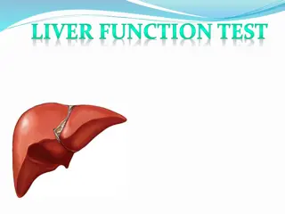
Delve into a comprehensive overview of the liver's anatomy, including ligaments, blood supply, and structure in the hilum. Explore the vital functions performed by the liver, such as maintaining body temperature, synthesizing clotting factors, and drug metabolism. Discover liver function tests and the implications of liver failure, whether acute or chronic. Lastly, learn about imaging studies used to evaluate liver health, including ultrasound, CT scans, and MRI.

Download Presentation

Please find below an Image/Link to download the presentation.
The content on the website is provided AS IS for your information and personal use only. It may not be sold, licensed, or shared on other websites without obtaining consent from the author.If you encounter any issues during the download, it is possible that the publisher has removed the file from their server.
You are allowed to download the files provided on this website for personal or commercial use, subject to the condition that they are used lawfully. All files are the property of their respective owners.
The content on the website is provided AS IS for your information and personal use only. It may not be sold, licensed, or shared on other websites without obtaining consent from the author.
E N D
Presentation Transcript
li liver ver
Anatomy ligaments and peritoneal reflection 1- lf triangular lig. fixed lf lobe of liver to the diaphragm ---division ant. and post. Leaf allow to mobilize lf lobe of liver from lf lat. wall of ivc ivc 2- RT triangular ligament. fixed RTlobeof liver to the RT Hemidiaghragm 3- falciform ligament which it s a remnant of umbilical vein runs from umbilicus to the liver 4- lesser omentumconnect liver to the stomach Blood supply of liver 80% from portal vein ,20% from hepatic artery which branch from caelic trunk which come from aorta. Hepatic artery dividing to RT and LF hepatic artery to supply the RTand lLFlobeof
Structure in hilum of liver Hepatic artery (above and medial),portal vein (posterior)and cbd(above and lat.),presents with free aged of lesser omintum , Venous drainage of the liver Via hepatic veins in to the inferior vena cava Anatomy of liver ( internl); liver divided to rtand lf lobe .rt lobe has segment from V to VIII SUPPLY by rt hepatic artery and rt branch of portal vein and drain via rt hepatic duct. Lf lobe of liver contain from 1 to 1v segments supply by lf hepatic artery and lf branch of portal vein and drain via lf hepatic duct.
Main function of liver 1- maintain core body temperature 2- PH balance and correction of lactic acidosis 3-SYNTHESIS OF CLOTTING FACTOR 4- glucose metabolism glycolysis and gluconeogensis 5- urea formation from protein catabolism 6- bilirobin formation from hg segregation 7-drug and hormone metabolism 8- removal of gut endotoxinand foreign antigen.
Liver function test 1-TSB DIRECT AND INDIRECT 2- ALP(alkalin phosphatase) 35-130 IU 3- AST(ASPERTATE TRANSAMINASE) 4-40 IU 4- ALT(ALANIN TRANSAMINASE) 5-40 5-GAMMA GLUTAMYL TRANSPEPTUDASE (GGT)10- 48IU 6- SERIEM ALBUMIN 35-50 GM 7- PROTHROMPIN TIMIME 12-16 SECOND
Liver failure Either acute or chronic Main feature of chronic liver failure are Lethargy ,fever, jaundice, wasting(protein catabolism), coagulopathy ,hyper dynamic circulation, hepatic encephalopathy , portal hypertension ,ascitis , esophageal varecies , splenomegaly , coetaneous --( spider naive , palmer erythema)
Imaging study of the liver U/S , SPIRAL CT SCAN (give information for lesion down to less than 1 cm in diameter and give information on there nature) ,MRI , ERCP (very useful in obstructive cause of liver failure but most check coagulation) , PTC ,ANGIOGRAPHY ,NUCLEAR MEDICIN SCANING radioisotope scanning , LAPARASCOPY AND LAPARASCOPIC U/S (is useful in staging of hepatopancreatobiliarycancer
Liver trauma Liver injury serious and associated with significant morbidity and mortality even with prompt and appropriate management. Because its highly vascular. Liver trauma divided to blunt(avulsion , laceration, contusion) and penetrating trauma9 stab wound or gun shoot). Diagnosis of liver injury; All lower rtchest and upper abdominal stab wound should be suspect liver injury and may be associated with other intra abdominal organ injury especially if there is sign and symptoms of blood loss. If pt haemodianamiclyunstable because of blood loss or pt with penetrating trauma needs urgent laparotomy ,if pt stable can do ct scan with contrast enhanced.---- peritoneal lavage if haemopertoneal---- laparoscopy by which can see if there is diaphragmatic rapture.
Managements ; First manage any pt with trauma as abcde pt with abnormal circulation ; 2 large bore canulae send blood for blood group and cross match 10 pints .sample for full blood count ,urea and electrolyte liver function test clotting screening ,glucose and amylase . Then start with fluid replacement start with colloid or o negative blood (what the difference between colloid and crystalloid??) Arterial blood gas should obtain and the pt intubate and ventilate if gas exchange in adequate. Chest tube if there is haemoor pnemothorax . abcde . in case of
Surgical approach to the liver; Rooftop incision provide excellent visualization of the liver and spleen. (What is Pringle maneuver ) a traumatic clump across foramen of Winslow to control on bleeding from liver. Packing of liver may be essential if there is diffuse bleeding from parenchyma of liver .pressure from below usually packs for 48 hours in most of pts no further treatment need.
Complication of liver trauma 1- profuse bleeding 2-liver abscess due to infection of liver hematoma 3- bile collection and sometime biliary fistula Late complication 1- hepatic artery aneurysm 2- a.v fistula or a. biliary fistula 3- rarly biliary tract stricture
Esophageal Esophageal varices Dilated tortuous veins of lower part of esophagus ,it s a complication of portal hypertension (what other site of potocystemicconnection). USUALLY present with acute onset of a large amount of haematamesis Diagnosis usually from history of pt with liver cirrhosis , Liver function test ,coagulating factor assessment Management mainly blood replacement ,frb to correct caogulopathy, vitamin k im. Platelet replacement if count less than 50x109/l-1 Sengstaken tube may be inserted if there is a profuse bleeding to provide temporally homeostasis in tube (gastric balloon inflated 250 ml of air -- esophageal balloon inflated at 40 mmgh pressure .there is a 2 remaining channel for gastric and esophageal aspiration. The balloon should temporally deflated after 12 hours to prevent pressure necrosis of esophagus. varices; ;
Drugs treatment vasopressin most widlyused for initial control of variceal hemorrhage (20 unit in 10 ml of g/w iv in 10 mints) Octreotide long acting somatostatin may equally effective. Endoscopiclly treatment By endoscopic a- seclerotherapy using ethanolamine oleate b- panding other treatments of oesophageal vareces are TIPSS(transjugular intrahepatic portocystemic stent shunt) Surgical procedure *portcystemic shunt *esophageal transaction *splenactomy and gastric devascolrization
Hydatid Hydatid cyst ; Causative agent is , echinoccousgranulosus . Humans become infected by ingested egg of the adult tapeworm which have been pass from the doge .the egg penetrate small bowel mucosa and enter blood stream from which distribute to various part of the body , ( why more common in the liver??) c. c.f f many cyst are asymptomatic and become discover incidentally .if become large it cause swelling in rt hypoch. Area. Some time pain because of pressure or infection of cyst. FEVER DUE TO INFACTION. Sequel of the cyst *enlarge in size producing pressure effect on surrounding structure *rapture producing anaphylactic shock *rapture to lung cause dyspneaand cough or to biliary passage producing obstructive jaundice cyst ;
Investigation 1- plain x ray of abdomen may show calcification or clear zone producing by the cyst 2- u/s and c.tscan 3- serological test use to detect antibodies in the serum as ELISA TEST &COMPLEMENT FIXATION , Cyst containing 3 layer outer layer due to reaction of host tissue to the parasite ,ectocyst & endocyst (containing germinal layers which containing scoleses and hydatid fluid)
Treatment Medical treatment albendazole 10 -15 mg /kg ( about 400 mg twice dialy) or mebandezol 40-50 mg /kg continue for 3 months without interruption the reassess the patient and decide whether continue on medical treatment or go to syrgical procedure ,also can use praziquantel 40 mg /kg/day. Post operatively 2 wks albendazol + praziquantel should be given .why?? Surgical treatment indication in infected cyst ,or in rapture to biliary tract
Types of surgery 1- ct scan or u/s guidance PAIR 2- marciplaizationand tube drainage or omentoplasty 3- radical surgical resection 4- partial bepatactomy Scolesydal agents used 1- 20 % hyper tonic slain 2- 0.5 silver nitrate 3- 95 % sterile ethanol (pair) 4- absolute alcohol (pair) . Whateare the indication ,contraindication and complication of pair.?
Indication for hepatic surgery; 1- large cyst with suspected multiple doughtercyst 2- super facial cyst with risk of rapture 3- 2ndbacterial infection of the cyst 4- cystobiliarycomplication 5- pressure effect on adjacent organs
Liver tumour Benign ; haemangiomas most common lesions it contain abnormal plexus of vessels and there nature ,diagnosed by u/s or c.tscan ,by ct scan with delayed contrast enhancement show the characteristic appearance of slow contrast enhancement due to small vessels uptake in the haemangioma. they varies in size. Hepatic adenoma ;rare, treatment is by surgical resection because they have potential malignant risk and there is no test by which can differentiate it from malignant tumor. it have major correlation with contraceptive piles. Focal nodular hyperplasia ;unknown etiology ,there is a focal over growth of functioning liver tissue supported by fibrous stroma ,contrast c.tscan may show central scaring and evidence of awell vasulirized lesion (not specific). Fnh contain both hepatocyteand kupffercells .kupffercells take up the colloid by which differentiated from either benign adenoma and 1 * or metastatic cancer ,non of which not containing significant number of kupffercells.
Hepatocellular carcinoma It s the most common primary malignant tumor of liver account more than 75 % of primary malignant tumor . Associated risk factor ;1-cirrhosis (chronic liver disease) 2- hepatitis b&cvirus 3- alcohol abuse 4- hemochromatosis , schestomiasis 5- aflatoxin (fungi) Any patient presented with withchronic liver disease must be secreaning for hcc by 1- us 2- c.tscan 3- measuring alfa feto protein ( afp) Signs and symptoms as patients with chronic liver disease or pt complaining from anorexia, loss of wt mass in rt hypochondriam
Staging of disease Depend on 1- general condition of the pt 2- child classification 3- size and site of tumor 4- chest c.tscan and bone scaning why?? Surgical treatment Based on resection of tumor with 1 -2 cm of normal edge and we minimize tissue resection in pt with liver cirrhosis to decrease incidence of post operative liver failure . Large tumor or multi focal treated by liver transplant Fallow up and adjuvant therapy There is a little evidence that adjuvant chemotherapy will improve the prognosis of the patients fallowing resection of hccand it may damage the function of liver in those underlying chronic liver disease . Alfa fetoprotein is clinically useful tumor marker.