Iron Overload: A Camp Sunshine Tale
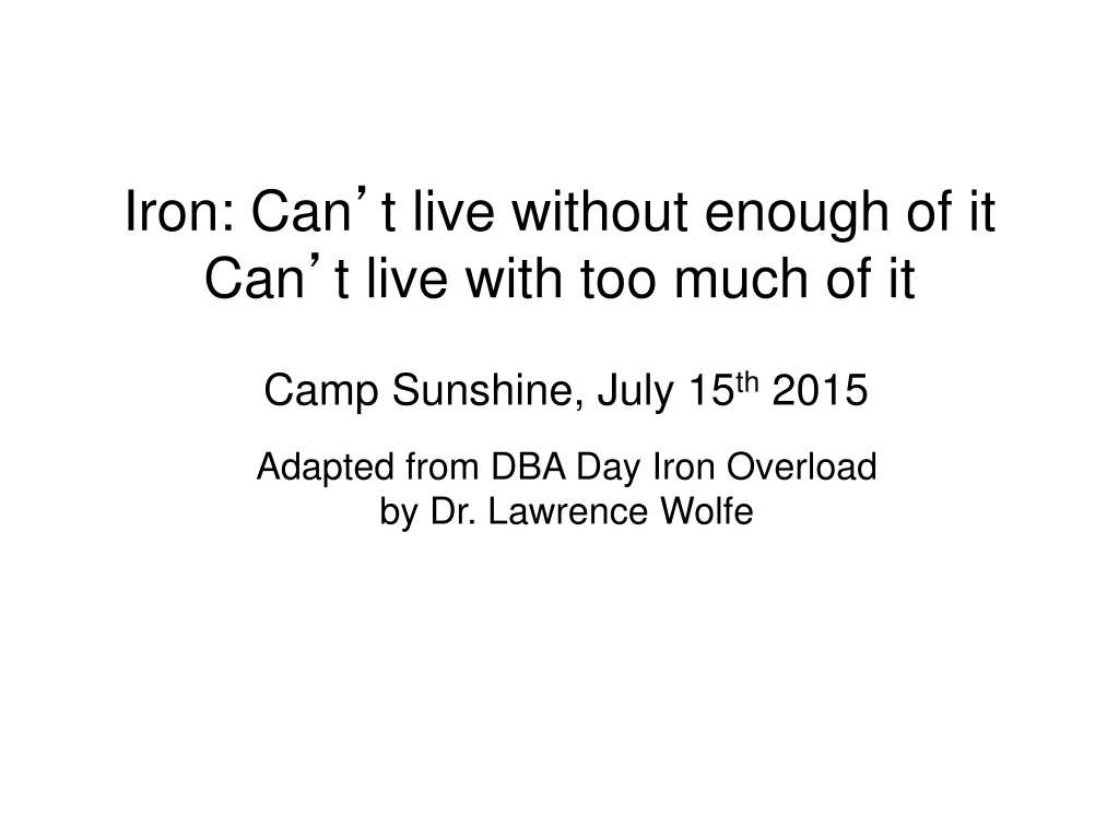

Iron: Can
’
t live without enough of it
Can
’
t live with too much of it
Camp Sunshine, July 15
th
2015
Adapted from DBA Day Iron Overload
by Dr. Lawrence Wolfe
Oxygen solubility
Plasma
2.3 ml/L
Whole Blood
200 ml/L
Hemoglobin and Myoglobin
Reduce oxygen
’
s reactivity
O
2
X
H
2
O
OxX
Oxygen Transport Proteins – Hemoglobin/Myoglobin
Protected
environment
provided by
Mb and Hb
The Heme Prosthetic Group
The heme iron has two
oxidation states: Fe
2+
,
ferrous; Fe
3+
, ferric
Ferrous iron can
form up to 6 bonds
Ferric iron doesn
’
t bind oxygen
O
2
Fe
2+
H
2
O
Fe
3+
protected
environment of
globin chains
pathogenic
variants
protoporphyrin IX
When bound to proteins both oxygen and iron are in
protected states and bad things don’t happen
Iron Metabolism – Distribution in the Human Body
Iron is an essential, but also potentially highly toxic nutrient. Its uptake,
transport, and storage in the body are highly regulated
Iron Distribution in Humans
Fe
2+
+ H
2
O
2
Fe
3+
+ OH
-
+ OH
proteins
nucleic acids
lipids
Fenton Reaction
mutation
macrophage
bone marrow
reactive oxygen
species (ROS)
chain reaction more ROS production
excess
AKA: nontransferrin
bound iron (NTBI)
ferrihydrite
Fe
3+
OH/PO
4
Adapted from Casiday and Frey,
Washington University St. Louis
F
e
3
+
F
e
2
+
Iron Storage - Ferritin
Ferritin enters serum by an unknown mechanism under normal conditions (values
proportional to cellular content) and is used as a non-invasive measure of iron stores.
Measurements of serum ferritin can be used in the diagnosis of disorders of iron
metabolism or tissue damage. Normal values: men 12-300 ng/ml; women 10-150
ng/ml.
Ferritin can also be released to serum by damage to cells of the liver, spleen,
or bone marrow and other pathogenic states
L subunits (iron binding)
H subunits (ferrioxidase
activity)
(hemosiderin)
Iron Transport - Transferrin
Fe
2+
Fe
2+
Fe
3+
ceruloplasmin
+
transferrin
Fe
3+
-transferrin-Fe
3+
transferrin
receptor
internalization
33%
67%
enterocytes
liver
macrophages
transferrin saturation
Iron Uptake from Diet
ingested iron
Fe
3+
Fe
2+
R
DMT1
Fe
2+
ferriportin
ferritin
not absorbed
Fe
2+
Fe
3+
transferrin
e
n
t
e
r
o
c
y
t
e
daily requirement
men 10 mg/1mg
menstruating women
20mg/2mg
vitamin C, ethanol
poor bioavailablity
GUT
CIRCULATION
macrophages play an important
role in regulating circulating iron
using transporters similar or
identical to those found on
enterocytes
DMT1
circulating iron
ferriportin
circulating iron
ceruloplasmin
macrophages/
ferriportin
macrophages/
DMT1
Replace iron lost by sloughing of intestinal and
skin cells and by bleeding
ingested iron
Fe
3+
Fe
2+
R
DMT1
Fe
2+
ferriportin
ferritin
internalization,
degradation
not absorbed
hepcidin
HFE
TfR2
HJV
Fe
2+
e
n
t
e
r
o
c
y
t
e
loss with
cellular slough
circulatory system
Regulation of Iron Absorption
ingested iron
Fe
3+
Fe
2+
R
DMT1
Fe
2+
ferriportin
ferritin
internalization,
degradation
not absorbed
hepcidin
HFE
TfR2
HJV
Fe
2+
e
n
t
e
r
o
c
y
t
e
loss with
cellular slough
circulatory system
Regulation of Iron Absorption
Iron Overload: Hereditary Hemochromatosis
Transfusion therapy results in
iron overload
•
1 blood unit contains 200 mg
iron
•
A 60 kg patient with thalassemia
receiving 45 units of blood
annually has transfusional iron
intake of 9 g iron/year
–
0.4 mg iron/kg body wt/day
•
In addition, up to 4 mg/day may
be absorbed from the gut
–
Up to 1.5 g iron/year
•
Overload can occur after 10–20
transfusions
2
0
0
–
2
5
0
m
g
i
r
o
n
:
W
h
o
l
e
b
l
o
o
d
:
0
.
4
7
m
g
i
r
o
n
/
m
L
‘
P
u
r
e
’
r
e
d
c
e
l
l
s
:
1
.
1
6
m
g
i
r
o
n
/
m
L
Porter JB.
Br J Haematol
2001;115:239–252
Iron overload is an inevitable
consequence of multiple blood
transfusions
E
r
y
t
h
r
o
n
2
g
Hershko C
et al
.
Ann NY Acad Sci
1998;850:191–201
Normal distribution and turnover
of body iron
Iron balance is achieved in the normal state
P
a
r
e
n
c
h
y
m
a
R
e
t
i
c
u
l
o
e
n
d
o
t
h
e
l
i
a
l
m
a
c
r
o
p
h
a
g
e
s
G
u
t
E
r
y
t
h
r
o
n
T
r
a
n
s
f
e
r
r
i
n
T
r
a
n
s
f
e
r
r
i
n
R
e
t
i
c
u
l
o
e
n
d
o
t
h
e
l
i
a
l
m
a
c
r
o
p
h
a
g
e
s
P
a
r
e
n
c
h
y
m
a
NTBI, non-transferrin-bound iron
Hershko C
et al
.
Ann NY Acad Sci
1998;850:191–201
Imbalance of distribution and turnover
of body iron with transfusion therapy
Iron balance is disturbed by blood transfusion because the body cannot
remove the excess iron
Iron overload leads to formation
of NTBI
Uncontrolled iron loading
of organs
Subsequent formation
of NTBI in plasma
Fe
Fe
Fe
Fe
Fe
Fe
Fe
100%
30%
Normal: no NTBI
produced
Iron overload
Transferrin saturation due to:
•
Frequent blood transfusions, or
•
Ineffective erythropoiesis leading to
increased iron absorption
Transferrin saturation
T
r
a
n
s
f
e
r
r
i
n
i
r
o
n
C
o
n
t
r
o
l
l
e
d
u
p
t
a
k
e
L
a
b
i
l
e
I
r
o
n
Storage
iron
Uncontrolled uptake of labile iron
leads to cell and organ damage
Porter JB.
Am J Hematol
2007;82:1136
–113
9
Labile iron
Cell death
Fibrosis
O
r
g
a
n
e
l
l
e
d
a
m
a
g
e
T
G
F
-
β
1
F
r
e
e
r
a
d
i
c
a
l
g
e
n
e
r
a
t
i
o
n
L
i
p
i
d
p
e
r
o
x
i
d
a
t
i
o
n
L
y
s
o
s
o
m
a
l
f
r
a
g
i
l
i
t
y
E
n
z
y
m
e
l
e
a
k
a
g
e
C
o
l
l
a
g
e
n
s
y
n
t
h
e
s
i
s
TGF, transforming growth factor
Cohen AR and Porter JB. In Disorders of Hemoglobin: Genetics, Pathophysiology, and Clinical Management, Steinberg MH
et
al.
(Eds); 2001:979–1027
Iron overload negatively affects
organ function
L
i
v
e
r
c
i
r
r
h
o
s
i
s
/
f
i
b
r
o
s
i
s
/
c
a
n
c
e
r
D
i
a
b
e
t
e
s
m
e
l
l
i
t
u
s
E
n
d
o
c
r
i
n
e
d
i
s
t
u
r
b
a
n
c
e
s
→
g
r
o
w
t
h
f
a
i
l
u
r
e
C
a
r
d
i
a
c
f
a
i
l
u
r
e
I
n
f
e
r
t
i
l
i
t
y
Excess iron is deposited in multiple
organs, resulting in organ damage
Iron overload
Capacity of serum transferrin to bind
iron is exceeded
NTBI circulates in the plasma; some forms of NTBI
(eg LPI) load tissues with excess iron
Excess iron promotes the generation of free
hydroxyl radicals, propagators of oxygen
-
related
tissue damage
Insoluble iron complexes are deposited in body
tissues and
end
-
organ toxicity occurs
Delve into the world of iron and its essentiality, exploring the fine line between necessity and excess. Join the journey on July 15th, 2015 at Camp Sunshine with insightful information adapted from "DBA Day: Iron Overload" by Dr. Lawrence Wolfe.

Download Presentation

Please find below an Image/Link to download the presentation.
The content on the website is provided AS IS for your information and personal use only. It may not be sold, licensed, or shared on other websites without obtaining consent from the author.If you encounter any issues during the download, it is possible that the publisher has removed the file from their server.
You are allowed to download the files provided on this website for personal or commercial use, subject to the condition that they are used lawfully. All files are the property of their respective owners.
The content on the website is provided AS IS for your information and personal use only. It may not be sold, licensed, or shared on other websites without obtaining consent from the author.
E N D
Presentation Transcript
Iron: Cant live without enough of it Can t live with too much of it Camp Sunshine, July 15th 2015 Adapted from DBA Day Iron Overload by Dr. Lawrence Wolfe
Oxygen Transport Proteins Hemoglobin/Myoglobin Oxygen solubility Plasma 2.3 ml/L 200 ml/L Whole Blood Hemoglobin and Myoglobin Reduce oxygen s reactivity O2 X Protected environment provided by Mb and Hb H2O OxX
The Heme Prosthetic Group The heme iron has two oxidation states: Fe2+, ferrous; Fe3+, ferric Ferrous iron can form up to 6 bonds Ferric iron doesn t bind oxygen O2 Fe2+ protected environment of globin chains H2O Fe3+ protoporphyrin IX pathogenic variants When bound to proteins both oxygen and iron are in protected states and bad things don t happen
Iron Metabolism Distribution in the Human Body Iron is an essential, but also potentially highly toxic nutrient. Its uptake, transport, and storage in the body are highly regulated Iron Distribution in Humans Compartment Iron Content (mg) Total Body Iron (%) hemoglobin iron 1500(W)-2000(M) 67 storage iron (ferritin, hemosiderin) 1000 27 macrophage bone marrow myoglobin iron 130 3.5 other tissue iron (cytochromes, etc.) 8 0.2 excess transport iron (transferrin) 3 0.08 labile pool 80 2.2 reactive oxygen species (ROS) AKA: nontransferrin bound iron (NTBI) proteins nucleic acids lipids chain reaction more ROS production Fe2+ + H2O2 Fe3+ + OH- + OH mutation Fenton Reaction
Iron Storage - Ferritin L subunits (iron binding) H subunits (ferrioxidase activity) Fe3+ OH/PO4 Fe2+ Fe3+ ferrihydrite (hemosiderin) Ferritin enters serum by an unknown mechanism under normal conditions (values proportional to cellular content) and is used as a non-invasive measure of iron stores. Measurements of serum ferritin can be used in the diagnosis of disorders of iron metabolism or tissue damage. Normal values: men 12-300 ng/ml; women 10-150 ng/ml. Ferritin can also be released to serum by damage to cells of the liver, spleen, or bone marrow and other pathogenic states Adapted from Casiday and Frey, Washington University St. Louis
Iron Transport - Transferrin enterocytes liver macrophages transferrin saturation Fe2+ Fe2+ ceruloplasmin 33% Fe3+ + Fe3+-transferrin-Fe3+ transferrin 67% transferrin receptor internalization
Iron Uptake from Diet macrophages/ ferriportin macrophages/ DMT1 Replace iron lost by sloughing of intestinal and skin cells and by bleeding poor bioavailablity ingested iron not absorbed vitamin C, ethanol daily requirement Fe3+ Fe2+ GUT men 10 mg/1mg menstruating women 20mg/2mg R DMT1 Fe2+ ferritin enterocyte macrophages play an important role in regulating circulating iron using transporters similar or identical to those found on enterocytes ferriportin circulating iron DMT1 Fe3+ Fe2+ ferriportin circulating iron ceruloplasmin CIRCULATION transferrin
Regulation of Iron Absorption ingested iron not absorbed Fe3+ Fe2+ loss with cellular slough R DMT1 Fe2+ ferritin internalization, degradation enterocyte ferriportin circulatory system hepcidin Fe2+ HFE HJV TfR2
Regulation of Iron Absorption ingested iron not absorbed Fe3+ Fe2+ loss with cellular slough R DMT1 Fe2+ ferritin internalization, degradation enterocyte ferriportin circulatory system hepcidin Fe2+ HFE HJV TfR2
Iron Overload: Hereditary Hemochromatosis frequency gene hepcidin severity clinical findings symptoms start after 4th decade: chronic fatigue, hepatic fibrosis and cirrhosis, cardiomyopathy, diabetes mellitus, infertility, joint pain Heterozygous frequency: 1/10 North Americans classic ++ HFE Symptoms start after first decade: abdominal pain, hypogonadism, heart failure, diabetes mellitus juvenile Rare ++++ HJV Symptoms similar to HJV-related HH juvenile very rare ++++ HAMP Symptoms similar to HFE-related HH very rare +++ TfR2 SLC40A1 (ferriportin) Symptoms similar to HFE-related HH rare +
Transfusion therapy results in iron overload 1 blood unit contains 200 mg iron A 60 kg patient with thalassemia receiving 45 units of blood annually has transfusional iron intake of 9 g iron/year 0.4 mg iron/kg body wt/day In addition, up to 4 mg/day may be absorbed from the gut Up to 1.5 g iron/year Overload can occur after 10 20 transfusions Iron overload is an inevitable consequence of multiple blood transfusions 200 250 mg iron: Whole blood: 0.47 mg iron/mL Pure red cells: 1.16 mg iron/mL Porter JB. Br J Haematol 2001;115:239 252
Normal distribution and turnover of body iron Erythron 2 g 20 30 mg/day 20 30 mg/day Reticuloendothelial macrophages 0.6 g Parenchyma 0.3 g Liver 1 g Transferrin 20 30 mg/day 1 2 mg/day 2 3 mg/day Gut Iron balance is achieved in the normal state Hershko C et al. Ann NY Acad Sci 1998;850:191 201
Imbalance of distribution and turnover of body iron with transfusion therapy Transfusions Erythron 20 40 mg/day NTBI Reticuloendothelial Reticuloendothelial macrophages macrophages Transferrin Transferrin Parenchyma Parenchyma Gut Iron balance is disturbed by blood transfusion because the body cannot remove the excess iron NTBI, non-transferrin-bound iron Hershko C et al. Ann NY Acad Sci 1998;850:191 201
Iron overload leads to formation of NTBI Transferrin saturation due to: Frequent blood transfusions, or Ineffective erythropoiesis leading to increased iron absorption Subsequent formation of NTBI in plasma Uncontrolled iron loading of organs Pituitary Parathyroid Thyroid Normal: no NTBI produced Iron overload Heart Liver Pancreas 100% Fe Transferrin saturation Fe Fe Gonads Fe Fe Fe Fe 30%
Uncontrolled uptake of labile iron leads to cell and organ damage Storage iron Non-transferrin iron Uncontrolled uptake Functional iron Transferrin iron Labile Iron Controlled uptake Free-radical generation Organelle damage Porter JB. Am J Hematol 2007;82:1136 1139
Iron overload negatively affects organ function Labile iron Free radical generation Lipid peroxidation Organelle damage TGF- 1 Lysosomal fragility Collagen synthesis Enzyme leakage Fibrosis Cell death TGF, transforming growth factor Cohen AR and Porter JB. In Disorders of Hemoglobin: Genetics, Pathophysiology, and Clinical Management, Steinberg MH et al. (Eds); 2001:979 1027
Excess iron is deposited in multiple organs, resulting in organ damage Iron overload Capacity of serum transferrin to bind iron is exceeded NTBI circulates in the plasma; some forms of NTBI (eg LPI) load tissues with excess iron Excess iron promotes the generation of free hydroxyl radicals, propagators of oxygen-related tissue damage Insoluble iron complexes are deposited in body tissues and end-organ toxicity occurs Endocrine disturbances growth failure Liver cirrhosis/ fibrosis/cancer Diabetes mellitus Cardiac failure Infertility