Insights into Tyzzer's Disease: An Overview of a Bacterial Infection in Laboratory Animals
Tyzzer's disease is an acute bacterial infection affecting rodents and rabbits, caused by Clostridium piliforme. Discovered in 1917 by Ernest Tyzzer, the disease is characterized by necrotic lesions in the caecal mucosa, liver, and heart. Initially known as Bacillus piliformis, it was later renamed C. piliforme. The disease can be transmitted horizontally through the fecal-oral route, with laboratory animals being more susceptible. Learn more about the history, etiology, and transmission of this disease.

Download Presentation

Please find below an Image/Link to download the presentation.
The content on the website is provided AS IS for your information and personal use only. It may not be sold, licensed, or shared on other websites without obtaining consent from the author.If you encounter any issues during the download, it is possible that the publisher has removed the file from their server.
You are allowed to download the files provided on this website for personal or commercial use, subject to the condition that they are used lawfully. All files are the property of their respective owners.
The content on the website is provided AS IS for your information and personal use only. It may not be sold, licensed, or shared on other websites without obtaining consent from the author.
E N D
Presentation Transcript
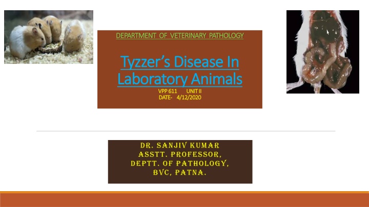
DEPARTMENT OF VETERINARY PATHOLOGY DEPARTMENT OF VETERINARY PATHOLOGY Tyzzer s Disease Tyzzer s Disease In Laboratory Animals Laboratory Animals VPP 611 UNIT II VPP 611 UNIT II DATE DATE- - 4/12/2020 4/12/2020 In DR. SANJIV KUMAR ASSTT. PROFESSOR, DEPTT. OF PATHOLOGY, BVC, PATNA.
Definition Definition Tyzzer s disease Tyzzer's disease is an acute epizootic bacterial disease of rodents and rabbits caused by spore-forming bacterium Clostridium piliforme, formerly known as Bacillus piliformis characterized by segmental necrosis of caecal mucosa and focal necrotic lesions on the liver and heart. The disease name has been given in his honour of the scientist who first recorded it.
History History Tyzzer's disease was first discovered by parasitologist Ernest Tyzzer in 1917 when his entire colony of Japanese waltzing mice suddenly died. Upon closer observation, Tyzzer discovered necrotic lesions and spore-forming bacillus in the livers of the deceased mice. This led Tyzzer to name the bacteria Bacillus piliformis and deem it the cause of this new disease. Later on, B. piliformis was renamed C. piliforme.
In the 1940s, a biologist named Paul Errington found a fatal condition in Iowa muskrat populations that he believed to be a new disease. Dead muskrats were found with blood around their anus and internal bleeding, therefore Errington called this new condition hemorrhagic disease After Errington's death, the name hemorrhagic disease was changed to Errington s disease . It was not until 1971 that the two diseases were discovered to be the same, upon which the name was reverted to Tyzzer s disease .
Etiology Etiology Clostridium piliforme is an obligate intracellular Gram-negative bacteria and have variable morphology in tissue section. Most often, it appears as a long, slender (i.e. hair-like or piliform) bacillus, approximately 8 10 mm long and 0.5 mm wide, in the cytoplasm of infected cells, although shorter, thicker, cigar- shaped forms may also occasionally be seen.
Transmission Transmission Tyzzer's disease is transmitted horizontally through the fecal-oral route. Bacterial spores within infected fecal matter can contaminate soil or feed and become orally ingested by a viable host. Success of the disease is determined by the amount and virulence of the bacteria and the resistance of the host. Laboratory animals are more susceptible to this disease, as bacteria spores from infected feces can survive in bedding at room temperature for over one year. Animals do occasionally transfer the disease asymptomatically, acting as carriers. Infected animals are more likely to develop the disease when subjected to stressful conditions.
Pathogenesis Pathogenesis 1. Spores are shed in the feces and it can survive in contaminated 6. Produce exotoxin lead to necrosis of intestinal epithelium 2. bedding for at least 1 year, and in the natural environment for at least 5 years 7. Bacteria are either deposited back into the lumen, or sometimes find their way 3. Infection occurs by ingestion of spores from environment or feces of infected animals 8. deeper into the intestinal wall, where they may infect smooth muscle cells 9. or gain access to the portal circulation 4. After ingestion of the spores, the bacterium is phagocytosed by intestinal epithelial cells. 10. From the portal vein bacteria may infect the liver and/or heart 5. Inside the cell, the vegetative form escapes the phagosome and begins replication in the cytoplasm 11. Hepatitis and occasionally myocarditis
Clinical Signs Clinical Signs Most infected mice are asymptomatic. If disease occurs, it is usually observed in recently weaned mice or mice with genetic or induced immune deficits. Although weanling rats with Tyzzer s disease have a distended abdomen due to megaloileitis, this has not been reported in mice. Mice with Tyzzer s disease may have diarrhoea and perianal staining, or may seem thin and unkempt for a short period. Sudden deaths without premonitory signs may also occur.
Pathology Pathology The target organs in animal infections are the intestine (distal ileum, caecum and proximal colon), the liver and, less often the heart. Ileum, caecum and colon may be slightly enlarged and reddened by hyperaemia or mild haemorrhage. Microscopically, foci of degeneration, inflammation, edema, and necrosis in the intestinal mucosa and muscularis mucosae with clusters of organisms are apparent in enterocytes and smooth muscle Miliary pale foci or larger umbilicated foci are usually visible throughout the parenchyma of the liver.
Lesions are characterized by multifocal coagulation to caseation hepatic necrosis, with polymorphonuclear leukocyte infiltration. Gray foci in the ventricular myocardium with myocyte degeneration, myocarditis, and intracellular bacteria. In tissue sections stained with the Warthin Starry, Giemsa, or PAS methods, typical intracytoplasmic bundles of bacilli are usually readily found in enterocytes, hepatocytes adjacent to necrotic foci, and myocardial cells
Characteristic lesions are seen in the liver, myocardium, and intestinal tract. In the liver, white, gray, or yellowish foci of necrosis, 2 mm in diameter, are few to disseminated. The hepatic necrosis is most marked and disseminated in foals in which the multiple necrotic foci with slightly depressed hemorrhagic centers appear to infect almost every hepatic lobule. In addition, there is marked hepatomegaly, and the hepatic lymph nodes are edematous and hyperplastic. In rabbits, severe lesions develop in the intestines and heart. The terminal ileum, cecum, and proximal colon are diffusely reddened. Diffuse ( paint-brush ) hemorrhage is frequently seen on the serosa of the cecum.
Patchy areas of mucosal necrosis are present in the cecum and colon, together with marked edema of the wall of the cecum. Mesenteric lymph nodes may be enlarged and edematous. White streaks in the myocardium may be present, especially near the apex. Intestinal and heart lesions are generally milder or absent in other animal species.
Microscopically, there are numerous widespread multifocal areas of necrosis and hepatitis. In foals, the hepatic lesions are more pronounced than in other animals. Often the necrotic foci are so numerous that two or more coalesce. The hepatocytes in the center of the necrotic foci are destroyed and replaced by a mixture of mononuclear cells, neutrophils, and red blood cells. The causative bacteria are found in a crisscross pattern in viable hepatocytes at the periphery of the necrotic foci.
In the cecum and colon of rabbits, patchy areas of necrosis extend as deep as the muscularis externa with associated mucosal and submucosal infiltrates of neutrophils. Organisms may be found within the epithelium, muscularis mucosa, and muscularis externa of the affected intestine. When cardiac lesions are present, they consist of foci of fiber fragmentation, vacuolation, loss of cross-striations, and minimal inflammatory cell infiltration.
Diagnosis Diagnosis Serology of the blood and PCR assay of the feces of suspect infected animals may be used clinically to test for C piliforme. However, clinical signs in addition to the commonly available diagnostic methods must be interpreted together for a presumptive clinical diagnosis. In postmortem specimens, a diagnosis is based on demonstration of organisms in tissue sections with special stains. C piliforme stains poorly with H&E and Gram stains.
With Giemsa stain, the bacillus stains well in the liver and intestinal epithelium and in smears of infected organs but poorly in smooth muscles and cardiac muscle cells. The Warthin-Starry or Levaditi silver stains are preferable to other stains, because the bacillus stains well in the cytoplasm of all infected cells. In addition to special histochemical stains, the PCR assay can be used to detect C. piliforme gene sequences in liver tissues from infected animals.
Prevention Prevention In laboratory animals, prevention includes a low-stress environment, an adequate amount of nutritional feed, and appropriate sanitation measurements. Because animals likely ingest bacterial spores from contaminated bedding and feed, regular cleaning is a helpful method of prevention. No prevention methods are currently available for wild animal populations.
Treatment and Control Treatment and Control Currently, antibiotic drugs such as penicillin or tetracycline are the only effective methods for disease treatment. Within wild populations, disease control consists of reducing the amount of bacterial spores present in the environment. This can be done by removing contaminated carcasses and scat.