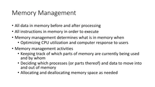
How Your Brain Works: Memory, Attention, and Processing

Memory, Attention and
Summing Up
How Your Brain Works - Week 13
Prof. Jan Schnupp
wschnupp@cityu.edu.hk
HowYourBrainWorks.net
Types of
Memory
Short Term or
“Working”
Memory
Long Term
Procedural
Declarative
Episodic
Semantic
Visuo-Spatial
Phonological
Working Memory
Working memory is thought to be
mediated by sustained activity of
neurons in prefrontal cortex (PFC) as
illustrated in the experiment by
Freedman et al discussed over the
next few slides.
D
 
J
 
F
r
e
e
d
m
a
n
 
e
t
 
a
l
.
 
S
c
i
e
n
c
e
 
2
0
0
1
;
2
9
1
:
3
1
2
-
3
1
6
,
 
F
i
g
u
r
e
 
2
:
T
a
s
k
 
d
e
s
i
g
n
 
a
n
d
 
b
e
h
a
v
i
o
r
.
 
M
o
n
k
e
y
s
 
w
e
r
e
 
t
r
a
i
n
e
d
 
t
o
 
c
a
t
e
g
o
r
i
z
e
 
c
o
m
p
u
t
e
r
 
i
m
a
g
e
s
 
o
f
c
a
t
s
 
o
r
 
d
o
g
s
 
i
n
 
a
 
d
e
l
a
y
e
d
 
m
a
t
c
h
-
t
o
-
s
a
m
p
l
e
 
t
a
s
k
.
 
T
o
 
v
a
r
y
 
d
i
f
f
i
c
u
l
t
y
,
 
t
h
e
 
i
m
a
g
e
s
 
c
o
u
l
d
 
b
e
m
o
r
p
h
e
d
 
t
o
 
b
e
,
 
s
a
y
,
 
7
0
%
 
d
o
g
 
a
n
d
 
3
0
%
 
c
a
t
.
 
(
A
)
 
A
 
s
a
m
p
l
e
 
w
a
s
 
f
o
l
l
o
w
e
d
 
b
y
 
a
 
d
e
l
a
y
a
n
d
 
a
 
t
e
s
t
 
s
t
i
m
u
l
u
s
.
 
I
f
 
t
h
e
 
s
a
m
p
l
e
 
a
n
d
 
t
e
s
t
 
s
t
i
m
u
l
u
s
 
w
e
r
e
 
t
h
e
 
s
a
m
e
 
c
a
t
e
g
o
r
y
 
(
a
 
m
a
t
c
h
)
,
m
o
n
k
e
y
s
 
h
a
d
 
t
o
 
r
e
l
e
a
s
e
 
a
 
l
e
v
e
r
 
b
e
f
o
r
e
 
t
h
e
 
t
e
s
t
 
d
i
s
a
p
p
e
a
r
e
d
.
 
O
t
h
e
r
w
i
s
e
,
 
t
h
e
r
e
 
w
a
s
a
n
o
t
h
e
r
 
d
e
l
a
y
 
f
o
l
l
o
w
e
d
 
b
y
 
a
 
m
a
t
c
h
.
 
E
q
u
a
l
 
n
u
m
b
e
r
s
 
o
f
 
m
a
t
c
h
 
a
n
d
 
n
o
n
-
m
a
t
c
h
 
t
r
i
a
l
s
 
w
e
r
e
r
a
n
d
o
m
l
y
 
i
n
t
e
r
l
e
a
v
e
d
.
 
(
B
)
 
A
v
e
r
a
g
e
 
p
e
r
f
o
r
m
a
n
c
e
 
o
f
 
b
o
t
h
 
m
o
n
k
e
y
s
.
 
R
e
d
 
a
n
d
 
b
l
u
e
 
b
a
r
s
i
n
d
i
c
a
t
e
 
p
e
r
c
e
n
t
a
g
e
s
 
o
f
 
s
a
m
p
l
e
s
 
c
l
a
s
s
i
f
i
e
d
 
a
s
 
d
o
g
 
a
n
d
 
c
a
t
,
 
r
e
s
p
e
c
t
i
v
e
l
y
.
D
 
J
 
F
r
e
e
d
m
a
n
 
e
t
 
a
l
 
(
2
0
0
1
)
F
i
g
u
r
e
 
3
B
:
The average activity of a single neuron in response to stimuli at the six morph
blends. The vertical lines correspond (from left to right) to sample onset, offset,
and test stimulus onset. The inset shows the neuron's delay activity in response to
stimuli along each of the nine between-class morph lines. The prototypes (C1, C2,
C3, D1, D2, and D3) are represented in the outermost columns; each appears in
three morph lines. A color scale indicates the activity level.
Comparing ITC
 and PFC
Neurons in the infratemporal cortex (ITC) had learned to
distinguish the picture categories. They are active when
the stimulus is present.
Neurons in the PFC hold the last seen picture in
memory. Their activities are different from the end of
stimulus presentation until the monkey responds.
Forming Long Term Memories
with Synaptic Plasticity
 
Long-term Memory
For “procedural” type learning and
memory, see last lecture.
For “declarative” and spatial memories,
welcome to the world of “patient H.M.”:
Patient HM
Henry Molaison had his hippocampus
removed bilaterally in 1953 to treat severe
epilepsy.
He died in 2008.
The hippocampus
 
Patient HM
The surgery successfully cured his epilepsy, but
left him with severe 
anterograde amnesia.
He could no longer form new episodic
memories. For example, he would not be able to
remember people he met just hours before.
He could not learn new landmarks to help him
orient in a new environment.
But his ability for procedural learning remained
in tact. He learned to play ping-pong after his
surgery. Could not remember ever having played
it, but became quite good at it.
What does the hippocampus do?
The hippocampus probably has “limbic” roles
related to anxiety and depression too, but in this
lecture we focus on episodic and spatial memory
roles.
It is generally thought that Long Term
Potentiation (LTP) following “Hebb’s Rule” is an
essential ingredient for the hippocampus’ key
role in memory formation.
Hebb’s Rule
“Cells that fire together, wire together”
Psychologist Donald Hebb first suggested that
connections between neurons that are simultaneously
active might be strengthened.
This is advantageous for “associative learning”.
(Associations form between things that are
simultaneously signalled in the brain.)
Strengthening (or “long-term potentiation” - LTP) of
simultaneously activated synapses has been observed
in hippocampus (and later many other brain
structures).
Structure of the hippocampus
Hippocampus receives high level multisensory
information via enthorinal cortex (EC)
Inputs go to dentate gyrus (DG), then cornus ammonis
(CA) region 3 then CA1 and then back to EC via the
subiculum.
The synapses are glutamatergic and plastic.
Inputs
and
outputs
from
hippo-
campus
EPSPs recorded in hippocampal CA1 cell.
100 Hz stimulus bursts applied to “Schaeffer collateral” inputs,
either under voltage clamp or with simultaneous
depolarisation.
If the input bursts are paired with depolarisation, the EPSPs
are “potentiated” (i.e. larger).
 
An Example of Hippocampal LTP
The NMDA Receptor
NMDA receptors appear to
be critically involved in LTP
at glutamatergic synapses.
NMDA receptor channels
open only if glutamate binds
AND depolarisation removes
a Mg++ from the channel’s
pore. This implements
Hebb’s rule. The
postsynaptic neuron must be
active already for the
synapse to be modified.
Drugs that block the NMDA
receptor (AP-5, MK-801,
ketamine) prevent LTP.
NMDA receptor activation lets Ca
++
 in
Dendrite filled with Ca
++
indicator “calcium
green” emits a flash of
fluoresecent light at
synaptic spine when
synapse is activated.
The fluorescence is
inhibited by NMDA
receptor blocker AP5
Fig 7 of Lisman et al Nat Rev Neurosci 2002 Vol 3 p 175
LTP increases AMPA currents
Ca++ activates Calcium/Calmodulin Kinase II (CaMKII)
CaMKII increases AMPA currents in 3 ways:
It phosphoryaltes AMPA channels
It anchors AMPA channels at the postsynaptic
membrane
It favours the insertion of further AMPA receptors in the
membrane
Fig 7 of Lisman et al Nat RevNeurosci 2002 Vol 3 p 175
Why might Hebb’s Rule be useful?
Let’s consider how “associative learning” via the Hebb
Rule in a neural network might support “recognition
memory” where seeing only a part of something (e.g.
your friend’s favourite t-shirt) might remind you of the
whole.


Auto-associative nets
 
 
Computer
simulations using
artificial neural
networks illustrate
the pattern
completion and noise
robustness
properties that can
be achieved with
auto-associative
memory networks.
Source: Hertz, Krogh and
Palmer “Theory of Neural
Computation”
What does this remind you of
A Rorschach Blot
Break
 
“Distributed representations”
In artificial neural networks trained to recognize or recall
images, the information is not “stored” in any one place,
but distributed widely across the connection pattern
between the artificial neurons.
No individual synapse or neuron plays a particularly
important role, the activity patterns of individual neurons
in the network can be very hard to interpret, and in fact a
fair proportion of neurons can be removed without
obvious loss of performance (“graceful degeneration”).
So you don’t need, or expect, so called “grandmother
cells”: single neurons which “represent” or “recognize”
highly specific concepts or objects.
So who asked for Jennifer Aniston neurons?
“Jennifer Aniston
neurons” were
discovered by
Rodrigo Quan-
Quiroga in
hippocampal
recordings obtained
from human epilepsy
sufferers in the clinic
of Yitzak Fried.
Note that Quan-
Quiroga does not
think of his neurons
as “one-ofs”.
A “Halle Berry” neuron
 
Episodic memories
It is probably best not to think of “Jenifer Aniston”
neurons as proof that the brain works with “grandmother
cells”. Rather, they show that the hippocampus receives
“sparse, high level, multisensory feature representations”
of the environment, and it can combine these with spatial
information to form memories of what happened when
and with whom.
Hippocampal “place cells” are thought to represent
spatial location. They were discovered by John O’Keefe,
using “tetrode” recordings from the hippocampus of freely
moving rats.
The discovery won him the Nobel prize.
Combining Objects with Places as a
Memory-Trick
In the “memory palace” or “method of loci” technique,
people imagine a list of objects that they want to
remember as placed along a path through a familiar
environment, such as their family home.
In your imagination you walk through the chosen place
and imagine the objects in prominent locations. When
you later wish to recall the list, just imagine walking
through the same route and “see” the objects where you
had placed them in your imagination.
Tetrode
recordings
 
Place
cells
Place cells were discovered by John O’Keefe and Bruce
McNaughton in the early 70s. John O’Keefe won the Nobel Prize for
this discovery.
The video shows recordings of rat hippocampal place cells made in
Matt Wilson’s lab at MIT.
Remarkably, recordings from sleeping rats from Wilson’s lab
suggest that rats “revisit” places they have explored in their dreams,
as place cells fire in sequence when they sleep. This may be related
to memory consolidation during sleep.
Testing Spatial Memory with a
“Morris Water Maze”
The Morris Water Maze is a popular technique to test
spatial memory in rodents (rats or mice).
The “maze” consists of a basin filled with “milky” water
(water that has a dye in it to make it opaque).
The water is too deep for the animals to stand, except at
one point where a small platform is hidden, submerged
just below the water surface.
If made to swim repeatedly in the basin, animals with
good memory usually learn to remember quickly where
the platform is hidden and will search for the platform in
the appropriate quadrant. Animals with poor memory will
continue to swim aimlessly through the basin even after
repeated experience.
NMDA receptor antagonists can impair the
ability to learn spatial landmarks
Rat brains injected
with either saline
(control) or NMDA
antagonist AP5.
R
a
t
s
 
t
r
a
i
n
e
d
 
i
n
M
o
r
r
i
s
 
w
a
t
e
r
 
m
a
z
e
t
a
s
k
.
Control rats learn to
remember where the
submerged platform
is, AP5 treated rats
don’t.
Morris et al 
Nature 319, 774 - 776 (1986)
Sleep and memory consolidation
Participants in a motor sequence finger-tapping task show similar
sleep-dependent improvement, correlated with late-night stage 2
non-REM sleep.
From Stickgold (2005) Nature
Sleep phases and memory
Procedural memory (such as finger sequence
tasks) benefits from slow wave and REM sleep.
Declarative maze running or water maze
performance benefits particularly from REM
sleep.
The role of sleep in learning declarative items
such as vocabulary is less clear.
Forgetting
Memory is due to widely distributed
patterns of changed synaptic connectivity.
Memories can be lost either through
degradation 
or through 
interference.
Some degradation is normal, but certain
pathological conditions can hasten
memory loss and cause retrograde
amnesia or dementia.
Korsakoff’s Syndrome
Between 10% and 24% of cases of
dementia in the UK are estimated to be
alcohol related (Kopelman et al  Alcohol
and Alcoholism Jan 2009).
Alcohol can damage the brain directly as
well as by inducing thiamine (vitamin B1)
deficiency.
The mammillary bodies are often
particularly affected.
The Mammilary
Bodies
 
Alzheimer’s Disease
Thought to affect 10% of over 60 year olds and 20% of over 80 year
olds.
Cause unclear. Treatment accordingly extremely difficult.
How the Brain Works
(putting it all together)
 
Recapping from Previous Lectures
Electrical and chemical signalling in nerve cells is
used to link sensory input to motor output.
The link can be very simple (unconditioned
stretch reflex), moderately complex (conditioned
reflex) or highly complex (“cognitive” tasks).
Sensory
Input
CNS
Motor
Output
Recapping from Previous Lectures
The central
nervous system is
composed of
many subsystems
that are organized
in a hierarchical
manner.
Generally, more
complex the
“sensory input →
behaviour
mappings” require
more involvement
of “higher order
centres”.
Spinal Cord
Sensory
Input
Motor
Output
Brainstem
Sensory
Input
Motor
Output
Midbrain
Cortex
Cerebellum
Sensory
Input
Recapping from Previous Lectures
Synaptic connections along the
neural pathways can perform
computations by summation of
excitatory and inhibitory inputs
and divergent and convergent
connection patterns.
Many synapses are modifiable,
allowing connection patterns,
(and hence the function of
neurons) to be shaped by
experience.
Examples we considered
included early visual
development, reinforcement
learning and episodic memory
formation.
Recapping from Previous Lectures
Neurons in many parts of the
central nervous system are
highly spontaneously active,
and are parts of networks that
are wired up “recurrently” (i.e.
in loops).
In other words, nerve
impulses could in principle
come about for apparently no
good reason at all, keep going
round and around endlessly
through countless parallel
loops, and may trigger
spontaneous (pointless?!)
action.
Remember the dyskinetic
patient we saw in an earlier
lecture? Or the spinal pattern
generators?
Recapping from Previous Lectures
The “loops” through the brain provide key short- and
long term memory functions, and are subject to
regulation by “neuromodulator” (dopamine,
noradrenaline …) and hormonal (leptin, ghrelin,
oxytocin,…) systems. In this manner they link
experience and emotional and physiological states into
our action patterns.
CNS
Motor
Output
Sensory
Input
Internal State
“Memory”
Split-brain Patients and the
Conundrum of the Single “Me”
What “unifies” the massively parallel
and widely distributed brain activity
into an apparently single “mind”?
We don’t know for certain, but:
1.
the single, unified “self” is probably much
more of an illusion than we normally admit to
ourselves, and
2.
Being able to focus attention on “one thing
at a time” probably helps.
Competitive (“Winner Take All”)
Networks
Backprojections in
Sensory Pathways
Connections along
sensory pathways tend
to be two-way.
Descending connections
can outnumber
ascending connections.
In the case of hearing,
backprojections go all
the way back to the
cochlea.
Spinal Cord
Brainstem
Midbrain
Cortex
Attention Retunes Sensory
Receptive Fields :-
Experiments by Shamma and
Colleagues
Fritz et al.: Measuring STRFs in a Behaving Ferret
Ferrets drink from water spout while listening to sound stimuli. Broadband
“TORCs” signal that the animal can drink in comfort. Pure tones signal that a
mild but unpleasant electric voltage is about to be applied to the spout. The
animals quickly learn to interrupt drinking until the TORCs resume. The sound
frequency of the warning (“target”) tone is held constant throughout an
experimental session. A1 STRFs can be constructed by reverse correlation
with responses to TORC stimuli.
Attention Induced STRF Changes
F
r
o
m
 
F
r
i
t
z
 
e
t
 
a
l
 
N
a
t
u
r
e
 
N
e
u
r
o
s
c
i
e
n
c
e
 
 
6
,
 
1
2
1
6
 
-
 
1
2
2
3
 
(
2
0
0
3
)
Filter properties (STRFs) of A1 neurons change rapidly as the
animal attends to particular target frequencies.
Attention Has a High Metabolic Cost
:-
Experiments by David Heeger and
Colleagues
Stimulus:
Task:
yes
no
yes
Response:
present
present
absent
Stimulus:
Auditory cue:
Time (s)
0
20
40
30
10
Pattern Detection Task
Strong response when stimulus is present
fMRI response
(% BOLD signal)
Individual trial
time series
mean, std. error
Time (s)
0
2
4
6
8
10
12
14
16
-0.2
-0.1
0
0.1
0.2
0.3
0.4
0.5
average of 296 trials
subject: DBR
Base response when stimulus absent — attention?
Small increment when stimulus present — sensory
signal?
Most of the metabolic energy is spent trying to
see, rather than seeing.
fMRI response
(% BOLD signal)
Time (s)
Stimulus present
Stimulus
absent
0
2
4
6
8
10
12
14
16
subject: DBR
Base response
Increment
Attention
Is  mediated by feedback descending
pathways from higher order to lower
order sensory structures.
Is related to expectations.
Can reduce sensitivity to unattended
stimuli and enhance sensitivity to
attended ones.
Can require more “effort” (metabolic cost)
than mere feed-forward processing of
stimuli.
How the Brain Works:
CNS
Motor
Output
Sensory
Input
Internal State
“Memory”
“Attention”
It’s not that complicated
really.
We hope you enjoyed the course.
Please send us feedback!
Complete TLQ if you haven’t done
so already.
Slide Note
Embed
Share

Delve into the intricate workings of memory, attention, and cognitive processing in the brain as explored by Prof. Jan Schnupp. Discover the various types of memory, including short-term and long-term, procedural, declarative, and more. Explore the concept of working memory and its neuronal mediation as illustrated through experiments. Gain insights into forming long-term memories with synaptic plasticity and the distinction between ITC and PFC neurons. Unravel the mechanisms behind memory categorization, neuron activity in response to stimuli, and the storage of last seen images in memory. Dive deeper into the fascinating world of patient H.M. and the complexities of declarative and spatial memories.

  • Memory
  • Attention
  • Brain
  • Cognitive Processing
  • Neurology

Uploaded on Sep 12, 2024 | 0 Views


Download Presentation

Please find below an Image/Link to download the presentation.

The content on the website is provided AS IS for your information and personal use only. It may not be sold, licensed, or shared on other websites without obtaining consent from the author. Download presentation by click this link. If you encounter any issues during the download, it is possible that the publisher has removed the file from their server.

E N D

Presentation Transcript


  1. Memory, Attention and Summing Up How Your Brain Works - Week 13 Prof. Jan Schnupp wschnupp@cityu.edu.hk HowYourBrainWorks.net

  2. Types of Memory Memory Short Term or Working Long Term Procedural Declarative Visuo-Spatial Phonological Semantic Episodic

  3. Working Memory Working memory is thought to be mediated by sustained activity of neurons in prefrontal cortex (PFC) as illustrated in the experiment by Freedman et al discussed over the next few slides.

  4. D J Freedman et al. Science 2001;291:312-316, Figure 2: Task design and behavior. Monkeys were trained to categorize computer images of cats or dogs in a delayed match-to-sample task. To vary difficulty, the images could be morphed to be, say, 70% dog and 30% cat. (A) A sample was followed by a delay and a test stimulus. If the sample and test stimulus were the same category (a match), monkeys had to release a lever before the test disappeared. Otherwise, there was another delay followed by a match. Equal numbers of match and non-match trials were randomly interleaved. (B) Average performance of both monkeys. Red and blue bars indicate percentages of samples classified as dog and cat, respectively.

  5. D J Freedman et al (2001)Figure 3B: The average activity of a single neuron in response to stimuli at the six morph blends. The vertical lines correspond (from left to right) to sample onset, offset, and test stimulus onset. The inset shows the neuron's delay activity in response to stimuli along each of the nine between-class morph lines. The prototypes (C1, C2, C3, D1, D2, and D3) are represented in the outermost columns; each appears in three morph lines. A color scale indicates the activity level.

  6. Comparing ITC and PFC Neurons in the infratemporal cortex (ITC) had learned to distinguish the picture categories. They are active when the stimulus is present. Neurons in the PFC hold the last seen picture in memory. Their activities are different from the end of stimulus presentation until the monkey responds.

  7. Forming Long Term Memories with Synaptic Plasticity

  8. Long-term Memory For procedural type learning and memory, see last lecture. For declarative and spatial memories, welcome to the world of patient H.M. :

  9. Patient HM Henry Molaison had his hippocampus removed bilaterally in 1953 to treat severe epilepsy. He died in 2008.

  10. The hippocampus

  11. Patient HM The surgery successfully cured his epilepsy, but left him with severe anterograde amnesia. He could no longer form new episodic memories. For example, he would not be able to remember people he met just hours before. He could not learn new landmarks to help him orient in a new environment. But his ability for procedural learning remained in tact. He learned to play ping-pong after his surgery. Could not remember ever having played it, but became quite good at it.

  12. What does the hippocampus do? The hippocampus probably has limbic roles related to anxiety and depression too, but in this lecture we focus on episodic and spatial memory roles. It is generally thought that Long Term Potentiation (LTP) following Hebb s Rule is an essential ingredient for the hippocampus key role in memory formation.

  13. Hebbs Rule Cells that fire together, wire together Psychologist Donald Hebb first suggested that connections between neurons that are simultaneously active might be strengthened. This is advantageous for associative learning . (Associations form between things that are simultaneously signalled in the brain.) Strengthening (or long-term potentiation - LTP) of simultaneously activated synapses has been observed in hippocampus (and later many other brain structures).

  14. Structure of the hippocampus Hippocampus receives high level multisensory information via enthorinal cortex (EC) Inputs go to dentate gyrus (DG), then cornus ammonis (CA) region 3 then CA1 and then back to EC via the subiculum. The synapses are glutamatergic and plastic.

  15. Inputs and outputs from hippo- campus

  16. An Example of Hippocampal LTP Time (min) Time (min) EPSPs recorded in hippocampal CA1 cell. 100 Hz stimulus bursts applied to Schaeffer collateral inputs, either under voltage clamp or with simultaneous depolarisation. If the input bursts are paired with depolarisation, the EPSPs are potentiated (i.e. larger).

  17. The NMDA Receptor NMDA receptors appear to be critically involved in LTP at glutamatergic synapses. NMDA receptor channels open only if glutamate binds AND depolarisation removes a Mg++ from the channel s pore. This implements Hebb s rule. The postsynaptic neuron must be active already for the synapse to be modified. Drugs that block the NMDA receptor (AP-5, MK-801, ketamine) prevent LTP.

  18. NMDA receptor activation lets Ca++ in Dendrite filled with Ca++ indicator calcium green emits a flash of fluoresecent light at synaptic spine when synapse is activated. The fluorescence is inhibited by NMDA receptor blocker AP5 Fig 7 of Lisman et al Nat Rev Neurosci 2002 Vol 3 p 175

  19. LTP increases AMPA currents Ca++ activates Calcium/Calmodulin Kinase II (CaMKII) CaMKII increases AMPA currents in 3 ways: It phosphoryaltes AMPA channels It anchors AMPA channels at the postsynaptic membrane It favours the insertion of further AMPA receptors in the membrane Fig 7 of Lisman et al Nat RevNeurosci 2002 Vol 3 p 175

  20. Why might Hebbs Rule be useful? Let s consider how associative learning via the Hebb Rule in a neural network might support recognition memory where seeing only a part of something (e.g. your friend s favourite t-shirt) might remind you of the whole.

  21. Auto-associative nets

  22. Computer simulations using artificial neural networks illustrate the pattern completion and noise robustness properties that can be achieved with auto-associative memory networks. Source: Hertz, Krogh and Palmer Theory of Neural Computation

  23. What does this remind you of A Rorschach Blot

  24. Break

  25. Distributed representations In artificial neural networks trained to recognize or recall images, the information is not stored in any one place, but distributed widely across the connection pattern between the artificial neurons. No individual synapse or neuron plays a particularly important role, the activity patterns of individual neurons in the network can be very hard to interpret, and in fact a fair proportion of neurons can be removed without obvious loss of performance ( graceful degeneration ). So you don t need, or expect, so called grandmother cells : single neurons which represent or recognize highly specific concepts or objects. So who asked for Jennifer Aniston neurons?

  26. Jennifer Aniston neurons were discovered by Rodrigo Quan- Quiroga in hippocampal recordings obtained from human epilepsy sufferers in the clinic of Yitzak Fried. Note that Quan- Quiroga does not think of his neurons as one-ofs .

  27. A Halle Berry neuron

  28. Episodic memories It is probably best not to think of Jenifer Aniston neurons as proof that the brain works with grandmother cells . Rather, they show that the hippocampus receives sparse, high level, multisensory feature representations of the environment, and it can combine these with spatial information to form memories of what happened when and with whom. Hippocampal place cells are thought to represent spatial location. They were discovered by John O Keefe, using tetrode recordings from the hippocampus of freely moving rats. The discovery won him the Nobel prize.

  29. Combining Objects with Places as a Memory-Trick In the memory palace or method of loci technique, people imagine a list of objects that they want to remember as placed along a path through a familiar environment, such as their family home. In your imagination you walk through the chosen place and imagine the objects in prominent locations. When you later wish to recall the list, just imagine walking through the same route and see the objects where you had placed them in your imagination.

  30. Tetrode recordings

  31. Place cells Place cells were discovered by John O Keefe and Bruce McNaughton in the early 70s. John O Keefe won the Nobel Prize for this discovery. The video shows recordings of rat hippocampal place cells made in Matt Wilson s lab at MIT. Remarkably, recordings from sleeping rats from Wilson s lab suggest that rats revisit places they have explored in their dreams, as place cells fire in sequence when they sleep. This may be related to memory consolidation during sleep.

  32. Testing Spatial Memory with a Morris Water Maze The Morris Water Maze is a popular technique to test spatial memory in rodents (rats or mice). The maze consists of a basin filled with milky water (water that has a dye in it to make it opaque). The water is too deep for the animals to stand, except at one point where a small platform is hidden, submerged just below the water surface. If made to swim repeatedly in the basin, animals with good memory usually learn to remember quickly where the platform is hidden and will search for the platform in the appropriate quadrant. Animals with poor memory will continue to swim aimlessly through the basin even after repeated experience.

  33. NMDA receptor antagonists can impair the ability to learn spatial landmarks Rat brains injected with either saline (control) or NMDA antagonist AP5. Rats trained in Morris water maze task. Control rats learn to remember where the submerged platform is, AP5 treated rats don t. Morris et al Nature 319, 774 - 776 (1986)

  34. Sleep and memory consolidation Participants in a motor sequence finger-tapping task show similar sleep-dependent improvement, correlated with late-night stage 2 non-REM sleep. From Stickgold (2005) Nature

  35. Sleep phases and memory Procedural memory (such as finger sequence tasks) benefits from slow wave and REM sleep. Declarative maze running or water maze performance benefits particularly from REM sleep. The role of sleep in learning declarative items such as vocabulary is less clear.

  36. Forgetting Memory is due to widely distributed patterns of changed synaptic connectivity. Memories can be lost either through degradation or through interference. Some degradation is normal, but certain pathological conditions can hasten memory loss and cause retrograde amnesia or dementia.

  37. Korsakoffs Syndrome Between 10% and 24% of cases of dementia in the UK are estimated to be alcohol related (Kopelman et al Alcohol and Alcoholism Jan 2009). Alcohol can damage the brain directly as well as by inducing thiamine (vitamin B1) deficiency. The mammillary bodies are often particularly affected.

  38. The Mammilary Bodies

  39. Alzheimers Disease Thought to affect 10% of over 60 year olds and 20% of over 80 year olds. Cause unclear. Treatment accordingly extremely difficult.

  40. How the Brain Works (putting it all together)

  41. Recapping from Previous Lectures Electrical and chemical signalling in nerve cells is used to link sensory input to motor output. The link can be very simple (unconditioned stretch reflex), moderately complex (conditioned reflex) or highly complex ( cognitive tasks). Motor Output Sensory Input CNS

  42. Recapping from Previous Lectures The central nervous system is composed of many subsystems that are organized in a hierarchical manner. Generally, more complex the sensory input behaviour mappings require more involvement of higher order centres . Cortex Cerebellum Sensory Input Midbrain Motor Output Sensory Input Brainstem Motor Output Sensory Input Spinal Cord

  43. Recapping from Previous Lectures Cortex LGN Synaptic connections along the neural pathways can perform computations by summation of excitatory and inhibitory inputs and divergent and convergent connection patterns. Many synapses are modifiable, allowing connection patterns, (and hence the function of neurons) to be shaped by experience. Examples we considered included early visual development, reinforcement learning and episodic memory formation. + - +- +- - - + + - - - + - - - - + + +- -

More Related Content

giItT1WQy@!-/#giItT1WQy@!-/#giItT1WQy@!-/#giItT1WQy@!-/#giItT1WQy@!-/#giItT1WQy@!-/#giItT1WQy@!-/#