Quality Control and Electrophoresis Techniques
Exploring the process of analyzing product quality through techniques like polyacrylamide gel electrophoresis, evaluating purity and potency using various testing methods such as cell biology and chemistry. Understanding macromolecular charges, movement of molecules, and the use of agarose and polyacrylamide gels in DNA and protein analysis.

Download Presentation

Please find below an Image/Link to download the presentation.
The content on the website is provided AS IS for your information and personal use only. It may not be sold, licensed, or shared on other websites without obtaining consent from the author.If you encounter any issues during the download, it is possible that the publisher has removed the file from their server.
You are allowed to download the files provided on this website for personal or commercial use, subject to the condition that they are used lawfully. All files are the property of their respective owners.
The content on the website is provided AS IS for your information and personal use only. It may not be sold, licensed, or shared on other websites without obtaining consent from the author.
E N D
Presentation Transcript
Quality Control of Product Polyacrylamide Gel Electrophoresis
Analysis of Product Quality Control involves the entire process of obtaining a product that meets defined specificationsexpressing both its purity and potency Testing methods include cell biology, virology, chemistry, analytical chemistry, molecular biology & the potency of the product Different methods have different levels of detection ie, values can go from grams to nanograms
Electrophoresis and Movement of Molecules Molecules can have distinct charges Positive or Negative Net charge will cause different movement through gel Molecules can have different shapes Linear globular Alpha helix +
Macromolecular charge Macromolecules have a variable net charge that depends on pH pH at which net charge is zero = pI Electrical shielding of charge occurs when counterions are solvated V = V=
Electrophoresis Horizontal Agarose Gels Agarose forms a gel or molecular sieve that supports the movement of small materials in solution used for DNA Vertical Polyacrylamide Gels Made of Polyacrylamide Used for Protein molecular size, shape, charge IEF electrophoresis Western Blot technique
Horizontal Gels Gel Box set up frequently used in DNA analysis
Agarose gels Usually used in DNA analysis Made up of linear polysaccharide mol wt of 12,000 Basic repeating unit is agarobiose Gels are prepared at 1% to 3% providing tunnels for molecules to move through DNA can be much larger then most proteins
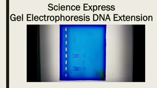
Agarose Gel with DNA Bands markers DNA is negatively charged Smaller sized DNA moves faster than Larger DNA Markers are used to determine relative sizes of DNA pieces
PAGE Native : Protein is prepared with little disturbance to the cellular material Proteins are associated Movement of samples through the gel can be inconsistent SDS : Sodium Dodecyl Sulfate Is a detergent Protein coated with a negative charge in proportion to its molecular weight Denatures and unfolds protein Reducing agents (DTT)break amino acid cross-links
PPolyacrylamide Gel Creates tunnels in gel for molecules to move through
Uses for PAGE Separates proteins from each other Proteins separated by size Isoelectric point Determines Molecular size of protein Quantifies the amount present Displays Impurities Used in western blot assays by antigen interactions
Determine Molecular Weight 1. Run standard molecular weight markers on gel 2. Run unknown protein on the same gel 3. Create a graph of the mol wt versus distance molecule has moved 4. Using the distance the unknown has moved determine the molecular weight from graph
Molecular Weight Markers Migration of molecular weight of standards are compared to unknown samplewt std vs unknown
Western Blot Analysis Identifies protein through antibody interaction Run proteins on denatured gel (SDS-PAGE) Transfer (blot) proteins onto membrane Probe the membrane with primary antibody Add secondary antibody (this antibody is linked to an enzyme) Substrate is added and color appears
SDS Effect on Protein Movement Sodium Dodecyl Sulfate denatures protein and covers it with negative charges : moves to + end Vertical gels are designed so the top of the gel box is attached to the negative power outlet The bottom of the gel box is attached to the positive power outlet Movement through the PAGE gel is proportional to mass not to charge
Movement of Proteins on an SDS Gel Protein Migration - Stacking of proteins at top of gel at start Highest Molecular Wt. protein Distribution of proteins in a charged field + Low weight molecular dye
% Polyacrylamide in Gel Gels can be made at different concentrations of polyacrylamide Example: gels made at 3%,6%,9% and 12% will produce different openings through which the molecule will migrate The larger the opening allows large molecules to move through the gel
Vertical Polyacrylamide Gel Electrophoresis
Gel Electrophoresis Equipment Mini-PROTEAN Tetra Cell
Open Gel Holder: Allows New Gel to be Inserted
Gel Holders Placed in Mini-Protean Tetra Cell
Procedure in Short LoadGe Place Buffer Equip
Electrophoresis of Samples Setting Up and Running Mini-PROTEAN TGX Precast Gels Samples: boiled 3 with loading dye (2x Laemmli buffer + running dye) Mini-PROTEAN tetra cell: Set up according to SOP given in workbook Power settings: 75 volts for 45 60 minutes Running dye should not run off the bottom of gel Youhttp://www.youtube.co m/watch?v=XnEdmk1SqvgT ube