Enzyme Assays in Biotechnology

March 8, 2016
Biotech 3
L
e
c
t
u
r
e
1
6
1. Enzyme Assays
2. ELISA
3. Protein Assays
Lecture Topics
Enzyme Assays
1. Technique used for
measuring enzymatic activity
2. Vital for the study of enzyme kinetics and enzyme
inhibition.
3. Amounts of enzymes can either be expressed as
a.
Molar amounts
– as with any other chemical
b.
Enzyme units
– measurement of activity
Enzyme Assays Cont.
•
Enzyme activity
–
moles of substrate converted per unit time
–
Expressed as
μmol min
−1
(Units)
–
enzyme activity is a measure of the quantity of active
enzyme present and is thus
dependent on conditions
,
which should be specified
•
Specific activity
–
another common unit used to describe enzymes
–
this is the activity of an enzyme per milligram of total
protein
–
Expressed in
μmol min
−1
mg
−1
(
or Units/mg
)
–
Specific activity gives a measurement of the purity of
the enzyme
Terminology
•
Rate of a reaction
is the concentration of substrate
disappearing (or product produced) per unit time
(
mol
L
−1
s
−1
)
•
% purity
is 100% × (specific activity of enzyme
sample / specific activity of pure enzyme)
–
The impure sample has lower specific activity because
some of the mass is not actually enzyme
–
If the specific activity of 100% pure enzyme is known, then
purity can be calculated
Types of Enzyme Assays
1.
Consumption of substrate
over time
2.
Production of product
over time
3.
A large number of different methods of
measuring the concentrations of substrates
and products exist and many enzymes can
be assayed in several different ways
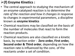
•
Biochemists usually study enzyme-catalyzed
reactions using four types of experiments:
a. Initial rate experiments
b. Progress curve
c. Relaxation
Types of Enzyme Assays Cont.
1.
Consumption of substrate
over time
2.
Production of product
over time
3.
A large number of different methods of
measuring the concentrations of substrates
and products exist and many enzymes can
be assayed in several different ways
•
Biochemists usually study enzyme-catalyzed
reactions using four types of experiments:
a. Initial rate experiments
b. Progress curve
c. Relaxation
Sampling methods
I. Continuous
II. Discontinuous
a. Initial Rate Experiments
•
When an enzyme is mixed with a large excess of the substrate, the enzyme-
substrate intermediate builds up in a fast initial transient
•
Then the reaction achieves a steady-state kinetics in which enzyme
substrate intermediates remains approximately constant over time and the
reaction rate changes relatively slowly
•
Rates are measured for a short period after the attainment of the quasi-
steady state, typically by monitoring the accumulation of product over time
•
Because the measurements are carried out for a very short period and
because of the large excess of substrate, the approximation free substrate is
approximately equal to the initial substrate can be made
•
The initial rate experiment is the simplest to perform and analyze, being
relatively free from complications, such as enzyme degradation
•
Most commonly used type of experiment in enzyme kinetics
Consumption of substrate
Production of product
Reactant/Substrate
Product
Enzyme Activity
N
A
D
H
•
Absorbs at
λ
340
•
Measure change in NADH concentration over time
•
Rate of NADH disappearance allows us to determine enzyme units/ml
•
The more enzyme, the faster the rate. The less enzyme, the slower the rate.
Lactate Dehydrogenase Enzyme Assay
Measuring a Reaction Rate of NADH Consumption
Enzyme is too dilute
Enzyme is too concentrated
Enzyme concentration
is just right
Measuring a Reaction Rate
L
a
m
b
e
r
t
-
B
e
e
r
L
a
w
A
=
ɛ
c
l
ɛ = M
-1
cm
-1
c = M
l = cm
-
Δ
A/ɛl
Δ
t
Initial reaction rate =
-
Δ
c
Δ
t
=
=
V
0
V
0
-
Δ
A/ɛl
Δ
t
=
-
Δ
A/
Δ
t
ɛl
=
-slope
ɛl
=
Measuring a Reaction Rate Cont.
•
One international unit (IU) of enzyme is defined as the amount of enzyme that
catalyses (at V
0
) the formation of one μmole of product per minute (or the loss
of one μmole of substrate per minute) under a specified set of conditions.
# of units of enzyme
in assay mixture
#
o
f
u
n
i
t
s
/
m
l
o
f
e
n
z
y
m
e
i
n
a
s
s
a
y
m
i
x
t
u
r
e
=
μ
mole of NADH consumed
minute
in the assay mixture
μmole
minute
IU
=
=
μ
m
o
l
e
/
m
l
o
f
N
A
D
H
c
o
n
s
u
m
e
d
minute
in the assay mixture
[Conc.]
Calculating the concentration of
enzyme in the assay mixture
#
o
f
u
n
i
t
s
/
m
l
o
f
e
n
z
y
m
e
i
n
a
s
s
a
y
m
i
x
t
u
r
e
=
μ
m
o
l
e
/
m
l
o
f
N
A
D
H
c
o
n
s
u
m
e
d
minute
μ
m
o
l
e
/
m
l
o
f
N
A
D
H
c
o
n
s
u
m
e
d
minute
=
in the assay mixture
-
Δ
[NADH]
Δ
t
=
-slope
ɛl
-
Δ
c
Δ
t
=
Calculating Enzyme Units
μ
m
o
l
e
/
m
l
o
f
N
A
D
H
c
o
n
s
u
m
e
d
minute
Units/ml of LDH
in assay mix
=
-slope (in sec
-1
)
(6220 M
-1
cm
-1
)(1cm)
60 sec
10
6
μmole 1 L
min mole 10
3
ml
Enzyme
concentration in
aliquot being
assayed
=
Units/ml of LDH
in assay mix
X
X
X
X
Volume of assay mix
Volume of aliquot being assayed
=
-
Δ
[NADH]
Δ
t
=
-slope
ɛl
-
Δ
c
Δ
t
=
Calculating Enzyme Units Cont.
b. Progress Curve Experiments
•
In these experiments, the kinetic parameters
are determined from expressions for the
species concentrations as a function of time
•
The concentration of the substrate or product
is recorded in time after the initial fast
transient and for a sufficiently long period to
allow the reaction to approach equilibrium
•
They are less common now, progress curve
experiments were widely used in the early
period of enzyme kinetics
c. Relaxation Experiments
•
An equilibrium mixture of enzyme, substrate,
and product is perturbed
•
For example: temperature, pressure, or pH
•
The return to equilibrium is monitored
•
The analysis of these experiments requires
consideration of the fully reversible reaction
Sampling Methods
Enzyme assays can be split into two groups
according to their sampling method:
I. Continuous assays
•
the assay gives a continuous reading of activity
•
continuous assays are most convenient, with one assay giving
the rate of reaction with no further work necessary
II. Discontinuous assays
•
samples are taken from an enzyme reaction at intervals
and the amount of product production or substrate
consumption is measured in these samples
I. Types of Continuous Enzyme Assays
•
Fluorometric
(Fluorescence) is when a
molecule emits light of
one wavelength after absorbing light of a different wavelength
•
Fluorometric assays use a difference in the fluorescence of
substrate from product to measure the enzyme reaction
•
These assays are in general much more sensitive than
spectrophotometric assays, but can suffer from interference
caused by impurities and the instability of many fluorescent
compounds when exposed to light
I. Types of Continuous Enzyme Assays Cont.
•
Chemiluminescent
(Chemiluminescence) is the emission of light by a
chemical reaction
•
Some enzyme reactions produce light and this can be measured to detect
product formation
•
These types of assay can be
extremely sensitive
, since the light produced
can be captured by photographic film over days or weeks, but can be hard
to quantify, because not all the light released by a reaction will be
detected
–
The detection of horseradish peroxidase by enzymatic chemiluminescence
(ECL) is a common method of detecting antibodies in western blotting.
–
Another example is the enzyme luciferase, this is found in fireflies and
naturally produces light from its substrate luciferin.
II. Types of Discontinuous Enzyme
Assays
•
Radiometric
assays measure the incorporation of radioactivity into
substrates or its release from substrates
•
The radioactive isotopes most frequently used in these assays are
14C, 32P, 35S and 125I
•
Since radioactive isotopes can allow the specific labeling of a single
atom of a substrate, these assays are both extremely sensitive and
specific
•
They are frequently used in biochemistry and are often the only
way of measuring a specific reaction in crude extracts (the complex
mixtures of enzymes produced when you lyse cells)
•
Radioactivity is usually measured in these procedures using a
scintillation counter
II. Types of Discontinuous Enzyme
Assays
•
Chromatographic
assays measure product formation by
separating the reaction mixture into its components by
chromatography
•
This is usually done by high-performance liquid
chromatography (HPLC), but can also use the simpler
technique of thin layer chromatography
•
Although this approach can need a lot of material, its
sensitivity can be increased by labeling the
substrates/products with a radioactive or fluorescent tag
•
Assay sensitivity has also been increased by switching
protocols to improved chromatographic instruments (e.g.,
ultra-high pressure liquid chromatography) that operate at
pump pressure a few-fold higher than HPLC instruments
Factors to control in Enzyme Assays
Salt Concentration
•
Most enzymes cannot tolerate extremely high salt
concentrations
•
The ions interfere with the weak ionic bonds of
proteins
•
Typical enzymes are active in salt concentrations of
1-500 mM
•
As usual there are exceptions, such as halophilic (salt
loving) algae and bacteria
Factors to control in Enzyme Assays Cont.
Temperature
•
All enzymes work within a range of temperature specific to the organism
•
Increases in temperature generally lead to increases in reaction rates
•
There is a limit to the increase because higher temperatures lead to a
sharp decrease in reaction rates
–
This is due to the denaturing (alteration) of protein structure resulting from the
breakdown of the weak ionic and hydrogen bonding that stabilize the three dimensional
structure of the enzyme
•
The "optimum" temperature for human enzymes is usually
between 35
and 40 °C
•
The average temperature for humans is 37 °C
•
Human enzymes start to denature quickly at temperatures above 40 °C
Factors to control in Enzyme Assays Cont. 2
Effects of pH
•
Most enzymes are
sensitive to pH
and have specific ranges of
activity
•
All have an
optimum pH
•
The pH can stop enzyme activity by denaturing (altering) the
three dimensional shape of the enzyme by breaking ionic, and
hydrogen bonds
•
Most enzymes function between a pH of 6 and 8
•
Pepsin in the stomach works best at a pH of 2
Factors to control in Enzyme Assays Cont. 3
Substrate Saturation
•
Increasing the substrate concentration increases the rate
of reaction (enzyme activity)
•
However, enzyme saturation limits reaction rates
•
An enzyme is saturated when the active sites of all the
molecules are occupied most of the time
•
At the saturation point, the reaction will not speed up, no
matter how much additional substrate is added
–
The graph of the reaction rate will plateau
Factors to control in Enzyme Assays Cont. 4
Level of crowding
•
Large amounts of macromolecules in a solution will alter the
rates and equilibrium constants of enzyme reactions, through
an effect called macromolecular crowding
E
L
I
S
A
A
S
S
A
Y
P
R
O
C
E
S
S
Immunoassay
•
A laboratory technique that makes use of the
binding between an antigen and its
homologous antibody in order to identify and
quantify the specific antigen or antibody in a
sample
Antibody and Antigens
Enzyme-Linked Immunosorbent Assay
•
A biochemical technique used mainly in
immunology to detect the presence of an
antibody or an antigen in a sample:
Three types of ELISA
1- Competitive ELISA
2- Sandwich ELISA (also called direct ELISA)
3- Indirect ELISA
1. Competitive ELISA
•
Antibody is bound to solid support
•
The
labelled antigen competes for primary antibody
binding sites with the sample antigen
(unlabeled)
•
The more antigen in the sample, the less labelled
antigen is retained in the well and the weaker the
signal
Solid phase
coated with
antibody
Add free
labeled
antigen
Free and
labelled
antigen are
captured
Color formation by
oxidation of substrate
into a colored
compound
1. Competitive ELISA Cont.
•
The ELISA plate is coated with an antibody
targeted against a specific antigen
2. Sandwich ELISA Process
2. Sandwich ELISA Steps
1. Prepare a surface
to which a known quantity
of capture antibody is bound.
2. Block
any non-specific binding sites on the
surface
3. Apply the antigen
-containing sample to the
plate.
4. Wash
the plate, so that unbound antigen is
removed
5. Apply enzyme linked primary antibodies
as
detection antibodies which also bind specifically
to the antigen
6. Wash
the plate, so that the unbound antibody-
enzyme conjugates are removed
(Alternatively can apply a secondary antibody after step 6)
2. Sandwich ELISA Steps Cont.
2. Sandwich ELISA Steps Cont. 2
7.
Apply a chemical substrate
which is converted
by the enzyme into a colored product
8.
Measure
the absorbency of the plate wells to
determine the presence and quantity of antigen
3. Indirect ELISA Steps
1.
Antigen
to be tested for is adhered to well
2.
Block
any plastic surface in the well that
remains uncoated by the protein antigen
3. Indirect ELISA Steps Cont.
3.
Serum is added
•
contains a mixture of the serum antibodies of
unknown concentration
•
some of which may bind specifically to the test
antigen that is coating the well
4.
Secondary antibody is added
, which will bind to
the antibody bound to the test antigen in the well
•
This secondary antibody often has an enzyme
attached to it
3.Indirect ELISA Steps Cont.2
5.
Substrate
for the enzyme is then added
–
Often this substrate changes color upon reaction
with the enzyme
–
The higher the concentration of the primary
antibody that was present in the serum, the
stronger the color change
6.
Measure
- A spectrophotometer is used to
give quantitative values for color strength
3. Indirect ELISA Example
•
This standard curve is used to determine the
unknown concentration of each sample using
the linear relationship between concentration
and absorbance
Standard Curve
•
First diagnosed in 1981
•
Over 20 million deaths worldwide,
over a half million in the United
States
•
Over 40 million currently infected,
over a million in the United States
•
Half of all new infections are in
people younger than 25
•
Education has been effective in
limiting the spread of HIV/AIDS
Human Immunodeficiency Virus (HIV)
HIV Biology: What do we know?
•
HIV is an RNA Retrovirus
•
Transmitted by exchange of body fluids, sharing needles, or blood
transfusion
•
Infects T-Cells in the immune system and thus destroys the immune system
•
Flu-like symptoms within 1-2 months followed by latent period of up to 10
years
•
HIV may have spread from an animal host to humans
•
Treated but not cured by drugs which inhibit the action of HIV enzymes
•
High error rate of replications (1/2000 nucleotides)
The Immune Response
ELISA-HIV Test: Detecting Antibodies in Serum
•
After 4-8 weeks of exposure to the HIV virus, the
body will have produced a detectable level
antibodies (immune response) against HIV
•
ELISA (HIV-Test) detects the presence of serum
antibodies against HIV protein antigens
•
This is how HIV is detected in clinical laboratories
•
Most common AIDS test
Ways The ELISA Kit Can Be Used
Protein determination
Proteins Colorimetric Assay
•
Hartree-Lowry and Modified Lowry Protein Assays:
The Lowry assay (1951)
is an often-cited general use protein assay. For some time it was the
method of choice for accurate protein determination for cell fractions,
chromatography fractions, enzyme preparations, and so on. The
bicinchoninic acid (BCA) assay is based on the same principle and can be
done in one step, therefore it has been suggested (Stoscheck, 1990) that
the 2-step Lowry method is outdated. However, the modified Lowry is done
entirely at room temperature. The Hartree version of the Lowry assay, a
more recent modification that uses fewer reagents, improves the
sensitivity with some proteins, is less likely to be incompatible with some
salt solutions, provides a more linear response, and is less likely to become
saturated.
•
Reaction Principle:
Under alkaline conditions the divalent copper ion forms
a complex with peptide bonds in which it is reduced to a monovalent ion.
Monovalent copper ion and the radical groups of tyrosine, tryptophan, and
cysteine react with Folin reagent to produce an unstable product that
becomes reduced to molybdenum/tungsten blue
Proteins Colorimetric Assay Cont.
•
Biuret Protein Assay:
The principle of the Biuret assay is
similar to that of the Lowry, however it involves a single
incubation of 20 min. There are very few interfering agents
(ammonium salts being one such agent), and Layne (1957)
reported fewer deviations than with the Lowry or ultraviolet
absorption methods. However, the biuret assay consumes
much more material. The biuret is a good general protein
assay for batches of material for which yield is not a problem.
The Bradford assay is faster and more sensitive.
•
Reaction Principle:
Under alkaline conditions substances
containing two or more peptide bonds form a purple complex
with copper salts in the reagent
Proteins Colorimetric Assay Cont.2
•
Bradford protein assay:
The Bradford assay is very fast and uses about the same
amount of protein as the Lowry assay. It is fairly accurate and samples that are out
of range can be retested within minutes. The Bradford is recommended for general
use, especially for determining protein content of cell fractions and assessing
protein concentrations for gel electrophoresis.
•
Assay materials including color reagent, protein standard, and instruction booklet
are available from Bio-Rad Corporation. The method described below is for a 100 µl
sample volume using 5 ml color reagent. It is sensitive to about 5 to 200 micrograms
protein, depending on the dye quality. In assays using 5 ml color reagent prepared in
lab, the sensitive range is closer to 5 to 100 µg protein. Scale down the volume for
the "microassay procedure," which uses 1 ml cuvettes. Protocols, including use of
microtiter plates are described in the flyer that comes with the Bio-Rad kit.
•
Reaction Principle:
The assay is based on the observation that the absorbance
maximum for an acidic solution of Coomassie Brilliant Blue G-250 shifts from 465
nm to 595 nm when binding to protein occurs. Both hydrophobic and ionic
interactions stabilize the anionic form of the dye, causing a visible color change. The
assay is useful since the extinction coefficient of a dye-albumin complex solution is
constant over a 10-fold concentration range.
Proteins Colorimetric Assay Cont.3
•
Bicinchoninic Acid (BCA) Protein Assay (Smith):
The bicinchoninic acid
(BCA) assay is available in kit form from Pierce (Rockford, Ill.). This
procedure is very applicable to microtiter plate methods. The BCA is used
for the same reasons the Lowry is used. Stoscheck (1990) has suggested
that the BCA assay will replace the Lowry because it requires a single step,
and the color reagent is stable under alkaline conditions.
•
Reaction Principle:
BCA serves the purpose of the Folin reagent in the
Lowry assay, namely to react with complexes between copper ions and
peptide bonds to produce a purple end product. The advantage of BCA is
that the reagent is fairly stable under alkaline conditions, and can be
included in the copper solution to allow a one step procedure. A
molybdenum/tungsten blue product is produced as with the Lowry.
Exploring enzyme assays in biotechnology, covering topics such as ELISA, protein assays, enzyme activity measurement techniques, specific activity, terminology related to enzyme assays, and different types of experiments conducted by biochemists to study enzyme-catalyzed reactions. The content delves into the significance of enzyme kinetics, inhibition, and purity measurements, providing an insightful overview for those interested in biotech.

Download Presentation

Please find below an Image/Link to download the presentation.
The content on the website is provided AS IS for your information and personal use only. It may not be sold, licensed, or shared on other websites without obtaining consent from the author.If you encounter any issues during the download, it is possible that the publisher has removed the file from their server.
You are allowed to download the files provided on this website for personal or commercial use, subject to the condition that they are used lawfully. All files are the property of their respective owners.
The content on the website is provided AS IS for your information and personal use only. It may not be sold, licensed, or shared on other websites without obtaining consent from the author.
E N D
Presentation Transcript
Lecture 16 March 8, 2016 Biotech 3
Lecture Topics 1. Enzyme Assays 2. ELISA 3. Protein Assays
Enzyme Assays 1. Technique used for measuring enzymatic activity 2. Vital for the study of enzyme kinetics and enzyme inhibition. 3. Amounts of enzymes can either be expressed as a. Molar amounts as with any other chemical b. Enzyme units measurement of activity
Enzyme Assays Cont. Enzyme activity moles of substrate converted per unit time Expressed as mol min 1 (Units) enzyme activity is a measure of the quantity of active enzyme present and is thus dependent on conditions, which should be specified Specific activity another common unit used to describe enzymes this is the activity of an enzyme per milligram of total protein Expressed in mol min 1 mg 1 (or Units/mg) Specific activity gives a measurement of the purity of the enzyme
Terminology Rate of a reaction is the concentration of substrate disappearing (or product produced) per unit time (molL 1s 1) % purity is 100% (specific activity of enzyme sample / specific activity of pure enzyme) The impure sample has lower specific activity because some of the mass is not actually enzyme If the specific activity of 100% pure enzyme is known, then purity can be calculated
Types of Enzyme Assays 1. 2. 3. Consumption of substrate over time Production of product over time A large number of different methods of measuring the concentrations of substrates and products exist and many enzymes can be assayed in several different ways Biochemists usually study enzyme-catalyzed reactions using four types of experiments: a. Initial rate experiments b. Progress curve c. Relaxation
Types of Enzyme Assays Cont. 1. 2. 3. Consumption of substrate over time Production of product over time A large number of different methods of measuring the concentrations of substrates and products exist and many enzymes can be assayed in several different ways Biochemists usually study enzyme-catalyzed reactions using four types of experiments: a. Initial rate experiments b. Progress curve c. Relaxation Sampling methods I. Continuous II. Discontinuous
a. Initial Rate Experiments When an enzyme is mixed with a large excess of the substrate, the enzyme- substrate intermediate builds up in a fast initial transient Then the reaction achieves a steady-state kinetics in which enzyme substrate intermediates remains approximately constant over time and the reaction rate changes relatively slowly Rates are measured for a short period after the attainment of the quasi- steady state, typically by monitoring the accumulation of product over time Because the measurements are carried out for a very short period and because of the large excess of substrate, the approximation free substrate is approximately equal to the initial substrate can be made The initial rate experiment is the simplest to perform and analyze, being relatively free from complications, such as enzyme degradation Most commonly used type of experiment in enzyme kinetics
Enzyme Activity Production of product Consumption of substrate Product Reactant/Substrate
Lactate Dehydrogenase Enzyme Assay LDH Enzyme NADH Absorbs at 340 Measure change in NADH concentration over time Rate of NADH disappearance allows us to determine enzyme units/ml The more enzyme, the faster the rate. The less enzyme, the slower the rate.
Measuring a Reaction Rate Enzyme is too dilute Enzyme concentration is just right Enzyme is too concentrated
Measuring a Reaction Rate Cont. Lambert-Beer Law - c - A/ l t A = cl = V0 = Initial reaction rate = t = M-1cm-1 c = M l = cm -slope l - A/ t l - A/ l t = V0 = =
Calculating the concentration of enzyme in the assay mixture One international unit (IU) of enzyme is defined as the amount of enzyme that catalyses (at V0) the formation of one mole of product per minute (or the loss of one mole of substrate per minute) under a specified set of conditions. mole minute IU = mole of NADH consumed minute # of units of enzyme in assay mixture in the assay mixture = mole/ml of NADH consumed minute # of units/ml of enzyme in assay mixture in the assay mixture = [Conc.]
Calculating Enzyme Units mole/ml of NADH consumed minute # of units/ml of enzyme in assay mixture in the assay mixture = - [NADH] -slope mole/ml of NADH consumed minute - c = = = t l t
Calculating Enzyme Units Cont. - [NADH] -slope - c mole/ml of NADH consumed minute = = = t l t -slope (in sec-1) 60 sec 106 mole 1 L X Units/ml of LDH in assay mix X X = (6220 M-1cm-1)(1cm) min mole 103 ml Enzyme concentration in aliquot being assayed Volume of assay mix Units/ml of LDH in assay mix = X Volume of aliquot being assayed
b. Progress Curve Experiments In these experiments, the kinetic parameters are determined from expressions for the species concentrations as a function of time The concentration of the substrate or product is recorded in time after the initial fast transient and for a sufficiently long period to allow the reaction to approach equilibrium They are less common now, progress curve experiments were widely used in the early period of enzyme kinetics
c. Relaxation Experiments An equilibrium mixture of enzyme, substrate, and product is perturbed For example: temperature, pressure, or pH The return to equilibrium is monitored The analysis of these experiments requires consideration of the fully reversible reaction
Sampling Methods Enzyme assays can be split into two groups according to their sampling method: I. Continuous assays the assay gives a continuous reading of activity continuous assays are most convenient, with one assay giving the rate of reaction with no further work necessary II. Discontinuous assays samples are taken from an enzyme reaction at intervals and the amount of product production or substrate consumption is measured in these samples
I. Types of Continuous Enzyme Assays Fluorometric (Fluorescence) is when a molecule emits light of one wavelength after absorbing light of a different wavelength Fluorometric assays use a difference in the fluorescence of substrate from product to measure the enzyme reaction These assays are in general much more sensitive than spectrophotometric assays, but can suffer from interference caused by impurities and the instability of many fluorescent compounds when exposed to light
I. Types of Continuous Enzyme Assays Cont. Chemiluminescent (Chemiluminescence) is the emission of light by a chemical reaction Some enzyme reactions produce light and this can be measured to detect product formation These types of assay can be extremely sensitive, since the light produced can be captured by photographic film over days or weeks, but can be hard to quantify, because not all the light released by a reaction will be detected The detection of horseradish peroxidase by enzymatic chemiluminescence (ECL) is a common method of detecting antibodies in western blotting. Another example is the enzyme luciferase, this is found in fireflies and naturally produces light from its substrate luciferin.
II. Types of Discontinuous Enzyme Assays Radiometric assays measure the incorporation of radioactivity into substrates or its release from substrates The radioactive isotopes most frequently used in these assays are 14C, 32P, 35S and 125I Since radioactive isotopes can allow the specific labeling of a single atom of a substrate, these assays are both extremely sensitive and specific They are frequently used in biochemistry and are often the only way of measuring a specific reaction in crude extracts (the complex mixtures of enzymes produced when you lyse cells) Radioactivity is usually measured in these procedures using a scintillation counter
II. Types of Discontinuous Enzyme Assays Chromatographic assays measure product formation by separating the reaction mixture into its components by chromatography This is usually done by high-performance liquid chromatography (HPLC), but can also use the simpler technique of thin layer chromatography Although this approach can need a lot of material, its sensitivity can be increased by labeling the substrates/products with a radioactive or fluorescent tag Assay sensitivity has also been increased by switching protocols to improved chromatographic instruments (e.g., ultra-high pressure liquid chromatography) that operate at pump pressure a few-fold higher than HPLC instruments
Factors to control in Enzyme Assays Salt Concentration Most enzymes cannot tolerate extremely high salt concentrations The ions interfere with the weak ionic bonds of proteins Typical enzymes are active in salt concentrations of 1-500 mM As usual there are exceptions, such as halophilic (salt loving) algae and bacteria
Factors to control in Enzyme Assays Cont. Temperature All enzymes work within a range of temperature specific to the organism Increases in temperature generally lead to increases in reaction rates There is a limit to the increase because higher temperatures lead to a sharp decrease in reaction rates This is due to the denaturing (alteration) of protein structure resulting from the breakdown of the weak ionic and hydrogen bonding that stabilize the three dimensional structure of the enzyme The "optimum" temperature for human enzymes is usually between 35 and 40 C The average temperature for humans is 37 C Human enzymes start to denature quickly at temperatures above 40 C
Factors to control in Enzyme Assays Cont. 2 Effects of pH Most enzymes are sensitive to pH and have specific ranges of activity All have an optimum pH The pH can stop enzyme activity by denaturing (altering) the three dimensional shape of the enzyme by breaking ionic, and hydrogen bonds Most enzymes function between a pH of 6 and 8 Pepsin in the stomach works best at a pH of 2
Factors to control in Enzyme Assays Cont. 3 Substrate Saturation Increasing the substrate concentration increases the rate of reaction (enzyme activity) However, enzyme saturation limits reaction rates An enzyme is saturated when the active sites of all the molecules are occupied most of the time At the saturation point, the reaction will not speed up, no matter how much additional substrate is added The graph of the reaction rate will plateau
Factors to control in Enzyme Assays Cont. 4 Level of crowding Large amounts of macromolecules in a solution will alter the rates and equilibrium constants of enzyme reactions, through an effect called macromolecular crowding
Immunoassay A laboratory technique that makes use of the binding between an antigen and its homologous antibody in order to identify and quantify the specific antigen or antibody in a sample
Enzyme-Linked Immunosorbent Assay A biochemical technique used mainly in immunology to detect the presence of an antibody or an antigen in a sample: Three types of ELISA 1- Competitive ELISA 2- Sandwich ELISA (also called direct ELISA) 3- Indirect ELISA
1. Competitive ELISA Antibody is bound to solid support The labelled antigen competes for primary antibody binding sites with the sample antigen (unlabeled) The more antigen in the sample, the less labelled antigen is retained in the well and the weaker the signal
1. Competitive ELISA Cont. Solid phase coated with antibody Add free labeled antigen Free and labelled antigen are captured Color formation by oxidation of substrate into a colored compound
2. Sandwich ELISA Process The ELISA plate is coated with an antibody targeted against a specific antigen
2. Sandwich ELISA Steps 1. Prepare a surface to which a known quantity of capture antibody is bound. 2. Block any non-specific binding sites on the surface 3. Apply the antigen-containing sample to the plate.
2. Sandwich ELISA Steps Cont. 4. Wash the plate, so that unbound antigen is removed 5. Apply enzyme linked primary antibodies as detection antibodies which also bind specifically to the antigen 6. Wash the plate, so that the unbound antibody- enzyme conjugates are removed (Alternatively can apply a secondary antibody after step 6)
2. Sandwich ELISA Steps Cont. 2 7. Apply a chemical substrate which is converted by the enzyme into a colored product 8. Measure the absorbency of the plate wells to determine the presence and quantity of antigen
3. Indirect ELISA Steps 1. Antigen to be tested for is adhered to well 2. Block any plastic surface in the well that remains uncoated by the protein antigen
3. Indirect ELISA Steps Cont. 3. Serum is added contains a mixture of the serum antibodies of unknown concentration some of which may bind specifically to the test antigen that is coating the well 4. Secondary antibody is added, which will bind to the antibody bound to the test antigen in the well This secondary antibody often has an enzyme attached to it
3.Indirect ELISA Steps Cont.2 5. Substrate for the enzyme is then added Often this substrate changes color upon reaction with the enzyme The higher the concentration of the primary antibody that was present in the serum, the stronger the color change 6. Measure - A spectrophotometer is used to give quantitative values for color strength
Standard Curve This standard curve is used to determine the unknown concentration of each sample using the linear relationship between concentration and absorbance
Human Immunodeficiency Virus (HIV) First diagnosed in 1981 Over 20 million deaths worldwide, over a half million in the United States Over 40 million currently infected, over a million in the United States Half of all new infections are in people younger than 25 Education has been effective in limiting the spread of HIV/AIDS
HIV Biology: What do we know? HIV is an RNA Retrovirus Transmitted by exchange of body fluids, sharing needles, or blood transfusion Infects T-Cells in the immune system and thus destroys the immune system Flu-like symptoms within 1-2 months followed by latent period of up to 10 years HIV may have spread from an animal host to humans Treated but not cured by drugs which inhibit the action of HIV enzymes High error rate of replications (1/2000 nucleotides)
ELISA-HIV Test: Detecting Antibodies in Serum After 4-8 weeks of exposure to the HIV virus, the body will have produced a detectable level antibodies (immune response) against HIV ELISA (HIV-Test) detects the presence of serum antibodies against HIV protein antigens This is how HIV is detected in clinical laboratories Most common AIDS test
Ways The ELISA Kit Can Be Used Type of ELISA Protocol Real-World Application Objectives Tracking outbreaks of disease HIV, Bird Flu and West Nile viruses, common cold, cholera, smallpox, anthrax, and STDs Pregnancy, drug, GMO and allergen tests Air food and water testing HIV, smallpox, West Nile and Bird Flu viruses HIV, Lyme disease, trichinosis, West Nile virus, and Bird Flu virus Epidemiology, disease spread, public health I Detecting antigens Uses for antibodies in research, medicine, and consumer goods II Detecting antibodies in serum Detecting exposure to disease causing agents III
Proteins Colorimetric Assay Hartree-Lowry and Modified Lowry Protein Assays: The Lowry assay (1951) is an often-cited general use protein assay. For some time it was the method of choice for accurate protein determination for cell fractions, chromatography fractions, enzyme preparations, and so on. The bicinchoninic acid (BCA) assay is based on the same principle and can be done in one step, therefore it has been suggested (Stoscheck, 1990) that the 2-step Lowry method is outdated. However, the modified Lowry is done entirely at room temperature. The Hartree version of the Lowry assay, a more recent modification that uses fewer reagents, improves the sensitivity with some proteins, is less likely to be incompatible with some salt solutions, provides a more linear response, and is less likely to become saturated. Reaction Principle: Under alkaline conditions the divalent copper ion forms a complex with peptide bonds in which it is reduced to a monovalent ion. Monovalent copper ion and the radical groups of tyrosine, tryptophan, and cysteine react with Folin reagent to produce an unstable product that becomes reduced to molybdenum/tungsten blue


")