Cytogenetics: The Study of Chromosomes and Genes
Cytogenetics is the study of chromosome number, structure, function, and behavior in relation to gene inheritance, organization, and expression. It plays a crucial role in detecting genetic abnormalities that can range from vitamin deficiencies to severe conditions like cancer and birth defects. By analyzing chromosomes and genes, researchers can uncover insights into human health and disease.

Download Presentation

Please find below an Image/Link to download the presentation.
The content on the website is provided AS IS for your information and personal use only. It may not be sold, licensed, or shared on other websites without obtaining consent from the author.If you encounter any issues during the download, it is possible that the publisher has removed the file from their server.
You are allowed to download the files provided on this website for personal or commercial use, subject to the condition that they are used lawfully. All files are the property of their respective owners.
The content on the website is provided AS IS for your information and personal use only. It may not be sold, licensed, or shared on other websites without obtaining consent from the author.
E N D
Presentation Transcript
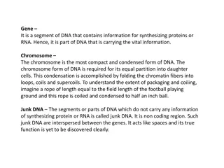
Cytogenetics = The study of chromosome number, structure, function, and behavior in relation to gene inheritance, organization and expression
Chromosome Chromo = colored in response to dye Some = body Chromosome of Eukaryotes have been the traditional subject for cytogenetic analysis because they are large enough to be examined with light microscope
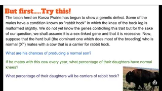
Why Analyse Chromosomes and Genes? Genetic errors arise from deletions or insertions of genetic material, abnormal numbers of whole chromosomes or genes, and even from misplacement of a single base in the DNA sequence. Genetic abnormalities can range from relatively harmless to severe: from vitamin deficiencies and food allergies to cancer, birth defects and infant mortality.
Cytogenetic methods to detect chromosomal abnormalities underlying human birth defects usually involve analysis of mitotic chromosome
What tissues are appropriate for chromosome study? A tissue that can be stimulated to undergo cell division in-vitro It is only during mitosis of the cell cycle that distinct chromosomes can be visualized with a light microscope After culturing, in-vitro, a proportion of cells are arrested in mitosis, and are then harvested for chromosome analysis After harvesting, the cell preparations are dropped onto glass slides and stained. For most chromosome analyses, a G-banding technique is utilized for staining. Metaphase spread
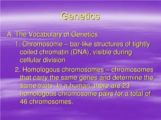
The chromosomes are so named as they may be stained by certain dyes Chromosomes are composed of chromatin, which is composed of protein and DNA When cells are not dividing, the genetic material is decondensed Chromosomes become visible as distinct structures when the cell divides
Chromosome Sister Chromatides
Chromosomes of different species differ in number and information content Humans and several other species of organisms have 46 chromosomes
Karyotyping Karyotype A pictorial display of metaphase chromosomes from a mitotic cell Homologous chromosomes- pairs
Karyotype Karyotyping is the analysis of chromosomes Cytogenetics is the study of chromosomes and inheritance Cytogenetics is based on studies of humans as well as Drosophila and other organisms
Preparing a karyotype 1. 2. Harvested cells are first cultured The cells are then treated with colchicine which arrests the cells in metaphase, and then treated and stained to observe the chromosomes Chromosomes can be photographed or visualized using a computer, and then analyzed Chromosomes are identified by size, position of the centromere, and banding and staining regions 3. 4.
The analysis involves comparing chromosomes for their length, the placement of centromeres (areas where the two chromatids are joined), and the location and sizes of G-bands.
Banding patterns on human mitotic chomosomes due to regions of condensed chomatin (darker - G bands) and less condensed chromatin (lighter - R bands) human chromosome 4 at varying resolutions due to exact mitotic stage, (or degrees of spreading - squashing - stretching)
Human chromosome number is determined by their length in mitotic figures"
International System for Cytogenetic Nomenclature, (ISCN,1995) Short arm of the chromosome = p Long arm of the chromosome = q Bands are numbered independently on the short and long arms Centromeres = p10,q10 Band numbers increase as move from the centromere to the telomere
Hundreds of genes are encompassed within a single G-band. Therefore, most constitutional chromosome abnormalities are associated with multiple congenital anomalies. Therefore, deletion of a single gene cannot be detected by G-banding.