Circulation and Gas Exchange in Living Organisms
Explore the diverse circulatory systems in invertebrates and vertebrates, from jellyfish to humans. Learn about open and closed circulatory systems, the structure and function of the heart, heart valves and sounds, as well as how heart rate and cardiac output are measured.

Download Presentation

Please find below an Image/Link to download the presentation.
The content on the website is provided AS IS for your information and personal use only. It may not be sold, licensed, or shared on other websites without obtaining consent from the author.If you encounter any issues during the download, it is possible that the publisher has removed the file from their server.
You are allowed to download the files provided on this website for personal or commercial use, subject to the condition that they are used lawfully. All files are the property of their respective owners.
The content on the website is provided AS IS for your information and personal use only. It may not be sold, licensed, or shared on other websites without obtaining consent from the author.
E N D
Presentation Transcript
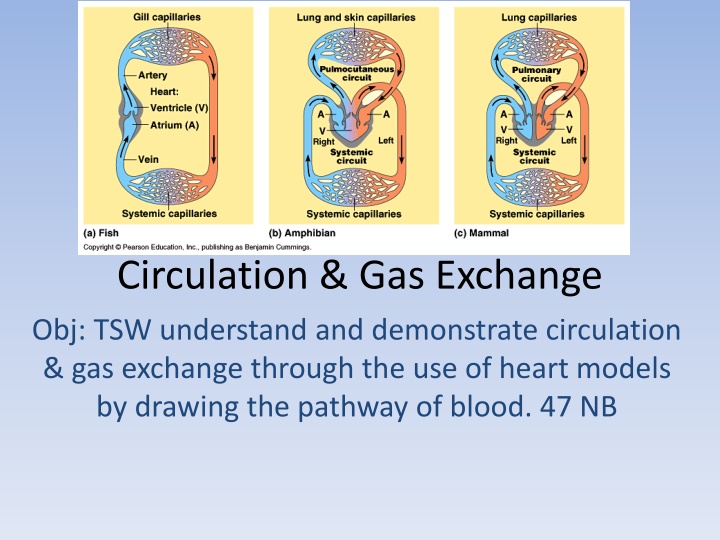
Circulation & Gas Exchange Obj: TSW understand and demonstrate circulation & gas exchange through the use of heart models by drawing the pathway of blood. 47 NB
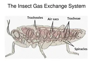
Internal Transport in Invertebrates Animals w/o a backbone Invertebrates Jellyfish (Cnidaria), planaria (flat worms), arthropods (grasshopper) Open Circulatory System No distinction between blood & interstitial fluid Hemolymph body fluid Chemical exchange happens at sinuses Heart (pumps) hemolymph in contact with body tissues to exchange respiratory gases: O2 & CO2 When the heart relaxes, hemolymph enters the heart through pores called ostia. O2 infiltrates insects body through air ducts called tracheae
Closed Circulatory System (Cardiovascular System) Annelids (segmented worms) Vertebrates & some Mollusks have closed circulatory system Blood is confined to vessels (capillaries, venules, veins, arterioles, arteries) Evolutionary Perspective of Vertebrate Circulatory System: Fish-2 chambered heart, Amphibians 3 chambered heart, Reptiles 3.5 chambered heart- partially divided septum. (crocodiles have a completely divided septum so the ventricle has 2 chambers)
The Heart Size of clenched fist, cardiac muscle Atria thin walled compared to ventricle, pumps blood only a short distance to the ventricles. Ventricles thicker and more powerful, especially the left ventricle. Heart Cycle (.8 sec) systolic & diastolic Systole the heart muscle contract (ventricle) and the chambers pump blood Diastole ventricles are filling with blood, relaxation Pulse Rate = 65 75 beats / minute
Heart Valves & Heart Sounds 4 Valves prevent backflow of blood when ventricles contract Atrioventricular Valves: Tricuspic & Mitral Valve Semilunar Valves: Exits of the heart Pulmonary & Aortic Valve lub-dupp, lub-dupp, lub-dupp First heart sound, lub is the forceful contraction of the ventricular valve Second heart sound, dupp is the recoil of blood against the semilunar valves
Heart Rate Pulse & Cardiac Output Pulse number of heartbeats / minute Count the pulsations of arteries in your wrist or neck. People who exercise regularly often have slower resting pulses than those who are less fit. Inverse relationship between size & pulse. Elephant= 25 beats/ minute Tiny Shrew = 600 beats / minute The metabolic rate per gram of tissue is proportionately grater for smaller mammals than for larger ones Enhances the delivery of oxygen for Cellular Respiration Cardiac Output volume of blood / minute, and is determined by heart rate and stroke volume
Excitation and control of the heart Heart cells are self- excitable (myogenic), they can contract w/o any signal from the nervous system. They have an intrinsic ability to contract Sinoatrial node (SA) or Pacemaker controls the rate of contraction of the heart & is located in the wall of the right atrium Initiate a wave of excitation that travels through the wall of the heart. EKG or ECG recorded electrical current of the cardiac muscle during the heart cycle.
Blood Pressure Hydrostatic pressure that blood exerts against the wall of a vessel.
Composition of Blood *Red Blood Cells transport Oxygen, biconcave disk (increases surface area) erythrocyte, no nuclei, no mitochondria (ATP anaerobic metabolism), small White Blood Cells (Leukocytes) (Immune system- defense) 5 major types: monocytes, neutrophils, basophils, eosinophils & lymphocytes Platelets no nucleus, not really a cell, help with blood clotting Pluripotent stem cells- come from Red marrow of bones (ribs, vertebrae, Breastbone, pelvis) dev. into any blood cells
Cardiovascular Disease Disease of the heart & blood vessels Heart Attack or Stroke Atherosclerosis blood clot plugging an artery Plaques growths develop on the inner walls of arteries LDL (bad Cholesterol) cholesterol travels in blood bound to protein and adds plaque to arteries. HDL (good Cholesterol) reduce the depositing of cholesterol in arterial plaques
Quick Write Describe some examples for structure and function concerning the circulatory system P. 50 NB. Veins valves Shape of heart Arteries flexible Shape-Red Blood Cells- Concave