Animal Circulation and Gas Exchange Systems Overview

1
Lecture #11 – Animal Circulation
and Gas Exchange Systems
2
Key Concepts:
Circulation and gas exchange – why?
Circulation – spanning diversity
Hearts – the evolution of double circulation
Blood circulation and capillary exchange
Blood structure and function
Gas exchange – spanning diversity
Breathing – spanning diversity
Respiratory pigments
3
Animals use O
2
 and produce CO
2
All animals are aerobic
L
o
t
s
 
o
f
 
o
x
y
g
e
n
 
i
s
 
r
e
q
u
i
r
e
d
 
t
o
 
s
u
p
p
o
r
t
 
a
c
t
i
v
e
m
o
b
i
l
i
t
y
Some animals use lots of oxygen to maintain
body temperature
All animals produce CO
2
 as  a byproduct of
aerobic respiration
Gasses must be exchanged
Oxygen must be acquired from the environment
Carbon dioxide must be released to the
environment
4
Except……breaking news!
http://www.biomedcentral.com/1741-7007/8/30
A
b
s
t
r
a
c
t
 
 
6
 
A
p
r
i
l
 
2
0
1
0
B
a
c
k
g
r
o
u
n
d
S
e
v
e
r
a
l
 
u
n
i
c
e
l
l
u
l
a
r
 
o
r
g
a
n
i
s
m
s
 
(
p
r
o
k
a
r
y
o
t
e
s
 
a
n
d
 
p
r
o
t
o
z
o
a
)
 
c
a
n
 
l
i
v
e
 
u
n
d
e
r
 
p
e
r
m
a
n
e
n
t
l
y
 
a
n
o
x
i
c
 
c
o
n
d
i
t
i
o
n
s
.
A
l
t
h
o
u
g
h
 
a
 
f
e
w
 
m
e
t
a
z
o
a
n
s
 
c
a
n
 
s
u
r
v
i
v
e
 
t
e
m
p
o
r
a
r
i
l
y
 
i
n
 
t
h
e
 
a
b
s
e
n
c
e
 
o
f
 
o
x
y
g
e
n
,
 
i
t
 
i
s
 
b
e
l
i
e
v
e
d
 
t
h
a
t
 
m
u
l
t
i
-
c
e
l
l
u
l
a
r
 
o
r
g
a
n
i
s
m
s
 
c
a
n
n
o
t
 
s
p
e
n
d
 
t
h
e
i
r
 
e
n
t
i
r
e
 
l
i
f
e
 
c
y
c
l
e
 
w
i
t
h
o
u
t
 
f
r
e
e
 
o
x
y
g
e
n
.
 
D
e
e
p
 
s
e
a
s
 
i
n
c
l
u
d
e
 
s
o
m
e
 
o
f
t
h
e
 
m
o
s
t
 
e
x
t
r
e
m
e
 
e
c
o
s
y
s
t
e
m
s
 
o
n
 
E
a
r
t
h
,
 
s
u
c
h
 
a
s
 
t
h
e
 
d
e
e
p
 
h
y
p
e
r
s
a
l
i
n
e
 
a
n
o
x
i
c
 
b
a
s
i
n
s
 
o
f
 
t
h
e
M
e
d
i
t
e
r
r
a
n
e
a
n
 
S
e
a
.
 
T
h
e
s
e
 
a
r
e
 
p
e
r
m
a
n
e
n
t
l
y
 
a
n
o
x
i
c
 
s
y
s
t
e
m
s
 
i
n
h
a
b
i
t
e
d
 
b
y
 
a
 
h
u
g
e
 
a
n
d
 
p
a
r
t
l
y
 
u
n
e
x
p
l
o
r
e
d
m
i
c
r
o
b
i
a
l
 
b
i
o
d
i
v
e
r
s
i
t
y
.
R
e
s
u
l
t
s
D
u
r
i
n
g
 
t
h
e
 
l
a
s
t
 
t
e
n
 
y
e
a
r
s
 
t
h
r
e
e
 
o
c
e
a
n
o
g
r
a
p
h
i
c
 
e
x
p
e
d
i
t
i
o
n
s
 
w
e
r
e
 
c
o
n
d
u
c
t
e
d
 
t
o
 
s
e
a
r
c
h
 
f
o
r
 
t
h
e
 
p
r
e
s
e
n
c
e
 
o
f
l
i
v
i
n
g
 
f
a
u
n
a
 
i
n
 
t
h
e
 
s
e
d
i
m
e
n
t
s
 
o
f
 
t
h
e
 
d
e
e
p
 
a
n
o
x
i
c
 
h
y
p
e
r
s
a
l
i
n
e
 
L
'
A
t
a
l
a
n
t
e
 
b
a
s
i
n
 
(
M
e
d
i
t
e
r
r
a
n
e
a
n
 
S
e
a
)
.
 
W
e
r
e
p
o
r
t
 
h
e
r
e
 
t
h
a
t
 
t
h
e
 
s
e
d
i
m
e
n
t
s
 
o
f
 
t
h
e
 
L
'
A
t
a
l
a
n
t
e
 
b
a
s
i
n
 
a
r
e
 
i
n
h
a
b
i
t
e
d
 
b
y
 
t
h
r
e
e
 
s
p
e
c
i
e
s
 
o
f
 
t
h
e
 
a
n
i
m
a
l
 
p
h
y
l
u
m
L
o
r
i
c
i
f
e
r
a
 
(
S
p
i
n
o
l
o
r
i
c
u
s
 
n
o
v
.
 
s
p
.
,
 
R
u
g
i
l
o
r
i
c
u
s
 
n
o
v
.
 
s
p
.
 
a
n
d
 
P
l
i
c
i
l
o
r
i
c
u
s
 
n
o
v
.
 
s
p
.
)
 
n
e
w
 
t
o
 
s
c
i
e
n
c
e
.
 
U
s
i
n
g
r
a
d
i
o
a
c
t
i
v
e
 
t
r
a
c
e
r
s
,
 
b
i
o
c
h
e
m
i
c
a
l
 
a
n
a
l
y
s
e
s
,
 
q
u
a
n
t
i
t
a
t
i
v
e
 
X
-
r
a
y
 
m
i
c
r
o
a
n
a
l
y
s
i
s
 
a
n
d
 
i
n
f
r
a
r
e
d
 
s
p
e
c
t
r
o
s
c
o
p
y
,
s
c
a
n
n
i
n
g
 
a
n
d
 
t
r
a
n
s
m
i
s
s
i
o
n
 
e
l
e
c
t
r
o
n
 
m
i
c
r
o
s
c
o
p
y
 
o
b
s
e
r
v
a
t
i
o
n
s
 
o
n
 
u
l
t
r
a
-
s
e
c
t
i
o
n
s
,
 
w
e
 
p
r
o
v
i
d
e
 
e
v
i
d
e
n
c
e
 
t
h
a
t
t
h
e
s
e
 
o
r
g
a
n
i
s
m
s
 
a
r
e
 
m
e
t
a
b
o
l
i
c
a
l
l
y
 
a
c
t
i
v
e
 
a
n
d
 
s
h
o
w
 
s
p
e
c
i
f
i
c
 
a
d
a
p
t
a
t
i
o
n
s
 
t
o
 
t
h
e
 
e
x
t
r
e
m
e
 
c
o
n
d
i
t
i
o
n
s
 
o
f
 
t
h
e
d
e
e
p
 
b
a
s
i
n
,
 
s
u
c
h
 
a
s
 
t
h
e
 
l
a
c
k
 
o
f
 
m
i
t
o
c
h
o
n
d
r
i
a
,
 
a
n
d
 
a
 
l
a
r
g
e
 
n
u
m
b
e
r
 
o
f
 
h
y
d
r
o
g
e
n
o
s
o
m
e
-
l
i
k
e
 
o
r
g
a
n
e
l
l
e
s
,
a
s
s
o
c
i
a
t
e
d
 
w
i
t
h
 
e
n
d
o
s
y
m
b
i
o
t
i
c
 
p
r
o
k
a
r
y
o
t
e
s
.
C
o
n
c
l
u
s
i
o
n
s
This is the first evidence of a metazoan life cycle that is spent entirely in permanently anoxic sediments.
Our findings allow us also to conclude that these metazoans live under anoxic conditions through an
obligate anaerobic metabolism that is similar to that demonstrated so far only for unicellular eukaryotes.
The discovery of these life forms opens new perspectives for the study of metazoan life in habitats
lacking molecular oxygen.
5
Animals use O
2
 and produce CO
2
Circulation systems move gasses (and other
essential resources such as nutrients,
hormones, etc) throughout the animal’s
body
Respiratory systems exchange gasses with
the environment
6
Circulation systems have evolved
over time
The most primitive animals exchange
gasses and circulate resources entirely by
diffusion
Process is slow and cannot support 3-D large
bodies
Sponges, jellies and flatworms use diffusion
alone
7
Critical Thinking
Why isn’t diffusion adequate for exchange
in a 3D large animal???
8
Critical Thinking
Why isn’t diffusion adequate for exchange
in a 3D large animal???
9
Critical Thinking
But…..plants rely on
diffusion for gas
exchange…..how do
they get so big???
10
Critical Thinking
But…..plants rely on
diffusion for gas
exchange…..how do
they get so big???
11
Circulation systems have evolved
over time
The most primitive animals exchange
gasses and circulate resources entirely by
diffusion
Process is slow and cannot support 3-D large
bodies
Surface area / volume ratio becomes too small
Sponges, jellies and flatworms use diffusion
alone
12
Diagram of sponge structure
Virtually every cell in a sponge is in direct
contact with the water – little circulation is
required
13
Diagram of jellyfish structure, and photos
Jellies and flatworms have thin bodies and
elaborately branched gastrovascular cavities
Again, all cells are very close to the external
environment
This facilitates diffusion
Some contractions help circulate (contractile
fibers in jellies, muscles in flatworms)
14
Diagram of open
circulatory system in a
grasshopper
Circulation systems have evolved
over time
Metabolic energy is
used to pump
hemolymph through
blood vessels into the
body cavity
Hemolymph is returned
to vessels via ostia –
pores that draw in the
fluid as the heart
relaxes
Most invertebrates (esp. insects) have an
open circulatory system
15
Diagram of a closed
circulatory system, plus
a diagram showing an
earthworm circulatory
system
Circulation systems have evolved
over time
Metabolic energy is
used to pump blood
through blood vessels
Blood is contained
within the vessels
Exchange occurs by
diffusion in capillary
beds
Closed circulatory systems separate blood
from interstitial fluid
16
Open vs. Closed…both systems
are common
Open systems….
Use less metabolic
energy to run
Use less metabolic
energy to build
Can function as a
hydrostatic skeleton
Most invertebrates
(except earthworms
and larger mollusks)
have open systems
Closed systems….
Maintain higher
pressure
Are more effective at
transport
Supply more oxygen
to support larger and
more active animals
All vertebrates have
closed systems
17
All vertebrates have a closed
circulatory system
Chambered heart pumps blood
Atria receive blood
Ventricles pump blood
Vessels contain the blood
Veins carry blood to atria
Arteries carry blood from ventricles
Capillary beds facilitate exchange
Capillary beds separate arteries from veins
Highly branched and very tiny
Infiltrate all tissues in the body
We’ll go over these
step by step
18
Diagram of a chambered heart
Chambered heart pumps blood
Atria receive blood
Ventricles pump
blood
One-way valves
direct blood flow
19
Critical Thinking
Atria receive blood; ventricles pump
Given that function, what structure would
you predict???
20
Critical Thinking
Atria receive blood; ventricles pump
Given that function, what structure would
you predict???
21
Diagram of a chambered heart
Chambered heart pumps blood
Atria receive blood
Soft walled, flexible
Ventricles pump
blood
Thick, muscular
walls
One-way valves
direct blood flow
22
Diagram showing
artery, vein and
capillary bed
Vessels contain the blood
Arteries carry blood
from
 ventricles
Always under pressure
Veins carry blood 
to
atria
One-way valves
prevent back flow
Body movements
increase circulation
Pressure is always low
23
Diagram of blood
circulation pattern
in humans
Note that blood vessel names reflect the
direction of flow, NOT the amount of
oxygen in the blood
A
r
t
e
r
i
e
s
 
c
a
r
r
y
 
b
l
o
o
d
A
W
A
Y
 
f
r
o
m
 
t
h
e
 
h
e
a
r
t
Arterial blood is always
under pressure
It is NOT always
oxygenated
V
e
i
n
s
 
c
a
r
r
y
 
b
l
o
o
d
 
T
O
t
h
e
 
h
e
a
r
t
24
Diagram showing
artery, vein and
capillary bed
Capillary beds facilitate exchange
Capillary beds separate arteries from veins
Highly branched and very tiny
Infiltrate all tissues in the body
More later
25
All vertebrates have a closed
circulatory system – REVIEW
Chambered heart pumps blood
Atria receive blood
Ventricles pump blood
Vessels contain the blood
Veins carry blood to atria
Arteries carry blood from ventricles
Capillary beds facilitate exchange
Capillary beds separate arteries from veins
Highly branched and very tiny
Infiltrate all tissues in the body
26
Diagram showing progression from a 1-
chambered heart to a 4-chambered heart.
This diagram is used in the next 12 slides.
Evolution of double circulation –
not all animals have a 4-chambered heart
27
Fishes have a 2-chambered heart
One atrium, one ventricle
A single pump of the heart
circulates blood through 2
capillary beds in a single circuit
Blood pressure drops as blood
enters the capillaries (increase in
cross-sectional area of vessels)
Blood flow to systemic capillaries
and back to the heart is very slow
Flow is increased by swimming
movements
28
Two circuits increases the efficiency of
gas exchange = double circulation
One circuit goes to exchange surface
One circuit goes to body systems
Both under high pressure – increases flow rate
29
Amphibians have a 3-chambered heart
Two atria, one ventricle
Ventricle pumps to 2 circuits
One circuit goes to lungs and skin
to release CO
2
 and acquire O
2
The other circulates through body
tissues
Oxygen rich and oxygen poor
blood mix in the ventricle
A ridge helps to direct flow
Second pump increases the
speed of O
2
 delivery to the body
30
Most reptiles also have a 3-chambered
heart
A partial septum further
separates the blood flow and
decreases mixing
Crocodilians have a complete
septum
Point of interest: reptiles have
two arteries that lead to the
systemic circuits
Arterial valves help direct blood
flow away from pulmonary circuit
when animal is submerged
31
Critical Thinking
What is a disadvantage of a 3 chambered
heart???
32
Critical Thinking
What is a disadvantage of a 3 chambered
heart???
33
Mammals and birds have
4-chambered hearts
Two atria and two ventricles
Oxygen rich blood is completely
separated from oxygen poor blood
No mixing 
 much more efficient
gas transport
Efficient gas transport is essential
for both movement and support of
endothermy
Endotherms use 10-30x more
energy to maintain body
temperatures
34
Mammals and birds have
4-chambered hearts
Mammals and birds are NOT
monophyletic
What does this mean???
35
Phylogenetic tree showing
the diversification of
vertebrates
Mammals and birds have
4-chambered hearts
Mammals and birds are NOT monophyletic
36
Mammals and birds have
4-chambered hearts
Mammals and birds are NOT
monophyletic
Four-chambered hearts evolved
independently
What’s this called???
37
Mammals and birds have
4-chambered hearts
Mammals and birds are NOT
monophyletic
Four-chambered hearts evolved
independently
38
Review: evolution of double circulation
39
Blood Circulation
Blood vessels are organs
Outer layer is elastic connective tissue
Middle layer is smooth muscle and elastic
fibers
Inner layer is endothelial tissue
Arteries have thicker walls
Capillaries have only an endothelium and
basement membrane
40
Critical Thinking
Arteries have thicker walls than veins
Capillaries have only an endothelium and
basement membrane
What is the functional significance of this
structural difference???
41
Critical Thinking
Arteries have thicker walls than veins
Capillaries have only an endothelium and
basement membrane
What is the functional significance of this
structural difference???
42
Diagram showing
artery, vein and
capillary bed
Form reflects function…
Arteries are
under more
pressure than
veins
Capillaries are
the exchange
surface
43
Graph showing relationships
between blood pressure,
blood velocity, and the cross-
sectional area of different
kinds of blood vessels –
arteries to capillaries to
veins.  This same graph is
on the next 3 slides.
Blood
pressure
and velocity
drop as
blood moves
through
capillaries
44
Total cross-
sectional area
in capillary
beds is much
higher than in
arteries or
veins; slows
flow
45
Velocity
increases as
blood passes
into veins
(smaller cross-
sectional
area);
pressure
remains
dissipated
46
One-way
valves and
body
movements
force blood
back to right
heart atrium
47
Critical Thinking
What makes rivers curl on the Coastal
Plain???
48
Critical Thinking
What makes rivers curl on the Coastal
Plain???
49
Emphasize the
difference
between velocity
and pressure!!!
Velocity
increases in the
venous system;
pressure does
NOT
50
Capillary Exchange
Gas exchange and other transfers occur in
the capillary beds
Muscle contractions determine which beds
are “open”
Brain, heart, kidneys and liver are generally
always fully open
Digestive system capillaries open after a meal
Skeletal muscle capillaries open during
exercise
etc…
51
Diagram showing
sphincter muscle
control over
capillary flow.
Micrograph of a
capillary bed.
Bed fully open
Bed closed, through-
flow only
Note scale – capillaries
are very tiny!!
52
Capillary Transport Processes:
Endocytosis 
 
exocytosis across membrane
Diffusion based on electrochemical gradients
Bulk flow between endothelial cells
Water potential gradient forces solution out at
arterial end
Reduction in pressure draws most (85%) fluid
back in at venous end
Remaining fluid is absorbed into lymph, returned
at shoulder ducts
53
Capillary Transport Processes:
Endocytosis 
 
exocytosis across membrane
Diffusion based on concentration gradients
Bulk flow between endothelial cells
Water potential gradient forces solution out at
arterial end
Reduction in pressure draws most (85%) fluid
back in at venous end
Remaining fluid is absorbed into lymph, returned
at shoulder ducts
54
Bulk Flow in Capillary Beds
Remember water potential: 
Ψ
 = P – s
Remember that in bulk flow P is dominant
No membrane
Plus, in the capillaries, s is ~stable (blood
proteins too big to pass)
P changes due to the interaction between
arterial pressure and the increase in cross-
sectional area
55
Diagram showing osmotic changes across a capillary bed
Bulk Flow in Capillary Beds
Remember: 
Ψ
 = P – s
56
Capillary Transport Processes:
Endocytosis 
 
exocytosis across membrane
Diffusion based on concentration gradients
Bulk flow between endothelial cells
Water potential gradient forces solution out at
arterial end
Reduction in pressure draws most (85%) fluid
back in at venous end
Remaining fluid is absorbed into lymph, returned
at shoulder ducts
57
Blood structure and function
Blood is ~55% plasma and ~45% cellular
elements
Plasma is ~90% water
Cellular elements include red blood cells, white
blood cells and platelets
58
Chart listing all blood components – both liquid and cellular
Blood Components
59
Plasma Solutes 
– 10% of plasma volume
Solutes
Inorganic salts that maintain osmotic balance, buffer pH
to 7.4, contribute to nerve and muscle function
Concentration is maintained by kidneys
Proteins
Also help maintain osmotic balance and pH
Escort lipids (remember, lipids are insoluble in water)
Defend against pathogens (antibodies)
Assist with blood clotting
Materials being transported
Nutrients
Hormones
Respiratory gasses
Waste products from metabolism
60
Cellular Elements
Red blood cells, white blood cells and
platelets
Red blood cells carry O
2
 and some CO
2
White blood cells defend against pathogens
Platelets promote clotting
61
Red Blood Cells
Most numerous of all blood cells
5-6 million per mm
3
 of blood!
25 trillion in the human body
Biconcave shape
No nucleus, no mitochondria
They don’t use up any of the oxygen they carry!
250 million molecules of hemoglobin per cell
Each hemoglobin can carry 4 oxygen molecules
More on hemoglobin later…
62
Critical Thinking
Tiny size and biconcave shape do what???
63
Critical Thinking
Tiny size and biconcave shape do what???
64
White Blood Cells
All function in defense against pathogens
We will cover extensively in the chapter on
immune systems
65
Platelets
Small fragments of cells
Formed in bone marrow
Function in blood clotting at wound sites
66
Diagram showing the clotting process
The Clotting Process
67
Diagram showing blood
cell production from stem
cells in bone marrow
Blood Cell Production
Blood cells are
constantly digested by
the liver and spleen
Components are re-
used
Pluripotent stem cells
produce all blood cells
Feedback loops that
sense tissue oxygen
levels control red blood
cell production
Fig 42.16, 7
th
 ed
68
Key Concepts:
Circulation and gas exchange – why?
Circulation – spanning diversity
Hearts – the evolution of double circulation
Blood circulation and capillary exchange
Blood structure and function
Gas exchange – spanning diversity
Breathing – spanning diversity
Respiratory pigments
Hands On
Dissect out the circulatory system of your
rat
Start by clearing the tissues around the
heart
Be especially careful at the anterior end of
the heart – this is where the major blood
vessels emerge
Trace the aorta, the vena cava, and as
many additional vessels as possible – use
your manual and lab handout for direction!
69
Hands On
Feel and describe the texture of the atria
vs. the ventricles
Take cross sections of the heart through
both the atria and the ventricles
Examine under the dissecting microscope
Do the same with aorta and vena cava
Try for a thin enough section to look at
under the compound microscope too
70
71
Gas Exchange
Gas Exchange 
≠ Respiration ≠ Breathing
Gas exchange = delivery of O
2
; removal of
CO
2
Respiration = the metabolic process that
occurs in mitochondria and produces ATP
Breathing = ventilation to supply the exchange
surface with O
2
 and allow exhalation of CO
2
72
Diagram showing indirect links between external environment,
respiratory system, circulatory system and tissues.
73
Gas Exchange Occurs at the
Respiratory Surface
Respiratory medium = the source of the O
2
 Air for terrestrial animals – air is 21% O
2
 by
volume
Water for aquatic animals – dissolved O
2
varies base on environmental conditions,
especially salinity and temperature; always
lower than in air
74
Gas Exchange Occurs at the
Respiratory Surface
Respiratory surface = the site of gas
exchange
Gasses move by diffusion across membranes
Gasses are always dissolved in the interstitial
fluid
Surface area is important!
75
Evolution of Gas Exchange Surfaces
Skin
Must remain moist – limits environments
Must maintain functional SA / V ratio – limits
3D size
Gills
Large SA suspended in water
Tracheal systems
Large SA spread diffusely throughout body
Lungs
Large SA contained within small space
76
Skin Limits
Sponges, jellies and flatworms rely on the
skin as their only respiratory surface
77
Evolution of Gas Exchange Surfaces
Skin
Must remain moist – limits environments
Must maintain functional SA / V ratio – limits
3D size
Gills
Large SA suspended in water
Tracheal systems
Large SA spread diffusely throughout body
Lungs
Large SA contained within small space
78
Diagrams and photos of gills
in different animals.
Invertebrate Gills
Dissolved oxygen
is limited
Behaviors and
structures
increase water
flow past gills to
maximize gas
exchange
Fig 42.20, 7
th
 ed
79
Diagram of countercurrent exchange in fish gills
Countercurrent Exchange in Fish Gills
Direction of blood flow allows for maximum
gas exchange – maintains high gradient
Fig 42.21, 7
th
 ed
80
Figure showing countercurrent vs co-current
flow effects on diffusion
How countercurrent flow maximizes diffusion
81
Evolution of Gas Exchange Surfaces
Skin
Must remain moist – limits environments
Must maintain functional SA / V ratio – limits
3D size
Gills
Large SA suspended in water
Tracheal systems
Large SA spread diffusely throughout body
Lungs
Large SA contained within small space
82
Diagram and micrograph of insect tracheal system.
Tracheal Systems in Insects
Air tubes diffusely penetrate entire body
Small openings to the outside limit
evaporation
Open circulatory system does not transport
gasses from the exchange surface
Body movements ventilate
83
Tracheal Systems in Insects
Rings of chitin
Look familiar???
84
Critical Thinking
Name 2 other structures that are held
open by rings
Name 2 other structures that are held
open by rings
85
Critical Thinking
86
Evolution of Gas Exchange Surfaces
Skin
Must remain moist – limits environments
Must maintain functional SA / V ratio – limits
3D size
Gills
Large SA suspended in water
Tracheal systems
Large SA spread diffusely throughout body
Lungs
Large SA contained within small space
87
Lungs in Spiders, Terrestrial
Snails and Vertebrates
Large surface area restricted to small part
of the body
Single, small opening limits evaporation
Connected to all cells and tissues via a
circulatory system
Dense capillary beds lie directly adjacent to
respiratory epithelium
In some animals, the skin supplements
gas exchange (amphibians)
88
Mammalian Lungs
Highly branched system of tubes – trachea,
bronchi, and bronchioles
Each ends in a cluster of “bubbles” – the
alveoli
Alveoli are surrounded by capillaries
This is the actual site of gas exchange
Huge surface area (100m
2
 in humans)
Rings of cartilage keep the trachea open
Epiglottis directs food to esophagus
89
Figure and micrograph of lung and alveolus structure.
90
Mammalian Lungs
Highly branched system of tubes – trachea,
bronchi, and bronchioles
Each ends in a cluster of “bubbles” – the
alveoli
Alveoli are surrounded by capillaries
This is the actual site of gas exchange
Huge surface area (100m
2
 in humans)
Rings of cartilage keep the trachea open
Epiglottis directs food to esophagus
91
Figure of vascularized alveolus
92
Mammalian Lungs
Highly branched system of tubes – trachea,
bronchi, and bronchioles
Each ends in a cluster of “bubbles” – the
alveoli
Alveoli are surrounded by capillaries
This is the actual site of gas exchange
Huge surface area (100m
2
 in humans)
Rings of cartilage keep the trachea open
Epiglottis directs food to esophagus
93
Breathing Ventilates Lungs
Positive pressure breathing – amphibians
Air is forced into trachea under pressure
Mouth and nose close, muscle contractions
force air into lungs
Relaxation of muscles and elastic recoil of lungs
force exhalation
94
Breathing Ventilates Lungs
Positive pressure breathing – amphibians
Air is forced into trachea under pressure
Mouth and nose close, muscle contractions
force air into lungs
Relaxation of muscles and elastic recoil of lungs
force exhalation
Negative pressure breathing – mammals
Air is sucked into trachea under suction
Circuit flow breathing – birds
Air flows through entire circuit with every breath
95
Diagram of negative pressure breathing
Negative Pressure Breathing
96
Breathing Ventilates Lungs
Positive pressure breathing – amphibians
Air is forced into trachea under pressure
Mouth and nose close, muscle contractions
force air into lungs
Relaxation of muscles and elastic recoil of lungs
forces exhalation
Negative pressure breathing – mammals
Air is sucked into trachea under suction
Circuit flow breathing – birds
Air flows through entire circuit with every breath
97
Diagram of circuit flow breathing in birds
Flow Through Breathing
No residual air left in lungs
Every breath brings fresh O
2
 past the exchange
surface
Higher lung O
2
 concentration than in mammals
98
Critical Thinking
What is the functional advantage of flow-
through breathing for birds???
99
Critical Thinking
What is the functional advantage of flow-
through breathing for birds???
100
Respiratory pigments – tying the
two systems together
Respiratory pigments are proteins that
reversibly bind O
2
 and CO
2
Circulatory systems transport the pigments
to sites of gas exchange
O
2
 and CO
2
 molecules bind or are
released depending on gradients of partial
pressure
101
Partial Pressure Gradients Drive
Gas Transport
Atmospheric pressure at sea level is
equivalent to the pressure exerted by a
column of mercury 760 mm high = 760 mm
Hg
This represents the total pressure that the
atmosphere exerts on the surface of the earth
Partial pressure is the percentage of total
atmospheric pressure that can be assigned
to each component of the atmosphere
102
Atmospheric pressure at
sea level is equivalent to
the pressure exerted by
a column of mercury 760
mm high = 760 mm Hg
(29.92” of mercury)
103
Partial Pressure Gradients Drive
Gas Transport
Atmospheric pressure at sea level is
equivalent to the pressure exerted by a
column of mercury 760 mm high = 760 mm
Hg
This represents the total pressure that the
atmosphere exerts on the surface of the earth
Partial pressure is the percentage of total
atmospheric pressure that can be assigned
to each component of the atmosphere
104
Partial Pressure Gradients Drive
Gas Transport
Each gas contributes to total atmospheric
pressure in proportion to its volume % in
the atmosphere
Each gas contributes a part of total pressure
That part = the partial pressure for that gas
The atmosphere is 21% O
2
 and 0.03%
CO
2
Partial pressure of O
2
 is 0.21x760 = 160 mm
Hg
Partial pressure of CO
2
 is 0.0003x760 = 0.23
mm Hg
105
Partial Pressure Gradients Drive
Gas Transport
Each gas contributes to total atmospheric
pressure in proportion to its volume % in
the atmosphere
Each gas contributes a part of total pressure
That part = the partial pressure for that gas
The atmosphere is 21% O
2
 and 0.03%
CO
2
Partial pressure of O
2
 is 0.21x760 = 160 mm
Hg
Partial pressure of CO
2
 is 0.0003x760 = 0.23
mm Hg
106
Partial Pressure Gradients Drive
Gas Transport
Atmospheric gasses dissolve into water in
proportion to their partial pressure and
solubility in water
Dynamic equilibriums can eventually develop
such that the PP in solution is the same as the
PP in the atmosphere
This occurs in the fluid lining the alveoli
107
Critical Thinking
If a dynamic equilibrium exists in the
alveoli, will the partial pressures be the
same as in the outside atmosphere???
108
Critical Thinking
If a dynamic equilibrium exists in the
alveoli, will the partial pressures be the
same as in the outside atmosphere???
109
Diagram showing
partial pressures of
gasses in various
parts of the body.
This diagram is used
in the next 3 slides.
Inhaled air PP’s =
atmospheric PP’s
Alveolar PP’s reflect
mixing of inhaled and
exhaled air
Lower PP of O
2
 and
higher PP of CO
2
 than in
atmosphere
110
O
2
 and CO
2
 diffuse
based on gradients of
partial pressure
Blood PP’s reflect supply
and usage
Blood leaves the lungs
with high PP of O
2
Body tissues have lower
PP of O
2
 because of
mitochondrial usage
O
2
 moves from blood to
tissues
111
Same principles with
CO
2
Blood leaves the lungs
with low PP of CO
2
Body tissues have
higher PP of CO
2
because of
mitochondrial production
CO
2
 moves from tissues
to blood
112
When blood reaches
the lungs the gradients
favor diffusion of O
2
into the blood and CO
2
into the alveoli
113
Diagram of hemoglobin structure and how it
changes with oxygen loading.  This diagram
is used in the next 3 slides.
Oxygen Transport
Oxygen is not very soluble in water (blood)
Oxygen transport and delivery are enhanced
by binding of O
2
 to respiratory pigments
Fig 42.28, 7
th
 ed
114
Oxygen Transport
Increase is 2 orders of magnitude!
Almost 50 times more O
2 
can be carried this
way, as opposed to simply dissolved in the
blood
115
Oxygen Transport
Most vertebrates and some inverts use
hemoglobin for O
2
 transport
Iron (in heme group) is the binding element
116
Oxygen Transport
Four heme groups per hemoglobin, each
with one iron atom
Binding is reversible and cooperative
117
Critical Thinking
Binding is reversible and cooperative
What does that mean???
118
Critical Thinking
Binding is reversible and cooperative
What does that mean???
119
Oxygen Transport
Reverse occurs during unloading
Release of one O
2
 induces shape change
that speeds up the release of the next 3
120
Graph showing how
hemoglobin oxygen saturation
changes with activity.
Oxygen Transport
More active
metabolism (ie:
during muscle
use) increases
unloading
Note steepness
of curve
O
2
 is unloaded
quickly when
metabolic use
increases
121
Graph showing the
Bohr Shift
Oxygen Transport –
the Bohr Shift
More active metabolism
also increases the
release of CO
2
Converts to carbonic
acid, acidifying blood
pH change stimulates
release of additional O
2
Fig 42.29, 7
th
 ed
122
Figure showing
how carbon
dioxide is
transported from
tissues to lungs.
This figure is used
in the next 3
slides.
Carbon Dioxide
Transport
Red blood cells also assist in
CO
2
 transport
7% of CO
2
 is transported
dissolved in plasma
23% is bound to amino groups of
hemoglobin in the RBC’s
70% is converted to bicarbonate
ions inside the RBC’s
123
Carbon Dioxide
Transport
CO
2
 in RBC’s
reacts with water
to form carbonic
acid (H
2
CO
3
)
H
2
CO
3
 dissociates
to bicarbonate
(HCO
3
-
) and H
+
124
Carbon Dioxide
Transport
Most H
+
 binds to
hemoglobin
This limits blood
acidification
HCO
3
-
 diffuses
back into plasma
for transport
125
Carbon Dioxide
Transport
Reverse occurs
when blood
reaches the lungs
Conversion back to
CO
2
 is driven by
diffusion gradients
as CO
2
 moves into
the lungs
126
REVIEW 
– Key Concepts:
Circulation and gas exchange – why?
Circulation – spanning diversity
Hearts – the evolution of double circulation
Blood circulation and capillary exchange
Blood structure and function
Gas exchange – spanning diversity
Breathing – spanning diversity
Respiratory pigments
Hands On
Dissect out the respiratory system of your
rat
Trace the trachea into the lungs
Examine trachea and lungs under the
dissecting microscope
Try for thin enough sections to also
examine with the compound microscope
127
Slide Note
Embed
Share

The lecture explores the importance of circulation and gas exchange systems in animals, covering topics such as the evolution of double circulation, blood structure and function, respiratory pigments, and the necessity of oxygen acquisition and carbon dioxide release in diverse animal species. It also highlights a unique discovery of metazoans that thrive in permanently anoxic conditions. Circulation systems play a crucial role in moving gases and essential resources throughout an animal's body, while respiratory systems facilitate gas exchange with the environment.

  • Animal Circulation
  • Gas Exchange
  • Evolution
  • Oxygen Acquisition
  • Carbon Dioxide Release

Uploaded on Sep 13, 2024 | 0 Views


Download Presentation

Please find below an Image/Link to download the presentation.

The content on the website is provided AS IS for your information and personal use only. It may not be sold, licensed, or shared on other websites without obtaining consent from the author. Download presentation by click this link. If you encounter any issues during the download, it is possible that the publisher has removed the file from their server.

E N D

Presentation Transcript


  1. Lecture #11 Animal Circulation and Gas Exchange Systems 1

  2. Key Concepts: Circulation and gas exchange why? Circulation spanning diversity Hearts the evolution of double circulation Blood circulation and capillary exchange Blood structure and function Gas exchange spanning diversity Breathing spanning diversity Respiratory pigments 2

  3. Animals use O2 and produce CO2 All animals are aerobic Lots of oxygen is required to support active mobility Some animals use lots of oxygen to maintain body temperature All animals produce CO2 as a byproduct of aerobic respiration Gasses must be exchanged Oxygen must be acquired from the environment Carbon dioxide must be released to the environment 3

  4. Exceptbreaking news! http://www.biomedcentral.com/1741-7007/8/30 Abstract 6 April 2010 Background Several unicellular organisms (prokaryotes and protozoa) can live under permanently anoxic conditions. Although a few metazoans can survive temporarily in the absence of oxygen, it is believed that multi- cellular organisms cannot spend their entire life cycle without free oxygen. Deep seas include some of the most extreme ecosystems on Earth, such as the deep hypersaline anoxic basins of the Mediterranean Sea. These are permanently anoxic systems inhabited by a huge and partly unexplored microbial biodiversity. Results During the last ten years three oceanographic expeditions were conducted to search for the presence of living fauna in the sediments of the deep anoxic hypersaline L'Atalante basin (Mediterranean Sea). We report here that the sediments of the L'Atalante basin are inhabited by three species of the animal phylum Loricifera (Spinoloricus nov. sp., Rugiloricus nov. sp. and Pliciloricus nov. sp.) new to science. Using radioactive tracers, biochemical analyses, quantitative X-ray microanalysis and infrared spectroscopy, scanning and transmission electron microscopy observations on ultra-sections, we provide evidence that these organisms are metabolically active and show specific adaptations to the extreme conditions of the deep basin, such as the lack of mitochondria, and a large number of hydrogenosome-like organelles, associated with endosymbiotic prokaryotes. Conclusions This is the first evidence of a metazoan life cycle that is spent entirely in permanently anoxic sediments. Our findings allow us also to conclude that these metazoans live under anoxic conditions through an obligate anaerobic metabolism that is similar to that demonstrated so far only for unicellular eukaryotes. The discovery of these life forms opens new perspectives for the study of metazoan life in habitats lacking molecular oxygen. 4

  5. Animals use O2 and produce CO2 Circulation systems move gasses (and other essential resources such as nutrients, hormones, etc) throughout the animal s body Respiratory systems exchange gasses with the environment 5

  6. Circulation systems have evolved over time The most primitive animals exchange gasses and circulate resources entirely by diffusion Process is slow and cannot support 3-D large bodies Sponges, jellies and flatworms use diffusion alone 6

  7. Critical Thinking Why isn t diffusion adequate for exchange in a 3D large animal??? 7

  8. Critical Thinking Why isn t diffusion adequate for exchange in a 3D large animal??? 8

  9. Critical Thinking But ..plants rely on diffusion for gas exchange ..how do they get so big??? 9

  10. Critical Thinking But ..plants rely on diffusion for gas exchange ..how do they get so big??? 10

  11. Circulation systems have evolved over time The most primitive animals exchange gasses and circulate resources entirely by diffusion Process is slow and cannot support 3-D large bodies Surface area / volume ratio becomes too small Sponges, jellies and flatworms use diffusion alone 11

  12. Virtually every cell in a sponge is in direct contact with the water little circulation is required Diagram of sponge structure 12

  13. Jellies and flatworms have thin bodies and elaborately branched gastrovascular cavities Again, all cells are very close to the external environment This facilitates diffusion Some contractions help circulate (contractile fibers in jellies, muscles in flatworms) Diagram of jellyfish structure, and photos 13

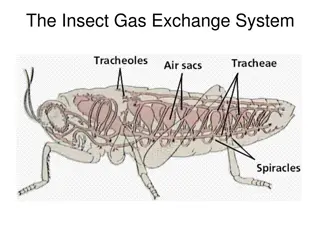
  14. Circulation systems have evolved over time Most invertebrates (esp. insects) have an open circulatory system Metabolic energy is used to pump hemolymph through blood vessels into the body cavity Hemolymph is returned to vessels via ostia pores that draw in the fluid as the heart relaxes Diagram of open circulatory system in a grasshopper 14

  15. Circulation systems have evolved over time Closed circulatory systems separate blood from interstitial fluid Metabolic energy is used to pump blood through blood vessels Blood is contained within the vessels Exchange occurs by diffusion in capillary beds Diagram of a closed circulatory system, plus a diagram showing an earthworm circulatory system 15

  16. Open vs. Closedboth systems are common Open systems . Use less metabolic energy to run Use less metabolic energy to build Can function as a hydrostatic skeleton Most invertebrates (except earthworms and larger mollusks) have open systems Closed systems . Maintain higher pressure Are more effective at transport Supply more oxygen to support larger and more active animals All vertebrates have closed systems 16

  17. All vertebrates have a closed circulatory system Chambered heart pumps blood Atria receive blood Ventricles pump blood Vessels contain the blood Veins carry blood to atria Arteries carry blood from ventricles Capillary beds facilitate exchange Capillary beds separate arteries from veins Highly branched and very tiny Infiltrate all tissues in the body We ll go over these step by step 17

  18. Chambered heart pumps blood Atria receive blood Ventricles pump blood Diagram of a chambered heart One-way valves direct blood flow 18

  19. Critical Thinking Atria receive blood; ventricles pump Given that function, what structure would you predict??? 19

  20. Critical Thinking Atria receive blood; ventricles pump Given that function, what structure would you predict??? 20

  21. Chambered heart pumps blood Atria receive blood Soft walled, flexible Ventricles pump blood Thick, muscular walls One-way valves direct blood flow Diagram of a chambered heart 21

  22. Vessels contain the blood Arteries carry blood from ventricles Always under pressure Veins carry blood to atria One-way valves prevent back flow Body movements increase circulation Pressure is always low Diagram showing artery, vein and capillary bed 22

  23. Note that blood vessel names reflect the direction of flow, NOT the amount of oxygen in the blood Arteries carry blood AWAY from the heart Arterial blood is always under pressure It is NOT always oxygenated Veins carry blood TO the heart Diagram of blood circulation pattern in humans 23

  24. Capillary beds facilitate exchange Capillary beds separate arteries from veins Highly branched and very tiny Infiltrate all tissues in the body More later Diagram showing artery, vein and capillary bed 24

  25. All vertebrates have a closed circulatory system REVIEW Chambered heart pumps blood Atria receive blood Ventricles pump blood Vessels contain the blood Veins carry blood to atria Arteries carry blood from ventricles Capillary beds facilitate exchange Capillary beds separate arteries from veins Highly branched and very tiny Infiltrate all tissues in the body 25

  26. Evolution of double circulation not all animals have a 4-chambered heart Diagram showing progression from a 1- chambered heart to a 4-chambered heart. This diagram is used in the next 12 slides. 26

  27. Fishes have a 2-chambered heart One atrium, one ventricle A single pump of the heart circulates blood through 2 capillary beds in a single circuit Blood pressure drops as blood enters the capillaries (increase in cross-sectional area of vessels) Blood flow to systemic capillaries and back to the heart is very slow Flow is increased by swimming movements 27

  28. Two circuits increases the efficiency of gas exchange = double circulation One circuit goes to exchange surface One circuit goes to body systems Both under high pressure increases flow rate 28

  29. Amphibians have a 3-chambered heart Two atria, one ventricle Ventricle pumps to 2 circuits One circuit goes to lungs and skin to release CO2 and acquire O2 The other circulates through body tissues Oxygen rich and oxygen poor blood mix in the ventricle A ridge helps to direct flow Second pump increases the speed of O2 delivery to the body 29

  30. Most reptiles also have a 3-chambered heart A partial septum further separates the blood flow and decreases mixing Crocodilians have a complete septum Point of interest: reptiles have two arteries that lead to the systemic circuits Arterial valves help direct blood flow away from pulmonary circuit when animal is submerged 30

  31. Critical Thinking What is a disadvantage of a 3 chambered heart??? 31

  32. Critical Thinking What is a disadvantage of a 3 chambered heart??? 32

  33. Mammals and birds have 4-chambered hearts Two atria and two ventricles Oxygen rich blood is completely separated from oxygen poor blood No mixing much more efficient gas transport Efficient gas transport is essential for both movement and support of endothermy Endotherms use 10-30x more energy to maintain body temperatures 33

  34. Mammals and birds have 4-chambered hearts Mammals and birds are NOT monophyletic What does this mean??? 34

  35. Mammals and birds have 4-chambered hearts Mammals and birds are NOT monophyletic Phylogenetic tree showing the diversification of vertebrates 35

  36. Mammals and birds have 4-chambered hearts Mammals and birds are NOT monophyletic Four-chambered hearts evolved independently What s this called??? 36

  37. Mammals and birds have 4-chambered hearts Mammals and birds are NOT monophyletic Four-chambered hearts evolved independently 37

  38. Review: evolution of double circulation 38

  39. Blood Circulation Blood vessels are organs Outer layer is elastic connective tissue Middle layer is smooth muscle and elastic fibers Inner layer is endothelial tissue Arteries have thicker walls Capillaries have only an endothelium and basement membrane 39

  40. Critical Thinking Arteries have thicker walls than veins Capillaries have only an endothelium and basement membrane What is the functional significance of this structural difference??? 40

  41. Critical Thinking Arteries have thicker walls than veins Capillaries have only an endothelium and basement membrane What is the functional significance of this structural difference??? 41

  42. Form reflects function Arteries are under more pressure than veins Capillaries are the exchange surface Diagram showing artery, vein and capillary bed 42

  43. Blood pressure and velocity drop as blood moves through capillaries Graph showing relationships between blood pressure, blood velocity, and the cross- sectional area of different kinds of blood vessels arteries to capillaries to veins. This same graph is on the next 3 slides. 43

  44. Total cross- sectional area in capillary beds is much higher than in arteries or veins; slows flow 44

  45. Velocity increases as blood passes into veins (smaller cross- sectional area); pressure remains dissipated 45

  46. One-way valves and body movements force blood back to right heart atrium 46

  47. Critical Thinking What makes rivers curl on the Coastal Plain??? 47

  48. Critical Thinking What makes rivers curl on the Coastal Plain??? 48

  49. Emphasize the difference between velocity and pressure!!! Velocity increases in the venous system; pressure does NOT 49

  50. Capillary Exchange Gas exchange and other transfers occur in the capillary beds Muscle contractions determine which beds are open Brain, heart, kidneys and liver are generally always fully open Digestive system capillaries open after a meal Skeletal muscle capillaries open during exercise etc 50

More Related Content

giItT1WQy@!-/#giItT1WQy@!-/#giItT1WQy@!-/#giItT1WQy@!-/#