Chronic liver disease
Chronic liver disease, including cirrhosis, can have varied clinical presentations and causes such as alcohol, viral hepatitis, and genetic factors. Clinical features include hepatomegaly, jaundice, and portal hypertension. Stigmata of CLD encompass muscle wasting, jaundice, petechiae, and more, making early diagnosis crucial for management and treatment.
Download Presentation

Please find below an Image/Link to download the presentation.
The content on the website is provided AS IS for your information and personal use only. It may not be sold, licensed, or shared on other websites without obtaining consent from the author.If you encounter any issues during the download, it is possible that the publisher has removed the file from their server.
You are allowed to download the files provided on this website for personal or commercial use, subject to the condition that they are used lawfully. All files are the property of their respective owners.
The content on the website is provided AS IS for your information and personal use only. It may not be sold, licensed, or shared on other websites without obtaining consent from the author.
E N D
Presentation Transcript
Chronic liver disease (CLD) Prepared by: Dr. Muntadher Abdulkareem Abdullah M.B.Ch.B,CABM,FIBMS,FIBMS(GE.&HEP.)
Liver cirrhosis: Cirrhosis: A consequence of CLD characterized by replacement of liver tissue by fibrosis and regenerative nodules, leads to irreversible loss of liver function and its complications It is the most common cause of portal hypertension and its complications Worldwide, the most common causes are chronic viral hepatitis, prolonged excessive alcohol consumption and NAFLD but any condition leading to persistent or recurrent hepatocyte death may lead to cirrhosis Cirrhosis is a histological diagnosis Cirrhosis can be classified histologically into: Micronodular cirrhosis, characterised by small nodules about 1 mm in diameter and typically seen in alcoholic cirrhosis. Macronodular cirrhosis, characterised by larger nodules of various sizes. Areas of previous collapse of the liver architecture are evidenced by large fibrous scars
Causes o o o o o o o o o o o Alcohol Chronic viral hepatitis (B or C) Non-alcoholic fatty liver disease Immune: Primary sclerosing cholangitis Autoimmune liver disease Biliary: Primary biliary cholangitis Secondary biliary cirrhosis Cystic fibrosis Genetic: Haemochromatosis Wilson s disease 1-antitrypsin defciency Cryptogenic (unknown 15%) Chronic venous outflow obstruction Any chronic liver disease
Clinical features : The clinical presentation is highly variable. Some patients are asymptomatic and the diagnosis is made incidentally at ultrasound or at surgery. Hepatomegaly (although liver may also be small) Jaundice Ascites Circulatory changes: spider telangiectasia, palmar erythema, cyanosis Endocrine changes: loss of libido, hair loss Men: gynecomastia, testicular atrophy, impotence Women: breast atrophy, irregular menses, amenorrhea Hemorrhagic tendency: bruises, purpura, epistaxis Portal hypertension: splenomegaly, collateral vessels, variceal bleeding Hepatic (portosystemic) encephalopathy Other features: pigmentation, digital clubbing, Dupuytren s contracture
Stigmata of CLD - Muscle wasting - Scratch marks - Pallor, jaundice - Parotid enlargement - Xanthelasma - Clubbing - Palmar erythema - Dupuytren s contracture - Spider nevi - Petechiae, purpura - Decreased body hair - Gynecomastia - Testicular atrophy - Caput medusa - Edema, ascites - Splenomegaly - Asterixis - Fetor hepaticus
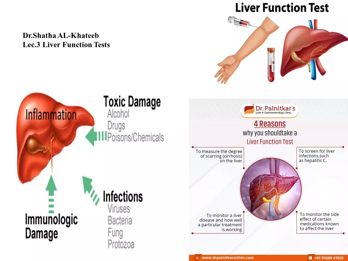
Investigations: Liver biopsy- gold standard, but not always necessary Fibroscan as non invasive tool for assessment of liver fibrosis - Deranged LFT- - elevated SGPT, alkaline phosphatase, GGT - Increased bilirubin - Low albumin, increased globulins - Increased PT/INR - Thrombocytopenia - Low sodium - Ultrasound- shrunken liver, portal HT/HCC - Endoscopy/UGIT- varices
Staging and prognosis of CLD Based on Child- Turcotte -Pugh scoring system includes- each given score of 1-3 - Ascites - Encephalopathy - Bilirubin - Albumin - PT/INR Class- total score Class A- 5-6 Class B- 7-9 Class C- 10-15
Manegments Aim of Management To retard progression and reduce complications - - Abstinence from alcohol Treat underlying cause accordingly Maintenance of nutrition and treatment of complications, including ascites, hepatic encephalopathy, portal hypertension and varices Vaccination : against HBV,HAV -
Complications 1) Ascites 2) Spontaneous bacterial peritonitis- SBP 3) Variceal bleed 4) Hepatic encephalopathy 5) Hepatorenal syndrome 6) Hepatocellular carcinoma- HCC 7) Hepatopulmonary syndrome and portopulmonary hypertension Ascites Diagnostic paracentesis- SAAG >=1.1, Mechanism of ascites: - Portal HT - Hypoalbuminemia - Raised renin-angiotensin-aldosterone level causing Na retention by kidneys Ascites- treatment - Salt fluid restriction - Diuretics- Spironolactone Furosemide - Large volume paracentesis-With massive or refractory ascites>5 litre fluid removed in one occasion - Albumin- is given 6-8 gm/litre fluid removed usually as 100 mL of 20% or 25% human albumin solution (HAS) for every 1.5 2 L of ascites drained) or another plasma expander to reduce the risk of hepatorenal syndrome - TIPS- transjugular intrahepatic portosystemic shunt.: For refractory ascites or refractory variceal bleed. Preferred for short duration, pending liver transplant, it increases risk of hepatic encephalopathy, due to shunt occlusion or infection
Spontaneous bacterial peritonitis: Symptoms and signs includes: abdominal pain, fever, worsened ascites and encephalopathy. Diagnosis Paracentesis showed polymorph neutrophil >=250/ microlitre. Ascitic fluid culture- bedside, commonly Gram ve bacteria, usually single microorganism Presence of multiorganism rise suspesion for secondary bacterial peritonitis(perforation) Treatment -i.v. antibiotics : like third generation cephalosporin Cefotaxime , ceftriaxone) or ciprofloxacin - Prophylaxis- Ciprofloxacin , levofloxacin or Co-trimoxazole - Prognosis- 30% mortality during hospital stay and 70% within 1 year
Variceal bleed - Varices- dilated submucosal veins, in esophagus or stomach - Cause- portal HT - Causes ~80% of UGI bleed in CLD - Risk factors for bleeding a) Size of varices b) Severity of liver disease c) Continued alcohol intake d) UGIE- red wale sign, hemostatic/ cherry red spots on avarix - Diagnosis Endoscopy Management - Acute : - Resuscitation - FFP, platelets, vitamin K - Terlipressin/octreotide - Lactulose -endoscopic Banding or sclerotherapy( banding preferred over sclerotherapy) - Balloon tamponade - TIPS - Surgery - Prevent rebreed - Band ligation- over repeated sessions - Non-selective - blockers- Propranolol or Nadolol - TIPS- for recurrent bleed or bleed from gastric varices - Surgery- portosystemic shunts - Liver transplantation
Hepatic encephalopathy - Constellation of neuropsychiatric disturbances range from confusion led to drowsiness led to stupor that result in coma - Ammonia is an identified measurable toxin Precipitants- - GI bleed - Constipation - Alkalosis, hypokalemia - Sedatives - Paracentesis hypovolemia - Infection - TIPS Diagnosis Based on clinical symptoms and signs of CLD with asterixis and altered sensorium. Management - Correct underlying precipitating factor - Avoid sedatives - Restrict dietary protein intake - Lactulose- 2-3 loose stools a day - Oral antibiotic- Metronidazole, Rifaximin, Neomycin - Correct hypoglycemia
Hepatorenal syndrome(HRS) - This occurs in 10% of patients with advanced cirrhosis complicated by ascites - Marked by renal impairment in the absence of any renal parenchymal disease or shock - Oliguria, hyponatremia and low urinary Na accompany raised creatinine - Two types of HRS : Type 1:This is characterised by progressive oliguria, a rapid rise of the serum creatinine and a very poor prognosis (without treatment, median survival is less than one month increase in serum creatinine, and has a better prognosis Type 2 : This usually occurs in patients with refractory ascites, is characterised by a moderate and stable - Albumin infusion, with vasoconstrictors (norepinephrine, terlipressin/ornipressin, octreotide) may help - Liver transplantation is treatment of choice.
Hepatocellular carcinoma - Associated with cirrhosis in ~80% - Suspect if- worsening of CLD, enlarged liver, hemorrhagic ascites, weight loss Diagnosis - CT/MRI with contrast- vascular space occupying lesion in cirrhotic liver : IS usually diagnostic in presence of characteristic vascular pattern - Raised AFP- -fetoprotein - Liver biopsy Treatment - Early-resection - Advanced- liver transplantation or local palliative treatment - Screening- US and AFP every 6 months N.B. Liver transplantation option in end stage liver disease , but limitation - Need donor - Cost - Technical expertise - GVHD - Recurrence
Hepatopulmonary syndrome : This condition is characterised by resistant hypoxemia (PaO2 < 9.3 KPa (70 mmHg)), intrapulmonary vascular dilatation in patients with cirrhosis, and portal hypertension. Clinical features: finger clubbing, cyanosis, spider naevi and a characteristic reduction in arterial oxygen saturation on standing. The hypoxia is due to intrapulmonary shunting through direct arteriovenous communications. Nitric oxide (NO) over- production may be important in pathogenesis Diagnosis is by echo study with intravenous microbubbles from agitated saline or 99mTc albumin aggregated Treatment :liver transplantation
Portopulmonary hypertension defined as pulmonary hypertension with increased pulmonary vascular resistance and a normal pulmonary artery wedge pressure in a patient with portal hypertension. Similar to idiopathic pulmonary arterial hypertension histologically The condition is caused by vasoconstriction and obliteration of the pulmonary arterial system and leads to breathlessness and fatigue Diagnosis : echo study followed by Rt. Heart catheterization Treatment : pulmonary vasodilator agents including : phosphodiesterase V inhibitor, endothelin receptor antagonist, prostacyclin analogue Definitive treatment is liver transplantation
