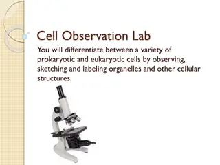
Cellular Structures: Microscopic Examination of Onion Cells
Learn how to prepare a microscope slide with onion cells for observation. Follow step-by-step instructions, including cutting and peeling onion sections, staining cells with iodine, and using a coverslip. Additionally, understand how to calculate magnification in microscopy to determine actual cell sizes.

Download Presentation

Please find below an Image/Link to download the presentation.
The content on the website is provided AS IS for your information and personal use only. It may not be sold, licensed, or shared on other websites without obtaining consent from the author.If you encounter any issues during the download, it is possible that the publisher has removed the file from their server.
You are allowed to download the files provided on this website for personal or commercial use, subject to the condition that they are used lawfully. All files are the property of their respective owners.
The content on the website is provided AS IS for your information and personal use only. It may not be sold, licensed, or shared on other websites without obtaining consent from the author.
E N D
Presentation Transcript
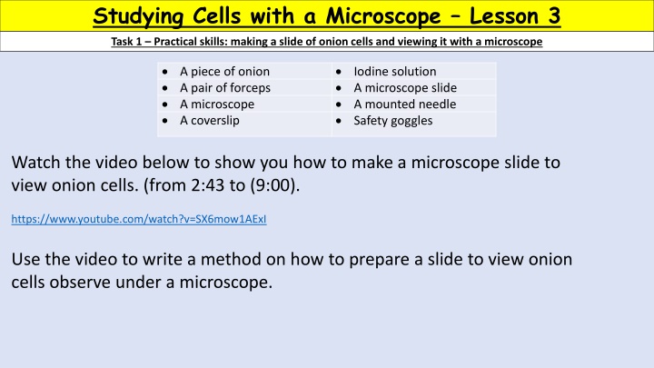
Studying Cells with a Microscope Lesson 3 Task 1 Practical skills: making a slide of onion cells and viewing it with a microscope A piece of onion A pair of forceps A microscope A coverslip Iodine solution A microscope slide A mounted needle Safety goggles Watch the video below to show you how to make a microscope slide to view onion cells. (from 2:43 to (9:00). https://www.youtube.com/watch?v=SX6mow1AExI Use the video to write a method on how to prepare a slide to view onion cells observe under a microscope.
Task 1 Practical skills: making a slide of onion cells and viewing it with a microscope Check your method and self assess! Cut a section of onion and then peel a thin layer of onion. Place the thin layer of onion onto a glass slide (be careful to only hold the slide by the edges to avoid fingerprints). Make the piece of onion skin as flat as you can on the slide to avoid cells overlapping. Add a few drops of iodine to stain your onion cells. This will make them easier to see. Carefully lower the coverslip over the onion at an angle. You can use a mounted needle to help with this. This reduces the chance of air bubbles forming.
Task 2 Numeracy - Magnification 1. Use the information given to work out the magnification. No calculators! Magnification = eye piece lens x objective lens Eye piece lens (x) Objective lens (x) Magnification (x) 10 40 100 25 55 10 20 30 7 12 100 800 3000 175 660
Task 2 Numeracy - Magnification Actual size = image size magnification or Actual size = image size magnification Image size = actual size x magnification Magnification = image size actual size Magnification = image size or Actual size Actual size = how big the cell is in real life (without being magnified) Image size = The size of the cell in a drawing or what you can now see down the microscope Magnification = How much bigger you ve made the cell look on your microscope
Task 2 Numeracy - Magnification If an onion cell was observed through a x100 magnification and it measured 17mm, what would be the actual size of the cell? Actual size = 17mm = 0.17 mm 100 2. Apply what you know to work out the actual size of the following. a. An animal cell measures 10mm under magnification x250. Actual size = image size magnification = 0.04 mm the size of the cell in real life (actual size) 10 250 a. A piece of muscle tissue measures 25mm under magnification x100. Actual size = image size magnification 25 100 = 0.25 mm A red blood cell measures 5mm under magnification x40. Actual size = image size magnification = 0.125 mm 40 5
Light microscope How to calculate total magnification? Eye piece lens 1. Eye piece magnification x objective lens magnification objective lens 2. What is the main limitation of a light microscope? Maximum magnification is quite low (x1000) compared to an electron microscope (x500 000 or more). What is the main limitation of an electron microscope? Can only be used to view dead tissues.
Preparing a slide to view cells Why do we use substances like iodine solution and methylene blue? They act as a stain to make the cells more visible. Why is a mounted needle used when preparing a glass slide? Prevents the formation of air bubbles.