Cell Membrane Structure and Function
 undefined


C
e
l
l
M
e
m
b
r
a
n
e
a
n
d
T
r
a
n
s
p
o
r
t
Physiology Team 436 – Foundation block lecture 2


Red: very important.
Green: only found in males’ slides.
Purple: only found in females’ slides.
Gray: notes.

Lecture: If work is intended for initial studying.
Review: If work is intended for revision.
1


2
Characteristics
:
1.
It covers the cell
2.
Thickness: 7.5-10 nm
3.
Selectively permeable
4.
It is fluid, not solid
5.
Composed of:
55%
proteins
42%
lipids
3%
other (Can be carbohydrates)
Phospholipid:
Head:
Phosphate
/
Glycerol
Tail: fatty acid
Interior of the
Phospholipid bi-layer
:
amphipathic
(Hydrophilic head+ hydrophobic tail)
Structure of the cell membrane (Plasma
membrane)
3
R
E
M
E
M
B
E
R
Cell membrane is composed of a lipid bilayer
Cell membrane = plasma membrane
4
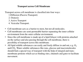
•
The membrane allows some substances to
cross it but not others.
A.
Through proteins: Water-soluble substances (Glucose,
ions)
B.
Directly through the bilayer: Fat-soluble substances (O2,
CO2, OH)
•
This controls the type & amount of
substances entering and leaving the cell.
•
It arises from the membrane structure.
What does Selective Permeability mean?
5
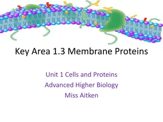
Integral
Span the thickness of the
membrane
Function:
1.
Channels (or pores)
2.
Carrier proteins
3.
Receptors
Peripheral
Only attach to the surface of
the membrane (or attached to
integral proteins)
Function:
Hormone
receptors
and
Enzymes
Membrane Proteins (Two categories)
6
Channel vs. Carrier Proteins
7
*Carbohydrates
CHO’s
* membrane carbohydrates are on
the surface bounded to lipids or proteins of the membrane
Function:
Function:
1.
Receptors
2.
Attach cells to each other
(Cell Adhesion)
3.
Immune reaction (They help
protect the cell by
differentiating host friendly
cells from enemy cells)
4.
Give most of cells overall
negative surface
Carbohydrates of the Cell Membrane
8
The Fluid Mosaic Model of Plasma
Membrane
9
Passive Transport:
Simple
diffusion
Active Transport:
Facilitated
diffusion
Osmosis
Secondary active
transport
Primary active
transport
Types of Membrane Movement:
10
Molecules move
against
their energy
gradient.
require energy.
The transport of material between body or cellular
compartments can be divided into:
Passive
Transport
Active
Transport
Molecules move
down or along
their energy
gradient.
Does not require
energy.
Transport Mechanisms:
11
O
s
m
o
s
i
s
:
Movement of water
from an area of
low
solute
concentration
(hypotonic)
to an
area of
high solute
concentration
(hypertonic)
Passive Transport (Osmosis)
12
Diffusion: Random movement
of substance either through the
membrane directly or in
combination with carrier
protein
down
concentration
gradient.
This gradient can be:
Concentration.
Electrochemical.
Pressure.
Passive Transport (Diffusion)
13
Simple diffusion
The movement of
molecules through
the intermolecular
spaces
or
membrane openings
(channels)
without
the necessity of
binding
to a
carrier protein
on
the membrane.
Facilitated diffusion
The transported molecule
binds to a
carrier
protein
which then
undergoes a
conformational change
allowing the molecule to
pass through to the other
side of the cell membrane.
The carrier
facilitates
passage of the molecule
through the CM.
Passive Transport ( Types of Diffusion
)
14
15
Amazing video summarizing cellular transport: https://m.youtube.com/watch?v=UgN76naeA1Q
Cross
freely
by
diffusion
Cross through
membrane proteins
Substances that can cross the Cell
Membrane
16
Cross
freely
by
diffusion
Cross through membrane proteins
Substances that can cross the Cell
Membrane
17
Achieved through a trans-membrane protein:
carrier/transporter/channel
Substances that can cross the Cell
Membrane
18
1
-
S
i
m
p
l
e
D
i
f
f
u
s
i
o
n
:
2- through the
channel
protein
1-
directly
through
the lipid bilayer
Pass through the
interstices of the
lipid bilayer
EX :
small lipid-
soluble substances
(uncharged
substances, O2, CO2,
alcohol, steroid and
general anesthetic).
Its require transport
protein (channel protein).
EX:
1- Large and
lipid-insoluble
substances
(charged
molecule).
2-
Water-soluble
substances (water, ions)
pass through channels
that penetrate through
the cell membrane.
Passive Transport (Simple Diffusion):
19
Passive Transport (Simple Diffusion):
20
Passive Transport (Simple Diffusion)
•
Non-carrier:
mediated transport
down an electrochemical gradient.
•
Diffusion of non-electrolytes:
(uncharged) from high
concentration to low concentration.
• Diffusion of electrolytes:
(charged) depends on both chemical
as will as electrical potential
difference.
21
Rate of simple diffusion
2-Number and sizes of opening in
the membrane for the substance
“pores”(selective gating system)
1-Amount of substance
available.
3-Chemical concentration
difference.
Net diffusion= P x A (Co-Ci)
4-Electrical potential difference.
EPD=± 61 log C1/C2
5-Molecular size of the
substance.
6-Lipid solubility
7-Temperature
Passive Transport (Simple Diffusion)
22
2-
Facilitated diffusion: also
called
(Carrier mediated
diffusion)
Diffusion of a substance is
“facilitated” by the use of a
specific carrier protein.
Diffusion continues until
equilibrium is reached or
terminated
.
Examples: Glucose, amino acids.
Passive Transport (Facilitated Diffusion)
23
Features Of
Carrier
Mediated
Transport:
(Facilitated
diffusion)
Saturation :
Stereospecificity :
Competition :
concentration binding
of protein.
If all proteins are occupied
we achieve
full saturation.
The binding site
recognizes a specific
substance
D-glucose but
not L-glucose .
Chemically similar
substances
can compete
for the same binding site
D- galactose / D-glucose.
Passive Transport (Facilitated Diffusion)
24
Passive Transport (Facilitated Diffusion)
Substance binding site substance protein
complex conformational changes release of
substance.
25
Simple diffusion
The rate of diffusion
increases
proportionately with the
concentration of the
diffusing substance.
Facilitated diffusion
The rate of diffusion
increases proportionately
with the concentration of
the diffusing substance
until
it reaches a maximum
Vmax.
At Vmax, an increase in the
concentration of the
diffusing substance
does
not increase the rate.
Passive Transport “rate of diffusion” (Simple Vs.
Facilitated)
26
The rate at which molecules can be transported by
facilitated diffusion
depends on the rate at
which the carrier protein molecule can
undergo conformational change
back and forth
between its bound and unbound state.
*كل ما زاد التركيز بيزيد
معدل دخول الجزيئات،
بمجرد يوصل للمرحلة
القصوى بيثبت المعدل، لأن
هذا النوع يعتمد على
الكارير فما راح يقدر يدخل
أكثر من طاقتة مهما كان
التركيز برا عالي.
27
Factors Affecting Net Rate of Diffusion:
Size.
Temperature.
Steepness of the gradient:
1-Concentration difference.
2-Membrane electrical difference.
3-Pressure difference.
Charge.
Pressure.
28
Active transport
Occurs
when
a
cell
membrane
moves
molecules
or
ions
“up-hill”
against
a
concentration
gradient
(or
“up-hill”
against
an
electrical
or
pressure
gradient).
•
Examples
include
:
➢
Ions
like:
sodium, potassium, calcium,
iron,
iodine,
hydrogen
ions.
➢
Amino
acids, glucose
and
other
sugars.
Requires
energy
and
a
carrier
protein
29
According to the source of energy used to
facilitate transport, it can be divided into;
30
•
The
energy
is
derived
directly
from
breakdown
of
(ATP)
to ( ADP)
this breakdown will release energy
Examples
include
:
Sodium-Potassium ATPase
pump
Calcium
ATPase
pump.
Hydrogen
ATPase
pump.
Primary Active
31
Pump Characteristics:
1- Carrier protein is made of alpha and beta subunits.
2- Na binding site is inside, K binding site is outside.
3- It has ATPase activity
In the first body fluid lecture we decided that the intercellular fluid
has more K and less Na, also extracellular fluid has more Na and less K.
If the cell have more Na inside and more K outside that the cell will burst,
therefore,
t
his
pump
functions
by
moving
3
molecules
of
sodium
OUT
and
2
molecules
of
potassium
INTO
the
cell
both
against
their
concentration
gradients to maintain the
body fluid balance.
Functions:
• Maintaining Na+ and
K+ concentration
difference.
• Establishes –ve
potential inside the
cell.
• Maintains a normal cell
volume.
• It is the basis of nerve
signal transmission.
32
More examples:
1- Ca+2
ATPase
Pump
•
Present
in:
A) Sarcoplasmic
reticulum
in
muscle
cells
B) Mitochondria
C) Some
cell
membranes
.
2- H+ ATPase (OR H+-K) Pump
Present in:
A) Parietal stomach cells
B) Intercalated cells of distal renal tubule
Function:
Maintains low Ca+2
concentrations in the cell
Function:
A) Secretes HCL in stomach
B) Excretes acids from the
body
Generally: Pumps H out of
the cell into lumen
H+-K ATPase inhibitors
treat ulcer disease
(omeprazol)
33
The
energy
is
derived
indirectly
by
using
the
concentration
or
electrochemical
gradient
generated
by
a
primary
active
transporter.
More Explanation (Co Transport) :
In primary NA-K pump, the concentration of sodium is more outside the cell, therefore the sodium will move
into the cell with it’s gradient, and goes back outside to maintain body fluid balance. When Na moves inside, the
cell will use energy from the concentration gradient using a carrier, but
the carrier has place for another
molecule (glucose, against its gradient) to pass with Na, sodium can not move alone.
Secondary Active Transport
34
Co-Transport
•
When
both
substances
are
transported
together
in
the
same
direction.
Examples: 1- Na+-Glucose
2- Na +-amino acid
3-
In the Kidney
Counter-Transport
When
one
substance
is
transported
in
the
opposite
direction
to
the
other
substance.
Examples: 1- Na+-H+
(Kidney)
2- Na+-Ca+2
(Many cell membranes)
35
Thank you!
Lina Alwakeel
Rana Barassain
Heba Alnasser
Munira Aldofayan
Sara Alshamrani
Sundus Alhawamda
Ruba Ali
Rehab Alanazi
Norah Alshabib
Nouf Alaqeeli
Buthaina Almajed
Alaa Alaqeel
Contact us:
Physiology436@gmail.com
@Physiology436
The Physiology 436 Team:
Team Leaders:
Qaiss Almuhaideb
Lulwah Alshiha
Fahad Al Fayez
Ibrahim Al Deeri
Hassan Al Shammari
Abdullah Al Otaibi
Abdullah Al Subhi
Ali Al Subaei
Omar Al Babteen
Foad Fathi
Faisal Al Fawaz
Muhammad Al Aayed
Muhammad Al Mutlaq
Nasser Abu Dujeen
Waleed Al Asqah
اعمل لترسم بسمة، اعمل لتمسح دمعة، اعمل و أنت تعلم أن الله لا يضيع أجر من أحسن عملا.
36
This educational content delves into the structure and function of the cell membrane, covering topics such as the fluid mosaic model, permeability, carrier-mediated processes, and membrane proteins. It explains the composition of the cell membrane, selective permeability, and the roles of integral and peripheral membrane proteins. Through detailed images and explanations, readers can grasp the fundamental aspects of cell membrane biology presented in a clear and concise manner.

Download Presentation

Please find below an Image/Link to download the presentation.
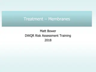
The content on the website is provided AS IS for your information and personal use only. It may not be sold, licensed, or shared on other websites without obtaining consent from the author.If you encounter any issues during the download, it is possible that the publisher has removed the file from their server.
You are allowed to download the files provided on this website for personal or commercial use, subject to the condition that they are used lawfully. All files are the property of their respective owners.
The content on the website is provided AS IS for your information and personal use only. It may not be sold, licensed, or shared on other websites without obtaining consent from the author.
E N D
Presentation Transcript
Cell Membrane and Cell Membrane and Transport Transport Red: very important. Green: only found in males slides. Purple: only found in females slides. Gray: notes. Physiology Team 436 Foundation block lecture 2 Lecture: If work is intended for initial studying. Review: If work is intended for revision. 1
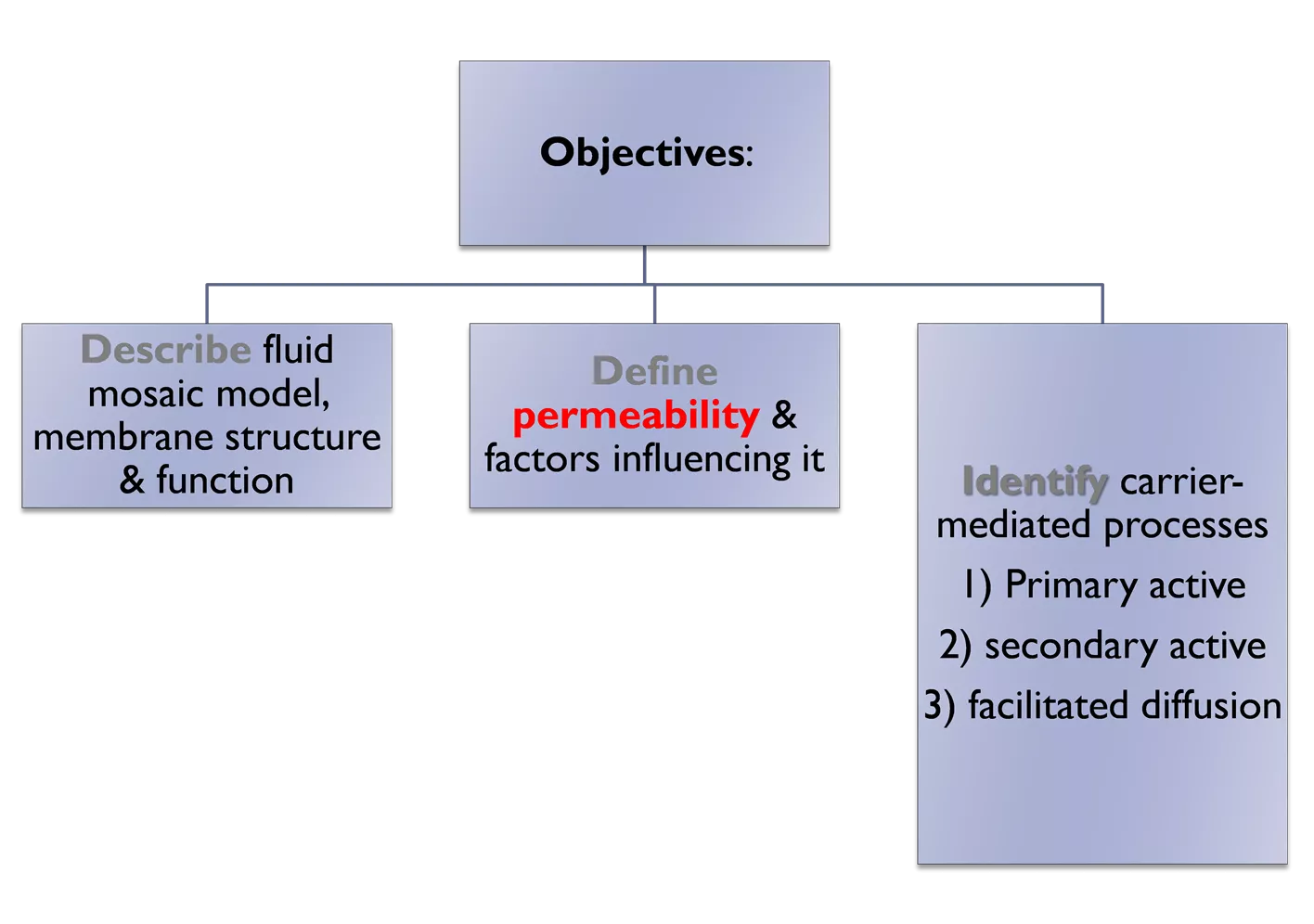
Objectives: Describe fluid mosaic model, membrane structure & function Define permeability & factors influencing it Identify carrier- mediated processes 1) Primary active 2) secondary active 3) facilitated diffusion 2
Structure of the cell membrane (Plasma membrane) Characteristics: It covers the cell Thickness: 7.5-10 nm Selectively permeable It is fluid, not solid Composed of: 55% proteins 42% lipids 3% other (Can be carbohydrates) 1. 1-Phospholipids (most abundant) 25% 2-Cholestrol 13% 3-Other 4% (Can be Glycolipid) 2. 3. 4. 5. Interior of the Phospholipid bi-layer: amphipathic (Hydrophilic head+ hydrophobic tail) Phospholipid: Head: Phosphate / Glycerol Tail: fatty acid 3
REMEMBER Cell membrane is composed of a lipid bilayer Cell membrane = plasma membrane 4
What does Selective Permeability mean? The membrane allows some substances to cross it but not others. A. Through proteins: Water-soluble substances (Glucose, ions) Directly through the bilayer: Fat-soluble substances (O2, CO2, OH) This controls the type & amount of substances entering and leaving the cell. It arises from the membrane structure. B. 5
Membrane Proteins (Two categories) Integral Peripheral Span the thickness of the membrane Function: 1. Channels (or pores) 2. Carrier proteins 3. Receptors Only attach to the surface of the membrane (or attached to integral proteins) Function: Hormone receptors and Enzymes 6
Channel vs. Carrier Proteins Form open pores that allow molecules of the appropriate size (e.g. Ions) to pass the membrane. Similar to a normal door. Channel proteins Selectively bind the small molecule to be transported and then undergo a conformational change to release the molecule on the other side of the membrane. Similar to electronic door. Carrier proteins 7
Carbohydrates of the Cell Membrane *Carbohydrates CHO s* membrane carbohydrates are on the surface bounded to lipids or proteins of the membrane If attached to Proteins Glycoproteins (mostly) Lipids Glycolipids If bound together by proteins Proteoglycans Function: 1. Receptors 2. Attach cells to each other (Cell Adhesion) 3. Immune reaction (They help protect the cell by differentiating host friendly cells from enemy cells) 4. Give most of cells overall negative surface 8 Glycocalyx: a loose coat of carbohydrate molecules outside the cell. Functions as a first layer of defense
The Fluid Mosaic Model of Plasma Membrane 9
Types of Membrane Movement: Passive Transport: Active Transport: Simple diffusion Primary active transport Facilitated diffusion Secondary active transport Osmosis 10
Transport Mechanisms: The transport of material between body or cellular compartments can be divided into: Passive Transport Active Transport Molecules move down or along their energy gradient. Does not require energy. Molecules move against their energy gradient. require energy. 11
Passive Transport (Osmosis) Osmosis : Movement of water from an area of low solute concentration (hypotonic) to an area of high solute concentration (hypertonic) 12
Passive Transport (Diffusion) Diffusion: Random movement of substance either through the membrane directly or in combination with carrier protein down concentration gradient. This gradient can be: Concentration. Electrochemical. Pressure. 13
Passive Transport ( Types of Diffusion) Simple diffusion The movement of molecules through the intermolecular spaces or membrane openings (channels) without the necessity of binding to a carrier protein on the membrane. Facilitated diffusion The transported molecule binds to a carrier protein which then undergoes a conformational change allowing the molecule to pass through to the other side of the cell membrane. The carrier facilitates passage of the molecule through the CM. 14
15 Amazing video summarizing cellular transport: https://m.youtube.com/watch?v=UgN76naeA1Q
Substances that can cross the Cell Membrane Cross freelyby diffusion Cross through membrane proteins 16
Substances that can cross the Cell Membrane Cross through membrane proteins Cross freely by diffusion 17
Substances that can cross the Cell Membrane Achieved through a trans-membrane protein: carrier/transporter/channel 18
Passive Transport (Simple Diffusion): 1 1- -Simple Simple Diffusion: Diffusion: 2- through the channel protein Its require transport protein (channel protein). EX: 1- Large and lipid-insoluble substances (charged molecule). 2-Water-soluble substances (water, ions) pass through channels that penetrate through the cell membrane. 1- directly through the lipid bilayer Pass through the interstices of the lipid bilayer EX : small lipid- soluble substances (uncharged substances, O2, CO2, alcohol, steroid and general anesthetic). 19
Passive Transport (Simple Diffusion) Non-carrier: mediated transport down an electrochemical gradient. Diffusion of non-electrolytes: (uncharged) from high concentration to low concentration. Diffusion of electrolytes: (charged) depends on both chemical as will as electrical potential difference. 21
Passive Transport (Simple Diffusion) 2-Number and sizes of opening in the membrane for the substance pores (selective gating system) 1-Amount of substance available. Rate of simple diffusion 3-Chemical concentration difference. Net diffusion= P x A (Co-Ci) 4-Electrical potential difference. EPD= 61 log C1/C2 5-Molecular size of the substance. 6-Lipid solubility 7-Temperature 22
Passive Transport (Facilitated Diffusion) 2- Facilitated diffusion: also called (Carrier mediated diffusion) Diffusion of a substance is facilitated by the use of a specific carrier protein. Diffusion continues until equilibrium is reached or terminated. Examples: Glucose, amino acids. 23
Passive Transport (Facilitated Diffusion) concentration binding of protein. If all proteins are occupied we achieve full saturation. Saturation : Features Of Carrier Mediated Transport: (Facilitated diffusion) The binding site recognizes a specific substance D-glucose but not L-glucose . Stereospecificity : Chemically similar substances can compete for the same binding site D- galactose / D-glucose. Competition : 24
Passive Transport (Facilitated Diffusion) Substance binding site substance protein complex conformational changes release of substance. 25
Passive Transport rate of diffusion (Simple Vs. Facilitated) Simple diffusion Facilitated diffusion The rate of diffusion increases proportionately with the concentration of the diffusing substance until it reaches a maximum Vmax. At Vmax, an increase in the concentration of the diffusing substance does not increase the rate. The rate of diffusion increases proportionately with the concentration of the diffusing substance. 26
* . The rate at which molecules can be transported by facilitated diffusion depends on the rate at which the carrier protein molecule can undergo conformational change back and forth between its bound and unbound state. 27
Factors Affecting Net Rate of Diffusion: Size. Temperature. Steepness of the gradient: 1-Concentration difference. 2-Membrane electrical difference. 3-Pressure difference. Charge. Pressure. 28
Active transport Occurswhenacellmembranemoves moleculesorions up-hill againsta concentrationgradient (or up-hill againstan electricalorpressuregradient). Examples include: Ions like: sodium, potassium, calcium, iron, iodine, hydrogen ions. Amino acids, glucose and other sugars. Requiresenergyandacarrierprotein 29
According to the source of energy used to facilitate transport, it can be divided into; 30
Primary Active Theenergyisderived directlyfrombreakdown of(ATP) to ( ADP) this breakdown will release energy Examplesinclude: Sodium-Potassium ATPasepump CalciumATPasepump. HydrogenATPasepump. 31
Functions: Maintaining Na+ and K+ concentration difference. Establishes ve potential inside the cell. Maintains a normal cell volume. It is the basis of nerve signal transmission. Pump Characteristics: 1- Carrier protein is made of alpha and beta subunits. 2- Na binding site is inside, K binding site is outside. 3- It has ATPase activity In the first body fluid lecture we decided that the intercellular fluid has more K and less Na, also extracellular fluid has more Na and less K. If the cell have more Na inside and more K outside that the cell will burst, therefore, this pump functions by moving 3 molecules of sodium OUT and 2 molecules of potassium INTO the cell both against their concentration gradients to maintain the body fluid balance. 32
More examples: Function: Maintains low Ca+2 concentrations in the cell 1- Ca+2ATPasePump Presentin: A) Sarcoplasmicreticuluminmusclecells B) Mitochondria C) Somecellmembranes. Function: A) Secretes HCL in stomach B) Excretes acids from the body 2- H+ ATPase (OR H+-K) Pump Present in: A) Parietal stomach cells B) Intercalated cells of distal renal tubule Generally: Pumps H out of the cell into lumen H+-K ATPase inhibitors treat ulcer disease (omeprazol) 33
Secondary Active Transport Theenergyisderived Type of secondary active transport indirectly byusingthe concentrationor electrochemicalgradient generatedbyaprimary 1-Counter- Transport 2-Co-Transport activetransporter. More Explanation (Co Transport) : In primary NA-K pump, the concentration of sodium is more outside the cell, therefore the sodium will move into the cell with it s gradient, and goes back outside to maintain body fluid balance. When Na moves inside, the cell will use energy from the concentration gradient using a carrier, but the carrier has place for another molecule (glucose, against its gradient) to pass with Na, sodium can not move alone. 34
Co-Transport Whenbothsubstances aretransported togetherinthesame direction. Examples: 1- Na+-Glucose 2- Na +-amino acid 3- In the Kidney Counter-Transport Whenonesubstanceis transportedinthe oppositedirectiontothe othersubstance. Examples: 1- Na+-H+ (Kidney) 2- Na+-Ca+2 (Many cell membranes) 35
Thank you! . The Physiology 436 Team: Lina Alwakeel Rana Barassain Heba Alnasser Munira Aldofayan Sara Alshamrani Sundus Alhawamda Ruba Ali Rehab Alanazi Norah Alshabib Nouf Alaqeeli Buthaina Almajed Alaa Alaqeel Fahad Al Fayez Ibrahim Al Deeri Hassan Al Shammari Abdullah Al Otaibi Abdullah Al Subhi Ali Al Subaei Omar Al Babteen Foad Fathi Faisal Al Fawaz Muhammad Al Aayed Muhammad Al Mutlaq Nasser Abu Dujeen Waleed Al Asqah Team Leaders: Qaiss Almuhaideb Lulwah Alshiha Contact us: Physiology436@gmail.com @Physiology436 36


")


")
")
")


:")
:")
")
")
")
")
")