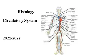
Capillary Circulation in the Vascular System
This content delves into the intricate components of capillary circulation, covering topics such as microcirculation, types of capillaries, regulation of flow in capillary beds, and the formation of interstitial fluid. It also discusses edema, lymphatics, diffusion, and filtration processes. The visuals aid in grasping the concepts, including the structure of capillaries, capillary wall composition, and variability of capillary filtration in different organs.

Download Presentation

Please find below an Image/Link to download the presentation.
The content on the website is provided AS IS for your information and personal use only. It may not be sold, licensed, or shared on other websites without obtaining consent from the author.If you encounter any issues during the download, it is possible that the publisher has removed the file from their server.
You are allowed to download the files provided on this website for personal or commercial use, subject to the condition that they are used lawfully. All files are the property of their respective owners.
The content on the website is provided AS IS for your information and personal use only. It may not be sold, licensed, or shared on other websites without obtaining consent from the author.
E N D
Presentation Transcript
Capillary Circulation ____________ Color Index: Main Text Important Girl s Slides Boy s Slides Dr s Notes Extra Info
Objectives Objectives To describe components of microcirculation To recognize different types of blood capillaries. To understand regulation of flow in capillary beds. To understand formation of the interstitial fluid. Define edema, state its causes, and understand its mechanism of formation. To understand the role of lymphatics. Diffusion and filtration.
The Vascular System & Components of Microcirculation The Vascular System & Components of Microcirculation Aorta : Elastic recoil. Arteries: muscular, low resistance vessels Arterioles : High resistance vessels Capillaries: Exchange vessels Venules :weaker muscular coat Veins : capacitance vessels
Distribution of Blood Within Aorta : Elastic recoil. The Circulatory Distribution of Blood Within Aorta : Elastic recoil. The Circulatory System System At Rest At Rest
Capillaries (Microcirculatory Capillaries (Microcirculatory Vessels) Vessels) Capillaries Cross Capillaries Cross- -Sectional Area Area Sectional Capillaries are the smallest blood vessels in the vascular system. There are large in number (over 10 billion) in the body. It consists ONLY ONLYof the Tunica Internawith a SINGLE SINGLELayer of endothelial cells Provide direct access to the cells. Most permeable. Site of exchange with tissue: Permits exchange of nutrients & waste products. At rest, 4-5% of circulating blood is present in capillaries. Capillary is of 0.5-1mm in length. Capillary is very small in diameter (4-9 microns). As the diameter of blood vessel decreases, the total cross-sectional area increases & the velocity of blood flow decreases. The total capillary surface area ranges from 700-1000 m2 of surface area (>3 tennis courts). The velocity of blood flow within each segment of the circulatory system is inversely proportional to the total cross-sectional area of the segment. Because the aorta has the smallest total cross-sectional area of all circulatory segments, it has the highest velocity of blood flow. V=Q/A V=velocity, Q=Flow, A=cross sectional area
Capillary Wall Capillary Wall Girls Slides Capillary wall is composed of a single layer of simple squamous epithelia. Capillary wall is of 0.5 micrometers in thickness. It regulates transfer of fluid from blood to the interstitial fluid space & vice versa. Organ Variability of Capillary Filtration Organ Variability of Capillary Filtration Girls Slides small Brain & Muscle Why is there a difference? Why is there a difference? Because of their permeability. moderate subcutaneous How does the permeability change? How does the permeability change? By alteration of clefts size/Pores between cells. large Intestines Liver & Kidneys extremely large
Types of Capillaries : Types of Capillaries : Capillaries are classified into (3) types according to their wall permeability and/or diameter. Wall permeability is affected by the size & diameter of the pores. Continuous Fenestrated Sinusoidal Do not have fenestrae. Allow only very small molecules to pass Found in brain, muscles, lung & adipose tissue. Have widerpores. Allow large substancesto pass but not plasma proteins. not plasma proteins.Found in kidney glomeruli, small intestine & endocrine glands. Large diameter with large fenestrae (wider gaps between the cells). Endothelium is discontinuous. Found in liver, spleen, bone marrow lymphoid tissue, & some endocrine glands.
Types of Capillaries .. CONT Types of Capillaries .. CONT
Girls Slides How does the permeability change? How does the permeability change? Factors that mediate pore size change: Factors that mediate pore size change: Histamine Histamine If little = will increase the flow (distends vessel). If a lot = will decrease the arterial blood pressure (anaphylaxis). Cytokines Cytokines Alters flow/pressure & permeability. Drugs Drugs Any drug influencing contraction or dilation of smooth muscle & endothelial permeability. Nerves Nerves Sympathetic Inflammation Inflammation Stretches the cells apart & increases the cleft size. If too much, can compress the vessels as well.
Girls Slides Functions of Capillaries Functions of Capillaries Play a role in temperature regulation Exchange vessels between blood & tissues Capillary tone Play a metabolic role Produce PGI2; Growth factors for blood cells: fibroblast GF, platelet GF; & in the lungs, angiotensin converting enzyme. Inactivation of intercellular messengers. Antithrombotic function. Blood vessel dilatation (vasodilatation), Increase heat loss across epidermis. Provide direct access to the cells. Most permeable. Transport nutrients & oxygen from blood to the tissues. Remove CO2 & cellular waste products from the tissues to the blood. Blood vessel constriction (vasoconstriction), heat conservation across epidermis. -
Capillary Beds (Network) / Capillary Beds (Network) / Component of Microcirculation Component of Microcirculation Capillaries are Capillaries are arranged arrangedin capillary beds Arterioles Arteriolesdivides into a number of metarterioles have a continuous smooth muscle coat. Blood flow through the metarteriole to enter capillary bed via precapillary sphincters. precapillary sphincters. Venules drain capillary network. Arteriolar smooth muscle, meta-arterioles, & precapillary sphincters regulate the blood flow in the capillary network. in capillary beds metarterioles, which donot Capillary beds consist of two types of vessels 1 1- -Vascular shunt Vascular shunt Directly connects an Arterioleto a Venule. 2 2- -True capillaries True capillaries Exchange vessels -O2 & nutrients cross to cells -CO2 & metabolic waste products cross into blood
Girls Slides Blood Flow Through Capillary Blood Flow Through Capillary Beds (Network) Beds (Network) Mechanisms of Trans Mechanisms of Trans- - Capillary Exchange Capillary Exchange Blood flows from arterioles through metarterioles, then to capillary network Simple diffusion Simple diffusion For lipids soluble gases (O2 and CO2) according to concentration gradient (passive diffusion) by cell membrane bilayer Filtration (bulk flow) Filtration (bulk flow) for fluid transfer by starlings force according to pressure gradient (hydrostatic pressure and osmotic pressure) Venules drain network. Vesicular transport Vesicular transport Transcytosis Arteriolar smooth muscle, metarterioles, and precapillary sphincters regulate regulatethe blood flow in capillary network. Mediated (membrane transport) Mediated (membrane transport) occurs only in capillaries of the brain secondary active transport secondary active transport,e.g.transport of glucose moves by co-transports in cell membrane brainand involves involves
Trans Trans- -Capillary Fluid Capillary Fluid Transfer Transfer Girls Slides Formation of Interstitial Formation of Interstitial Fluid (IF) Fluid (IF) The Interstitial Fluid (IF) formation is regulated by the net filtration pressure force at the two vascular ends (arteriolar & venular) of the capillary bed. Net filtration pressure Net filtration pressureis affected by affected bythe net balance the two forces (hydrostatic & oncotic pressures) found in the capillaries & in the interstitial fluid. Trans-capillary transfer Occurs by Diffusion or by vesicular transport. Diffusion occurs through, Diffusion occurs through, cells cells: for lipid soluble molecules. intercellular clefts between cells intercellular clefts between cells:for non-lipid soluble molecules. Filtration (Bulk Flow): Filtration (Bulk Flow): Fluid movement is affected & influenced by the concentration gradients, cleft size (permeability) & hydrostatic pressure influence the fluid movement (varies by organ & situation) net balance between Movement of fluid from Capillaries is affected by the: Movement of fluid from Capillaries is affected by the: Blood pressure Osmosis Capillary permeability Excessive fluid Excessive fluid gained by tissues is removed by the Lymphatic system. system. Lymphatic
Hydrostatic & Oncotic Pressures Hydrostatic & Oncotic Pressures Interstitial hydrostatic pressure (P IF) (Outward Force) : Capillary Hydrostatic Pressure (Outward Force): Interstitial hydrostatic pressure (PIF) is almost 0 mmHg. The interstitial fluid pressurewhich is caused by pumping of lymphatic system is negative 3 mmHgin most tissues. Encapsulated organs have positive interstitial pressures (+5 to +10 mmHg). PIF varies from one organ to another: Subcutaneous tissues: -2 mmHg. Liver, Kidney: +1mmHg. Brain: As high as +6mmHg. Normal Capillary hydrostatic pressure ranges from 30-35 mmHgat the arterial end, & from 10-15 mmHg at the venous end. Interstitial Oncotic (osmotic) Pressure (Inward Force): Capillary Oncotic (osmotic) Pressure (Inward Force): Colloid osmotic pressure is caused by presence of large proteins. There is almostno colloid osmotic pressurein the interstitial space. Colloid osmotic pressure is caused by presence of large proteins. High content of proteins in plasmaaccounts for + 10 mmHg its higher osmotic pressure compared to that of the Interstitial fluid (IF). Plasma osmotic pressure: Is high (25-28 mmHg) & will attract fluid & dissolved substances from tissue spaces into the circulation.
How Does Fluid Filtrate at The Capillary Bed? How Does Fluid Filtrate at The Capillary Bed? Girls Slides Fluid transfer depends on the relative balance & equilibrium between the hydrostatic & osmotic pressures between the hydrostatic & osmotic pressuresfound in capillary plasma & interstitial space balance & equilibrium Any loss of plasma colloids will result in an increase loss of fluid from the vasculature to the interstitial space.
Girls Slides How Do We Calculate Net How Do We Calculate Net Movement at Capillary Beds? Movement at Capillary Beds? Starling s Equation for Starling s Equation for Capillary Filtration Capillary Filtration Flux = Kf [(Pc Pif) ( p if)] Filtration rate of fluid at Capillary beds = Net Filtration Pressure (NFP) multiplied by Filtration Coefficient (Kf). Kf Kf= Capillary filtration coefficient (surface area & permeability) Pc Pc = Capillary hydrostatic pressure Pif Pif= Interstitial hydrostatic pressure = Capillary reflection coefficient (0 to 1) 1 = impermeable to proteins c c = Capillary colloid osmotic pressure if if= Interstitial colloid osmotic pressure Filtration Rate = Kf X NFP Filtration Coefficient (Kf) is a product of surface area times the hydraulic conductivity of membrane (membrane permeability). If NFP = positive, then fluid is lost from capillary. If NFP = negative, then fluid is gained by capillary. What Does this All Mean? What Does this All Mean? Fluid transfer depends on the Fluid transfer depends on the relative balance between relative balance between hydrostatic pressure & osmotic hydrostatic pressure & osmotic pressure. pressure.
Girls Slides Diffusion at Capillary Beds (Fluid Balance Diffusion at Capillary Beds (Fluid Balance Starling's Forces) Starling's Forces) Outward force out of the capillaries Inward force into the capillaries Capillary blood pressure Interstitial fluid pressure Interstitial hydrostatic pressure Interstitial fluid colloidal osmotic press Plasma colloidal osmotic pressure Total Pc= 30/ 35 to 15 mmHg PIF= 0 mmHg 38 -18 mmHg IF= 3mmHg c=25or 28mmHg PIF=0mmHg i is determined by interstitial fluid protein concentration. Normally, because there is little loss of protein from capillaries, there is little protein in interstitial fluid, making IF quite low. it s determined by protein concentration of capillary blood. Thus, in protein concentration causes in c Pc is determined by both arterial & venous pressure - - -
Girls Slides Starling s Forces & Filtration at Capillary Beds Starling s Forces & Filtration at Capillary Beds Fluid Balance: Net Filtration & Absorption of Fluid Across The Capillary Membrane Fluid Balance: Net Filtration & Absorption of Fluid Across The Capillary Membrane At arterial end of capillary At venous end of capillary Outward forces Value Outward forces Value Capillary blood pressure Pc= 30-35 mmHg Capillary blood pressure Pc= 10-15mmHg Negative interstitial free fluid pressure PIF= -3mmHg Negative interstitial free fluid pressure PIF= -3mmHg Total outward force 38mmHg Total outward force 18 mmHg Inwards forces Value Inwards forces Value Plasma colloidal osmotic pressure C= 25-28 mmHg Plasma colloidal osmotic pressure C= 25-28 mmHg Total inward force 25 mmHg Total inward force 25 mmHg Net outward force 38-25=+13mmHg Net outward force 18-25=-7mmHg
Girls Slides Diffusion at Capillary Beds Net Filtration & Absorption Diffusion at Capillary Beds Net Filtration & Absorption of Fluid Across The Capillary Membrane of Fluid Across The Capillary Membrane Net Filtration & absorption of fluid across the capillary membrane Net Filtration & absorption of fluid across the capillary membrane
Normal Forces a Arterial & Venous Ends of Capillaries Normal Forces a Arterial & Venous Ends of Capillaries Tissue hydrostatic pressure = 0 mmHg Negative interstitial free fluid pressure = 3 mmHg Interstitial Fluid Atrial blood Atrial blood Venous Blood Venous Blood Blood Capillary Blood Capillary Hydrostatic pressure = 35mmHg Hydrostatic pressure = 15mmHg Colloid Osmotic pressure = 25-28mmHg NFP = (+10+3) = +13 mmHg NFP = (-10+3) = -7mmHg At arterial end: At arterial end: Hydrostatic pressure dominates at the arterial end, as a net sum of pressure forces (blood hydrostatic pressure + Interstitial fluid (IF) osmotic pressure) flow fluid out of the circulation through capillary pores. Water moves outof the capillary with a net filtration pressure (NFP) of +13 mmHg. 13 mmHg NFP causes an average of 1/200 of plasma in flowing blood to filter out of arterial end of the capillary into the interstitial space. At venous end: At venous end: Oncotic pressure dominates at the venous end, as a net sum of pressure forces (blood osmotic pressure + Interstitial fluid (IF) hydrostatic pressure) flow fluid into the bloodstream. Water moves into the capillary with a NFP of -7 mmHg.
Clinical significance of capillary filtration Clinical significance of capillary filtration Blood loss: Blood loss: Vasoconstriction of arterioles lead to decrease in capillary hydrostatic pressure. Osmotic pressure of plasma proteins favours absorption of interstitial fluid lead to increase Blood volume. Blood volume. Congestive heart failure: Congestive heart failure:Venous pressure rises lead to build-up of blood in capillaries lead to increase capillary hydrostatic pressure lead to increase filtration lead to edema. Hypo Hypo- -proteinemia: proteinemia: (Starvation, liver disease) lead to decrease plasma protein colloid osmotic pressure lead to loss of fluid from capillaries edema. Inflammation: Inflammation: The gaps between the endothelial cells increase because of the inflammatory mediators increase movement of proteins into the interstitial space leading to edema.
Lymphatic Capillary System Lymphatic Capillary System Lymphatic capillaries are small, thin-walled , micro- vessels located in the spaces between cells except CNS. Serve to drain and process ECF. Lymphatic capillary carries lymph into lymphatic vessels, connects to a lymph node to the venous circulation . Lymphatic capillaries are slightly larger in diameter than blood capillaries, allow interstitial fluid to flow into them but not out. Begin as blind ended tubes. Parallel to the venous system. Collect the excess interstitial fluid & return it to blood vessels in the subclavian vein. Approximately 120 ml/day is returned to the blood vessels. Muscle activity pumps fluid in lymph vessels. Lymphatic vessels present between capillaries. 3 basic functions 3 basic functions Interstitial fluid enter the lymphatic capillaries through loose junctions between endothelial cells. Lymph flow back to the thoracic duct with the help of contraction of both the lymphatic vessel walls smooth muscle & the surrounding skeletal muscle. Failure of lymphatic drainage can lead to edema. Drain excess interstitial (tissue) fluid back to the blood, in order to maintain original blood volume. Transports absorbed fat from small intestine to the blood. Helps provide immunological defenses against pathogens.
Girls Slides Edema Edema Is the term used to describe unusual accumulation of interstitial fluid. Is the term used to describe unusual accumulation of interstitial fluid. Loss of fluid from the vascular space into the extravascular or interstitial space will result in : Low blood volume which will result in low blood pressure. Loss of fluid from capillaries into the tissue which will result in edema. Causes Causes 1. Occurs when an alteration in Starlings forces balance due to: a. Any decrease in Plasma protein(Albumin) concentration, will lead to a decrease in plasma osmolarity, allowing fluid to escape from circulation to the interstitial b. Any increase in Capillary hydrostatic pressure. Failure of Lymphatic drainage Increase in capillary permeability or pore size secondary to: a. Histamine b. Bradykinin or administration of certain drugs c. Inflammation & release of cytokines Involvement of some types of hormones. a. a. Activation Activation of Renin-Angiotensin-Aldosterone System which will results in secondary Hyperaldosteronism, leading to sodium (Na+) & water retention. b. b. Activation of Activation of Anti-diuretic hormone (ADH)/Vasopressin, leading to water retention. 2. 3. 4.
Remember! Remember! Hydrostatic Pressure (Outward Force) Hydrostatic Pressure (Outward Force) Oncotic (osmotic) Pressure (Inward Force) Oncotic (osmotic) Pressure (Inward Force) Average normal Capillary hydrostatic pressure: Average normal Capillary hydrostatic pressure: Is 17.3 mmHg. Pressure normally ranges from 30-35 mmHg on the arterial end arterial endto 10-15 mmHg on the venous end. venous end. Interstitial fluid pressure: Interstitial fluid pressure:In most tissues is negative 3. Encapsulated organs have positive interstitial pressures (+5 to +10 mmHg). Negative Interstitial fluid pressure Negative Interstitial fluid pressure: Is caused by pumping of lymphatic system. Capillary hydrostatic pressure: Capillary hydrostatic pressure:Is opposing the Capillary osmotic pressure & tends to force fluids out of the circulation into the tissue spaces. High content of proteins in plasma High content of proteins in plasma : accounts for its higher osmotic pressure compared to that of the interstitial fluid (IF). Note that the Osmotic pressure is determined by the protein concentration. Plasma osmotic pressure: Plasma osmotic pressure:Is high (25-28 mmHg) & will attract fluid & dissolved substances from tissue spaces into the circulation. Colloid osmotic pressure: Colloid osmotic pressure:Is caused by presence of large proteins. Equilibrium between osmotic and Equilibrium between osmotic and hydrostatic pressures is always hydrostatic pressures is always maintained. maintained.
Summary Summary Summary of Factors Affecting Capillary Filtration 1.Blood pressure 2. Permeability 3. Organ structure (encapsulated or not?) 4. Osmotic pressure Under normal circumstances, which of the following has the greatest influence on capillary fluid exchange ? (the order is important) 1.Blood pressure 1.Blood pressure(greatest effect) 2. Permeability 3. Osmotic pressure 4. Lymphatic pressure Which of the following is altered by the body to increase capillary filtration? 1.Blood pressure 1.Blood pressure 2. Permeability 3. Osmotic pressure 4. Lymphatic pressure
MCQs MCQs Answers: 1.A 2.D 3.C 4.B 5. A Q1 Q1:A type of capillary don t have fenestrae A-continuous B-fenestrated C-sinusoidal D-discontinuous Q2: Q2: Capillary plays a role in lung by produce: lung by produce: D-ACE A-growth factor B-fibroblast GF C-platelets GF Q3: Q3: The regulator of blood flow in capillary: A-valves B-no. of contractions D-left ventricular C- precapillary sphincter Q4: Q4: If the NFP negative this is means? A-lost fluid from capillary B-gain fluid from capillary C-nothing happen D-none of these Q5: Q5: Arterioles divides into a number of metarterioles, A-Do not have a continuous SMC B-Have a continuous SMC C-Both D-none
SAQs SAQs Q1 Q1: The Classification of Vascular System & Comparison of Blood Vessels? Answer slide 3 Q2: Q2: Define EDEMA? Answer: Is the term used to describe unusual accumulation of interstitial fluid. Q3: Q3: What are the types of capillary? And they re classified based on? Answer : 1-continues 2-Fenestrated 3-Sinusoidal And their classified according to their wall permeability
Team Leaders Team Leaders THANK YOU Saleh aldeligan Saleh aldeligan Qusai Alsultan Qusai Alsultan Raghad Alkhudair Raghad Alkhudair Raghad Alnadeef Raghad Alnadeef Team Members Team Members Nouf Aldhalaan Amani Alotaibi Shahed bukhari Shahad Aljeri Maha Alkoryshy Alhnouf Hadi Mayssam Aljaloud Hissa Alshiqari Mashael Albugami Renad Alayidh Ghada Binslmah Razan Alsulami Shaden albassam Razan Almanjomi Reem Alhazmi Dhai hamdan Shahad alaskar Haifa Almuddahi Reema alquraini deema aljuribah deema aljuribah Kadi Khalid aldossari Kadi Khalid aldossari Farah alhalafi Othman Aldraihem Abdulaziz Alqahtani Abdullah Alshahri Saad Al Hammad MOHAMMED ALDAWSARI Ahmad Almarshed Rayan Alahmari ABDULRAHMAN BINBAKHIT Abdulaziz Alymni Faisal Alkhunein Abdullah Alqarni Raid Almadi Mohammad Alrashed Abdulrahman bin Ateeq Amer Alghamdi Mishal Alsuwayegh Nawaf alturki Hussam Aleid Raid almadi Mohammed AlShehri Theme was done by Mohammad Alrashed Physio442@gmail.com Editing File


 /")