Artifact Sources in Head CT Imaging


Physics Case of the Day - Monday
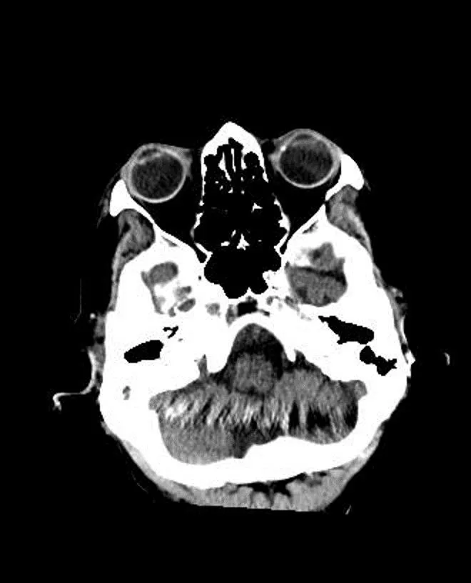
Head CT of patient wearing
cervical collar imaged at 120kVp
using 256 slice scanner with 80 x
0.625mm detector configuration.
Because the gantry can’t tilt,
images were acquired in helical
mode and then reconstructed
obliquely. What is the source of
the artifacts seen in the lower
cerebellum?
Author: David M. Gauntt, Ph.D.

Physics Case of the Day - Monday
The artifact in the image to the
right is a set of beam hardening
streaks streaks caused by dental
implants.
Author: David M. Gauntt, Ph.D.
Physics Case of the Day - Monday
The topogram (below) confirms
the presence of significant dental
amalgam, and a true axial view
(right) through the plane of the
amalgam shows significant beam
hardening streaking.
Physics Case of the Day - Monday
In a sagittal view, the artifact (yellow arrow) extends from
the dental amalgam back through the posterior fossa.
Physics Case of the Day - Monday
When the data are reformatted into oblique images, the
artifact appears in small sections of multiple planes.
Head CT images of a patient wearing a cervical collar show artifacts in the lower cerebellum caused by oblique reconstruction from helical scans due to the gantry's inability to tilt. Dental implants contribute to beam hardening streaks seen in the image, particularly evident in a sagittal view extending through the posterior fossa. These artifacts are exacerbated when data are reformatted into oblique images, highlighting the challenges in image interpretation posed by hardware limitations and patient characteristics.

Uploaded on Oct 06, 2024 | 0 Views
Download Presentation

Please find below an Image/Link to download the presentation.
The content on the website is provided AS IS for your information and personal use only. It may not be sold, licensed, or shared on other websites without obtaining consent from the author.If you encounter any issues during the download, it is possible that the publisher has removed the file from their server.
You are allowed to download the files provided on this website for personal or commercial use, subject to the condition that they are used lawfully. All files are the property of their respective owners.
The content on the website is provided AS IS for your information and personal use only. It may not be sold, licensed, or shared on other websites without obtaining consent from the author.
E N D
Presentation Transcript
Physics Case of the Day - Monday Head CT of patient wearing cervical collar imaged at 120kVp using 256 slice scanner with 80 x 0.625mm detector configuration. Because the gantry can t tilt, images were acquired in helical mode and then reconstructed obliquely. What is the source of the artifacts seen in the lower cerebellum? Author: David M. Gauntt, Ph.D. Page 1
Physics Case of the Day - Monday The artifact in the image to the right is a set of beam hardening streaks streaks caused by dental implants. Author: David M. Gauntt, Ph.D. Page 2
Physics Case of the Day - Monday The topogram (below) confirms the presence of significant dental amalgam, and a true axial view (right) through the plane of the amalgam shows significant beam hardening streaking. Page 3
Physics Case of the Day - Monday In a sagittal view, the artifact (yellow arrow) extends from the dental amalgam back through the posterior fossa. Page 4
Physics Case of the Day - Monday When the data are reformatted into oblique images, the artifact appears in small sections of multiple planes. Page 5