Anatomy of the Larynx, Trachea, and Bronchi: Respiratory System Overview
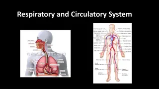
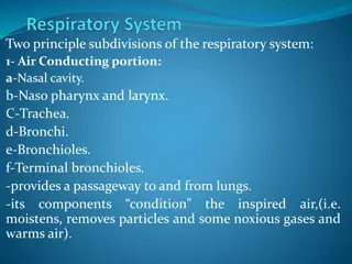
The larynx, trachea, and bronchi are vital structures of the respiratory system with distinct functions and anatomy. The larynx houses vocal cords, aids in breathing, voice production, and swallowing. It is interconnected with major arteries, veins, and nerves in the neck. The trachea extends from the larynx and branches into bronchi, facilitating air passage. The bronchial tree further divides into bronchioles, regulating airflow in the lungs. Understanding the intricate anatomy and functions of these structures is crucial in clinical practice.

Download Presentation

Please find below an Image/Link to download the presentation.
The content on the website is provided AS IS for your information and personal use only. It may not be sold, licensed, or shared on other websites without obtaining consent from the author.If you encounter any issues during the download, it is possible that the publisher has removed the file from their server.
You are allowed to download the files provided on this website for personal or commercial use, subject to the condition that they are used lawfully. All files are the property of their respective owners.
The content on the website is provided AS IS for your information and personal use only. It may not be sold, licensed, or shared on other websites without obtaining consent from the author.
E N D
Presentation Transcript
3 Anatomy of the Larynx, Trachea & Bronchi ____________ Respiratory Block Color Index: Main Text Male s Slides Female s Slides Important Doctor s Notes Extra Info Editing File
Objectives 1 Describe the Extent, structure and functions of the larynx 2 Describe the Extent, structure and functions of the trachea 3 Describe the bronchi and branching of the bronchial tree 4 Describe the functions of bronchi and their divisions. 5 Clinical anatomy Watch Video Before start studying this lecture, we highly recommend that you watch this video first! thanks to team 441
Larynx It is part of the respiratory tract which contains the vocal cords. In adults it is a 2-inch-long tube. It opens above into the laryngeal part of the pharynx. Below, it is continuous with the trachea. Respiration (breathing). Phonation (voice production) because of vocal cords. Deglutition (swallowing) Functions The larynx is related to major critical structures in the neck: Arteries: - Carotid arteries: 3 (common, external and internal) - Thyroid arteries: 3 (superior & inferior thyroid arteries) Relations Veins: Jugular veins (external & internal) Nerves: - Laryngeal nerves (Superior laryngeal & recurrent laryngeal) vagus nerve. Carotid sheath (deep cervical fascia) consists of: Carotid arteries (common and internal) & internal jugular vein & vagus nerve. - Lies in the anterior midline of the neck. From root of tongue to trachea, from laryngeal inlet up to lower border of cricoid cartilage. Opposite to C3-C6 vertebrae in men, Slightly higher in female & children. In males, after puberty thyroid cartilage becomes prominent- Adam s Apple (or laryngeal prominence). Situation & Extent
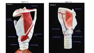
Structure: The larynx consists of four basic components: 1) Cartilaginous skeleton (Mainly) 2) Membranes and ligaments (connect the cartilage together) 3) Muscles (Intrinsic & extrinsic muscles) In other words (internal: within the larynx & external: surrounding the larynx)(they are helping to move of cartilage) 4) Mucosal lining (Lubrication) Functions: Protective sphincter at the air passage. Phonation. Regulates air passage in inspiration and expiration. Opens & closes during swallowing, coughing & sneezing 3 paired: (Small) 4. Arytenoid (pyramid shape) 5. Corniculate (very small) 6. Cuneiform 3 single: (Large) 1. Thyroid 2. Cricoid 3. Epiglottis The cartilages All the cartilages are hyaline except the epiglottis which is Elastic cartilage. (For more elasticity to open & close the inlet) The cartilages are connected by joints, ligaments, lined by membranes, and moved by muscles. posterior anterior
Membranes & Ligaments Quadrangular membrane (aryepiglottic membrane) 1 2 Thyrohyoid membrane The thyrohyoid membrane is thickened in the median plane to form median thyrohyoid ligament and on both sides to form lateral thyrohyoid ligaments. - It extends between the arytenoid and epiglottis. - Its lower free margin forms the vestibular ligament (it s form by quadrangular membrane) which forms the vestibular fold (false vocal cord). Cricothyroid membrane (conus elasticus) 3 4 Cricotracheal membrane - Its lower margin is attached to the upper border of cricoid cartilage. - Upper free margin forms Vocal ligament which forms the true vocal cord. Ligaments: 1- Hyoepiglottic ligament 2- Thyroepiglottic ligament In girls slides only Related to the previous slide The Arytenoid cartilage has two processes: the vocal process where the vocal ligament is attached and the muscular process where the cricoarytenoid dorsalis muscle( the laryngeal abductor muscle) inserts Hyo: U shape Oid: bone midsagittal
Laryngeal Inlet: It is the upper opening of the larynx. It faces upward and backward and opens into the laryngeal part of the pharynx, ( laryngopharynx). Bounded by: Anteriorly: by the upper margin of epiglottis (E) Posteriorly & below by: arytenoid cartilages (A), corniculate & cuneiform Laterally: by the Aryepiglottic folds (AEF) Closure by apposition of AEF. Larynx Laryngeal Cavity: Coronal plane Extends from laryngeal inlet to lower border of the cricoid cartilage. Narrow in the region of the vestibular folds (rima vestibuli). (False vocal cords) Narrowest in the region of the vocal folds (rima glottidis). (True vocal cords) Divided into three parts: A. Supraglottic part or vestibule: it is the part above the vestibular folds. B. Ventricle (glottic): it is the part between the vestibular folds & the vocal folds. C. Infraglottic (subglottic) part: the part below the vocal folds. NB. The ventricle has an upward invagination called saccule which is rich in goblet cells. lateral plane
Larynx Mucous Membrane Laryngeal muscles
Extrinsic Muscles Elevators of the larynx A) -The Suprahyoid (MSGD): Mylohyoid B) -The longitudinal muscles of pharynx: Stylopharyngeus Stylohyoid Salpingopharyngeus Geniohyoid Palatopharyngeus Digastric Depressors of the larynx The Infrahyoid Muscles: Sternohyoid Sternothyroid Omohyoid Omo: relating to the shoulder
Intrinsic Muscles Muscles controlling the vocal cords -Muscle decreasing the Length & Tension of Vocal Cords (relax vocal cords): Thyroarytenoid. (vocalis) the lower fibers of thyroarytenoid muscles - Muscle increasing the Length & Tension of Vocal Cords: Cricothyroid. *the only intrinsic muscle which found outside the larynx. - Adductors (close rima glottis): Lateral cricoarytenoid. Transverse arytenoid. - Abductor (open rima glottis): Posterior cricoarytenoid. Muscles Controlling the Laryngeal Inlet Close inlet. Open inlet. Oblique arytenoid. Aryepiglottic muscle. Thyro-epiglottics
Arteries Lower Half: Inferior Laryngeal Artery Upper Half: Superior Laryngeal Artery Branch of inferior thyroid artery from thyrocervical trunk of subclavian artery Branch of Superior thyroid artery Blood Supply of Larynx: Drain into Superior thyroid vein, which empty into Internal jugular vein. Drain into Inferior thyroid vein, which empty into Left brachiocephalic vein. Veins Superior Laryngeal Vein Inferior Laryngeal Vein The superior and inferior laryngeal veins drain the larynx and share the same course as the arteries (accompany corresponding arteries). Innervation Lymphatics Sensory: Motor: The lymph vessels drain into: the deep cervical lymph nodes Above the vocal cords: Internal laryngeal nerve, branch of the superior laryngeal of vagus nerve. All intrinsic muscles: Recurrent laryngeal nerve, except cricothyroid muscle. Below the vocal cords: Recurrent laryngeal of vagus nerve. Cricothyroid muscle: External laryngeal nerve, of superior laryngeal of vagus.
Semons law Damage to the Recurrent laryngeal nerve It indicates the different effect between damage (surgical trauma) & transection of Recurrent laryngeal nerve due to surgery in region of the neck. (e.g. thyroidectomy or parathyroidectomy) N.B: the nerve fibers supplying the abductors of the vocal folds lie in the periphery of the recurrent laryngeal nerve and any progressive lesion involves these fibers first before involving the deeper fibers that supply the adductors. Abductors of V.C are first to be paralysed & last to recover. THE RECURRENT LARYNGEAL NERVE (RLN) found in Girls Only R.L.N is responsible about coaptation of vocal cord so, produce Voice. The effect of injury of RLN as follows: For inspiration: complete abduction of cords is needed. Unilateral partial Dyspnea RLN has outer fibers for Abductor muscles and has Inner fibers for Adductor muscles. Bilateral partial stridor & suffocation So, with RLN injury: A.In partial injury, adductor only are acting so, cords in midline. B. In complete injury, abductors and adductors are paralysed so, cords placed midway position. Unilateral complete hoarseness of voice Bilateral complete cadaveric Aphonia
The windpipe Mobile, fibrocartilaginous tube, 5 inches long, 1 inch in diameter. Beginning: in the neck below the cricoid cartilage of the larynx (at lower border of cricoid cartilage at (C6). End: in the thorax at the level of sternal angle (lower border of T4), by dividing into right and left principal (main, primary) bronchi. The ridge at the bifurcation from inside is called Carina, It s the most sensitive part of the respiratory tract and is associated with the cough reflex. Trachea This picture is here to help you understand the Anatomy Remember: only the right side has Right structures, every thing else has Left Structures. This picture is here to help you understand the Relations Relations -Sternum. - Thymus (remains of thymus glands) - Left brachiocephalic vein. - Arch of Aorta. -Origin of: - Brachiocephalic artery - Left common carotid artery Anterior - Azygos vein Right side - Right vagus nerve - Pleura - Arch of Aorta - Left common carotid artery - Left subclavian artery - Left vagus nerve - Left phrenic nerve - Pleura Left side - Esophagus Posterior - Left recurrent laryngeal nerve
Blood Supply of Trachea: Arteries Veins Drain into the inferior thyroid veins Branches of: bronchial arteries From descending thoracic aorta inferior thyroid artery Innervation: Lymphatic Drainage: Branches of the vagus nerve and recurrent laryngeal nerve. Branches from the sympathetic trunks. Paratracheal Lymph Node Pretracheal Lymph Node Sensory fibers of Mucous membrane: Trachealis muscle and the blood vessels
Bronchi Right Principal Bronchus Left Principal Bronchus About one inch long. About two inches long. Wider, shorter and more vertical than the left. Anything that goes down the windpipe commonly gets stuck in it because it is wide. Narrower, longer and more horizontal than the right. Gives superior lobar bronchus before entering the hilum (hilas) of the right lung. Passes to the left below the aortic arch and in front of esophagus. On entering the hilum, it divides into middle and inferior lobar bronchi. On entering the hilum of the left lung, it divides into superior and inferior lobar bronchi. Within the lung, each bronchus divides to branches that can be classified into: Conduction zone branches Respiratory zone branches Primary (main) bronchi Secondary (lobar) bronchi Tertiary (segmental) bronchi (supply the bronchopulmonary segment) Smaller bronchi Bronchioles Terminal bronchioles - Respiratory bronchioles - Alveolar ducts - Alveolar sacs - Alveoli
Boys only slide Asthma Asthma is a chronic inflammatory condition of the airways characterized by airway obstruction. It causes bronchoconstriction, shortness of breath, wheezing, chest tightness, increased mucus production and coughing. There are Two Types of treatment for asthma: The symptoms can be triggered by inhaling allergens such as animal dander, dust mites, mould spores, pollens, certain chemicals and tobacco smoke. Used to prevent an attack Corticosteroid Other factors such as cold weather, exercise, stressful situations and respiratory infections can trigger an attack of asthma. Used as a Quick-Relief Drug for use During an attack During an asthma attack a person may experience tachycardia, difficulty breathing and severe anxiety. BronchoDilators Patients should try to avoid exposure to allergens or factors Customer that can trigger an attack
MCQs 1. Which one of the following is a paired cartilage? A.Thyroid B. Corniculate C.Epiglottis D.Cricoid 2. What is the most posterior structure of the laryngeal inlet? A.Arytenoid cartilage B.Epiglottis C. Conus elasticus D.Vocal folds 3. Wider, shorter and more vertical is the description of which of the following? A.Right Principal Bronchus B.Left Principal Bronchus C.Inferior thyroid artery D.Quadrangul ar membrane 4. Which one of the following muscles decreases the length and tension of the vocal cords? B.Oblique arytenoid C.Transverse arytenoid A. Cricothyroid D.Vocalis 5. Right side of the trachea? A.Sternum B.Right vagus nerve C.Pleura D.Both B and C Answers: 1)B ,2) A ,3) A , 4)D ,5) D
MCQs 6. Larynx below is continuous with? A.Pharynx B. Esophagus C. Bronchus D.Trachea 7. Which of the following is not a basic component of the Larynx? A.Mucosal lining B.Cartilaginous skeleton C.Membranes and ligaments D.Joints 8. It extends between the arytenoid and epiglottis? A.Cricoid cartilage B.Thyrohyoid membrane C.Quadrangular membrane D.Mylohyoid 9. Which of the following structures is covered with stratified squamous epithelium? A.Vestibular folds B.Aryepiglotti c folds C.Vocal folds D.Saccule 10. Veins of the trachea drain into? A.Inferior laryngeal Veins B.Bronchial veins D.Superior thyroid veins C. Inferior thyroid veins Answers: 6) D , 7) D , 8) C , 9) C , 10) C Click on the icon for more questions from Med442 s QBank
SAQs 1. Mention 2 ligaments in the larynx? 2- Thyroepiglottic ligament 1- Hyoepiglottic ligament 2. Mention 4 of the Extrinsic Suprahyoid muscles that elevate the Larynx (MSGD) Digastric Geniohyoid Stylohyoid Mylohyoid 3. Mention three of the conduction zone branches Tertiary bronchi (segmental) Secondary Bronchi (lobar) Primary Bronchi (main)
Team Leaders Najla Aldhbiban Mohammad Alrashed Team Members Faisal Alomar Nouf Aldalaqan Mishal Alsuwayegh Nouf Aldhalaan Hazem Almalki Atheer Alahmari Zyad Alodan Shatha Alshabani Salman Albader Aljazi AlBabtain Abdulaziz Alnasser Reema Alshehri Faris Alseraye Sarah Alzahrani Ahmad Alemam Maha Alkoryshy Alwaleed Faqihi Nada Albedaiwi Abdullah Alnajres Razan Almohanna Meshari Alshathri Atheer AlKanhal Nawaf Alturki Raneem AlWatban Nasser Alghaith