Alzheimer's Disease: Causes, Symptoms & Impact


Alzheimer's disease
•
It is a degenerative brain disorder of unknown etiology which is the
most common form of dementia.
•
usually starts in late middle age or in old age, results in progressive
memory loss, impaired thinking, disorientation, and changes in
personality and mood.
•
There is degeneration of brain neurons especially in the cerebral
cortex and presence of neurofibrillary tangles and plaques
containing beta-amyloid cells.
Origin of Alzheimer's Disease
The disease was first described
by Dr. Alois Alzheimer, a German
physician, in 1906. Alzheimer had a
patient named Auguste D, in her
fifties who suffered from what
seemed to be a mental illness. But
when she died in 1906, an autopsy
revealed dense deposits, now called
neuritic plaques
, outside and around
the nerve cells in her brain. Inside
the cells were twisted strands of
fiber, or
neurofibrillary tangles
.
Since Dr. Alois Alzheimer's was the
first person who discovered the
disease, AD was named after him.
Auguste D
•
Alzheimer’s disease is a chronic, irreversible disease
that affects the cells of the brain and causes impairment
of intellectual functioning.
•
Alzheimer's disease is a brain disorder which gradually
destroys the ability to reason, remember, imagine, and
learn.
INCIDENCE
•
About 3 percent of men and women ages 65 to 74
have AD, and nearly half of those age 85 and older
may have the disease.
•
About 360,000 new cases of Alzheimer’s are
diagnosed each year.
CAUSES
There is no precise cause of Alzheimer’s
disease known.
However, several factors are thought to be
implicated in this disease:
Neurochemical Factors
a) Acetylcholine.
b) Somatostatin.
c) Substance P.
d) Norepinephrine
Environmental factors
A number of environmental factors are associated with an increased risk of
AD, including:
•
Age
•
Decreased reserve capacity of the brain (reduced brain size, low
educational level, and reduced mental and physical activity in late life.
•
Head injury.
•
Risk factors for vascular disease (hypercholesterolemia, hypertension,
atherosclerosis, coronary heart disease, smoking, obesity, and diabetes).
•
Infections.
Genetic & immunological Factors
•
The majority and most aggressive early onset cases are
attributed to mutations of a gene located on chromosome 14,
which produces a protein called presenilin.
•
A structurally similar protein, presenilin 2, is produced by a
gene on chromosome 1.
•
Both presenilin 1 and presenilin 2 encode for membrane
proteins that may be involved in amyloid precursor protein
(APP) processing.
Risk Factors
a) Down's syndrome.
b) Family History.
c) Chronic high BP.
d) Head injuries.
e) Gender.
f) Smoking and Drinking
PATHOPHYSIOLOGY
•
Alzheimer's disease attacks nerves and brain cells as well as
neurotransmitters.
•
The destruction of these parts causes clumps of protein to form
around the brain's cells. These clumps are known as 'plaques'
and 'bundles'. The presence of the 'plaques' and 'bundles' start
to destroy more connections between the brain cells, which
makes the condition worse.
Pathophysiology
•
The signature lesions in AD are neuritic plaques
and neurofibrillary tangles (NFTs) located in the
cortical areas and medial temporal lobe structures
of the brain.
•
Along with these lesions, degeneration of neurons
and synapses, as well as cortical atrophy, occurs.
Amyloid Cascade
•
Neuritic plaques (also termed amyloid or senile plaques)
are extracellular lesions found in the brain and cerebral
vasculature. Plaques from AD brains largely consist of a
protein called
β-
amyloid peptide (
β
AP) βAP, which is
produced via processing of a larger protein, APP.
•
Specific APP physiologic roles are not entirely clear, but
in a general sense it is felt to contribute to proper neuronal
function and perhaps cerebral development.
Pathophysiology
Neurofibrillary Tangle
•
NFTs are commonly found in the cells of the hippocampus and
cerebral cortex in persons with AD and are composed of
abnormally hyperphosphorylated tau protein.
•
Tau protein provides structural support to microtubules, the
cell’s transportation and skeletal support system.
•
When tau filaments undergo abnormal phosphorylation at a
specific site, they cannot bind effectively to microtubules, and
the microtubules collapse.
Pathophysiology
•
Enzymes act on the APP (amyloid precursor protein) and cut it
into fragments. The beta-amyloid fragment is crucial in the
formation of senile plaques in AD
Tau proteins
are proteins that stabilize microtubules
Alzheimer's disease, are associated with tau proteins that have
become defective and no longer stabilize microtubules properly.
Inflammatory Mediators
Inflammatory or immunologic paradigms are often viewed as a
corollary of the amyloid cascade hypothesis. Certainly, brain
amyloid deposition associates with local inflammatory and
immunologic alterations.
This line of observation led some to propose that inflammation is
relevant to AD neurodegeneration.
Pathophysiology
The Cholinergic Hypothesis
•
Multiple neuronal pathways are destroyed in AD. There is variety of
neurotransmitter deficits, with cholinergic abnormalities being the most
prominent. Loss of cholinergic activity correlates with AD severity.
•
In late AD, the number of cholinergic neurons is reduced, and there is loss
of nicotinic receptors in the hippocampus and cortex. Presynaptic nicotinic
receptors control the release of acetylcholine, as well as other
neurotransmitters important for memory and mood, including glutamate,
serotonin, and norepinephrine
Pathophysiology
Other Neurotransmitter Abnormalities
•
Serotonergic neurons of the raphe nuclei and noradrenergic cells of the
locus ceruleus are lost, while monoamine oxidase type B activity is
increased. Monoamine oxidase type B is found predominantly in the brain
and in platelets, and is responsible for metabolizing dopamine.
•
In addition, abnormalities appear in glutamate pathways of the cortex and
limbic structures, where a loss of neurons leads to a focus on excitotoxicity
models as possible contributing factors to AD pathology.
Pathophysiology
DUE TO THE ETIOLOGICAL FACTORS
CHANGES OCCUR IN THE PROTIENS OF THE NERVE CELLS
OF THE CEREBRAL CORTEX
ACCUMULATION OF NEUROFIBRILLARY TANGLES AND PLAQUES
GRANULO VASCULAR DEGENERATION
LOSS OF CHOLINERGIC NERVE CELLS
LOSS OF MEMORY, FUNCTION AND COGNITION
SIGNS
Ten warning signs of Alzheimer's disease
1) Memory loss
2) Difficulty to performing familiar tasks
3) Problems with language
4) Disorientation to time and place
5) Poor or decreased judgment
6) Problems with abstract thinking
7) Misplacing things
8) Changes in mood or behavior
9) Changes in personality
10) Loss of initiative
SYMPTOMS
•
Confusion
•
disturbances in short-term memory
•
problems with attention and spatial orientation
•
personality changes
•
language difficulties
•
unexplained mood swings
Diagnostic tests
•
Psychiatric assessments.
•
Mental status examination and neuropsychological
assessment.
•
Laboratory tests.
•
Brain imaging
.
* CT
scan
* MRI
* PET (Positron emission tomography)
* SPECT (Single-photon emission computed tomography)
•
CSF Examination
•
Electro-encephalogram (EEG)
•
Electromyogram
PET scan
of the brain of a person with AD showing a
loss of function in the temporal lobe
Treatment
DESIRED OUTCOMES
•
The primary goal of treatment in AD is to symptomatically
treat cognitive difficulties and preserve patient function as
long as possible.
•
Secondary goals include treating the psychiatric and
behavioral sequelae that occur as a result of the disease.
•
Current AD treatments have not been shown to prolong life,
cure AD, or halt or reverse the pathophysiologic processes of
the disorder.
Pharmacological Intervention
•
In mild-moderate disease, consider therapy with a
cholinesterase inhibitor:
•
Acetylcholinesterase inhibitors: prevent the breakdown of
acetylcholine, a chemical messenger important for learning
and memory
eg. Donepezil (Aricept)
Rivastigmine (Exelon)
Galantamine (Razadyne)
•
In moderate to severe disease, consider adding
antiglutamatergic therapy:
N-Methyl d-aspartate Receptor Antagonist (NMDA)
E.g.: Memantine – blocks the NMDA receptor and inhibit their
overstimulation by glutamate (neurotransmitter)
•
Antidepressents.
•
Anxiolytics.
•
Antipsychotics.
•
Anticonvulsants
•
Cholinesterase Inhibitors
to enhance cholinergic activity in
patients with AD by inhibiting the hydrolysis of acetylcholine
through reversible inhibition of cholinesterase.
•
Tacrine was the first such drug. However, tacrine is fraught with
significant side effects, including hepatotoxicity, that severely
limit its usefulness. the use of tacrine has been replaced by the
use of safer, more tolerable cholinesterase inhibitors.
•
Newer cholinesterase inhibitors donepezil, rivastigmine, and
galantamine, show similar efficacy and are generally well
tolerated.
•
The most frequent adverse events associated with these agents
are mild to moderate gastrointestinal symptoms (nausea,
vomiting, and diarrhea).
•
Other cholinergic side effects are generally dose-related and
include urinary incontinence, dizziness, headache, syncope,
bradycardia, muscle weakness, salivation, and sweating.
•
Gradual dose titration over several months can improve
tolerability
Antiglutamatergic Therapy
•
Memantine is the only NMDA antagonist currently available.
•
Memantine blocks glutamatergic neurotransmission by
antagonizing NMDA receptors.
•
Glutamate is an excitatory neurotransmitter in the brain
implicated in long-term potentiation, a neuronal mechanism
important for learning and memory.
•
By blocking NMDA receptors, excitotoxic reactions, which
ultimately lead to cell death, may be prevented.
•
Memantine has been well tolerated in clinical trials. The most
common adverse events associated with memantine include
constipation, confusion, dizziness, headache, hallucinations,
coughing, and hypertension.
•
Memantine is likely to be used as monotherapy and also in
combination with cholinesterase inhibitors in patients with
moderate to severe AD.
•
Memantine should be initiated at 5 mg once a day and
increased weekly by 5 mg a day to the effective dose of 10 mg
twice daily.
Non-conventional Pharmacologic Treatment
•
Vitamin E
; The antioxidant vitamin E has been proposed as a treatment for
AD because of the association of oxygen free radicals with AD
•
Estrogen
; There is clinical evidence that loss of estrogen after menopause
is associated with subclinical impairment in some aspects of
neuropsychological function.
•
NASIDs
;
Antiinflammatory Agents
Epidemiologic studies suggest a
protective effect against AD in patients who have taken NSAIDs.
•
Statins
; pravastatin and lovastatin, but not simvastatin, were associated
with a lower prevalence of AD.
(3-hydroxy-3-methylglutaryl-CoA reductase inhibitors)
Non-conventional Pharmacologic Treatment
Ginkgo biloba
; mechanisms of action in AD include:
•
increasing blood flow, decreasing the viscosity of blood
•
antagonizing platelet activating factor receptors
•
increasing tolerance to anoxia
•
inhibiting monoamine oxidase
•
Anti-infective properties
•
preventing the damage of membranes caused by free radicals.
•
Ginkgo biloba may also inhibit catecholamine-
O
-methyl transferase
Medications Used for Noncognitive Symptoms
•
Antipsychotics
: Psychosis: (hallucinations, delusions, Disruptive behaviors:
agitation, aggression)
Haloperidol, Olanzapine, Quetiapine, Risperidone, Ziprasidone
•
Antidepressants
Depression: (poor appetite, insomnia, hopelessness,
anhedonia, suicidal thoughts, agitation, anxiety)
Citalopram, Escitalopram, Fluoxetine, Paroxetine, Sertraline, Venlafaxine,
Trazodone
•
Anticonvulsants
: (Agitation or aggression)
Carbamazepine and Valproic Acid
Psychosocial intervention
•
Behavioral approach
•
Emotion oriented approach
-Remnisence therapy
-Validation therapy
-supportive psychotherapy
-sensory integration
-stimulated presence therapy
•
Cognition oriented approach
•
Stimulation oriented approach
snoezelen
;
Caregiving
Since Alzheimer's has no cure and it gradually
renders people incapable of tending for their own
needs, caregiving essentially is the treatment and
must be carefully managed over the course of the
disease.
Alzheimer's disease is a degenerative brain disorder that affects memory, thinking abilities, personality, and mood. First described in 1906 by Dr. Alois Alzheimer, it is a chronic and irreversible condition with no known precise cause. Factors such as neurochemical imbalances and environmental influences may contribute to its development. Incidence rates increase with age, with approximately 3% of individuals aged 65-74 affected and a higher percentage among those over 85. Early detection and understanding are crucial in managing this prevalent form of dementia.

Download Presentation

Please find below an Image/Link to download the presentation.
The content on the website is provided AS IS for your information and personal use only. It may not be sold, licensed, or shared on other websites without obtaining consent from the author.If you encounter any issues during the download, it is possible that the publisher has removed the file from their server.
You are allowed to download the files provided on this website for personal or commercial use, subject to the condition that they are used lawfully. All files are the property of their respective owners.
The content on the website is provided AS IS for your information and personal use only. It may not be sold, licensed, or shared on other websites without obtaining consent from the author.
E N D
Presentation Transcript
Alzheimer's disease It is a degenerative brain disorder of unknown etiology which is the most common form of dementia. usually starts in late middle age or in old age, results in progressive memory loss, impaired thinking, disorientation, and changes in personality and mood. There is degeneration of brain neurons especially in the cerebral cortex and presence of neurofibrillary tangles and plaques containing beta-amyloid cells.
Origin of Alzheimer's Disease The disease was first described by Dr. AloisAlzheimer, a German physician, in 1906. Alzheimer had a patient named Auguste D, in her fifties who suffered from what seemed to be a mental illness. But when she died in 1906, an autopsy revealed dense deposits, now called neuritic plaques, outside and around the nerve cells in her brain. Inside the cells were twisted strands of fiber, or neurofibrillary tangles. Since Dr. AloisAlzheimer's was the first person who discovered the disease, AD was named after him. 180px-Auguste_D_aus_Marktbreit Auguste D
Alzheimers disease is a chronic, irreversible disease that affects the cells of the brain and causes impairment of intellectual functioning. Alzheimer's disease is a brain disorder which gradually destroys the ability to reason, remember, imagine, and learn.
INCIDENCE About 3 percent of men and women ages 65 to 74 have AD, and nearly half of those age 85 and older may have the disease. About 360,000 new cases of Alzheimer s are diagnosed each year.
CAUSES There is no precise cause of Alzheimer s disease known. However, several factors are thought to be implicated in this disease:
Neurochemical Factors a) Acetylcholine. b) Somatostatin. c) Substance P. d) Norepinephrine
Environmental factors A number of environmental factors are associated with an increased risk of AD, including: Age Decreased reserve capacity of the brain (reduced brain size, low educational level, and reduced mental and physical activity in late life. Head injury. Risk factors for vascular disease (hypercholesterolemia, hypertension, atherosclerosis, coronary heart disease, smoking, obesity, and diabetes). Infections.
Genetic & immunological Factors The majority and most aggressive early onset cases are attributed to mutations of a gene located on chromosome 14, which produces a protein called presenilin. A structurally similar protein, presenilin 2, is produced by a gene on chromosome 1. Both presenilin 1 and presenilin 2 encode for membrane proteins that may be involved in amyloid precursor protein (APP) processing.
Risk Factors a) Down's syndrome. b) Family History. c) Chronic high BP. d) Head injuries. e) Gender. f) Smoking and Drinking
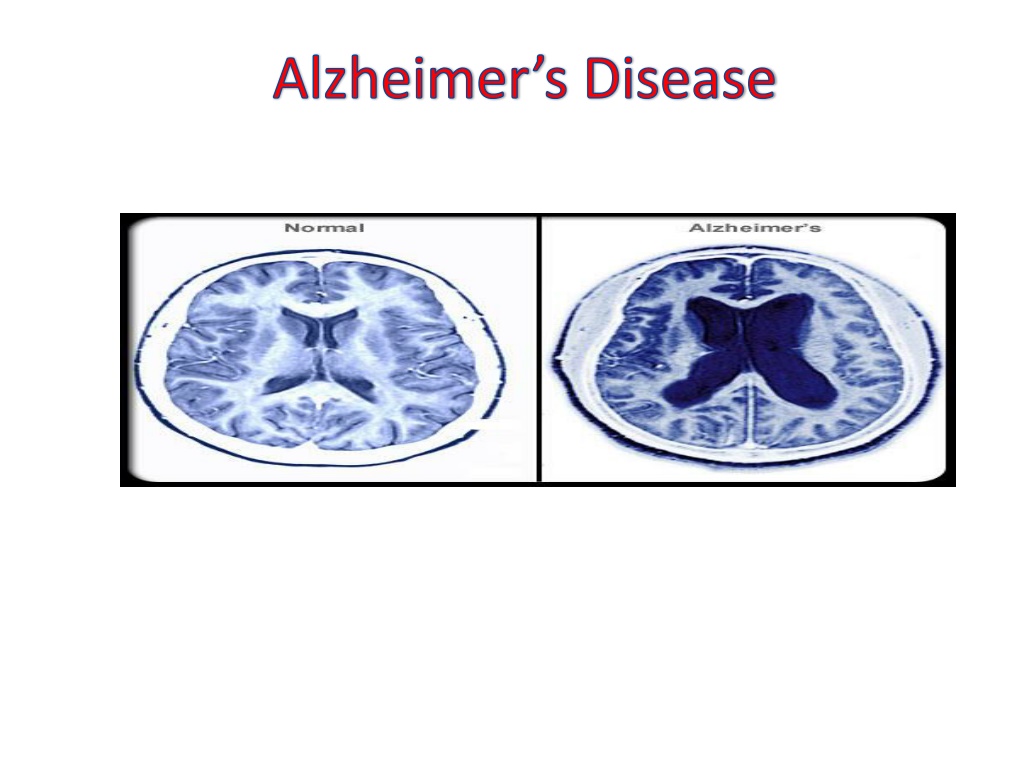
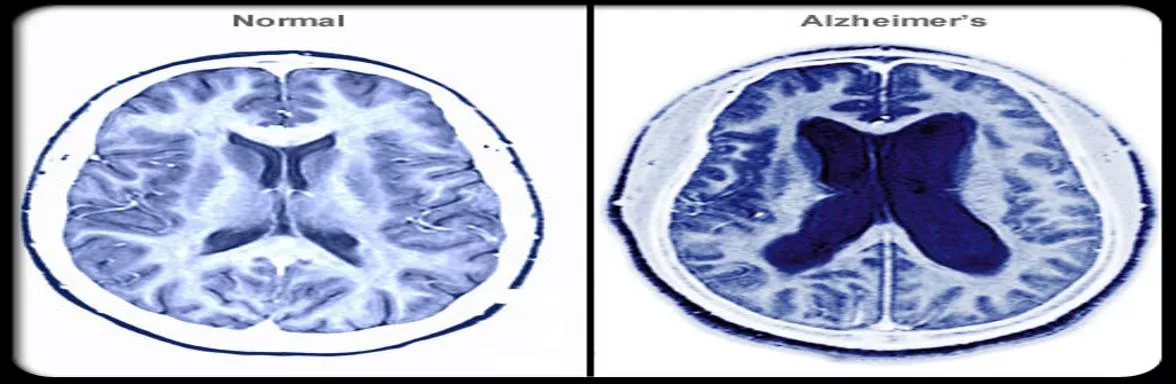
PATHOPHYSIOLOGY Alzheimer's disease attacks nerves and brain cells as well as neurotransmitters. The destruction of these parts causes clumps of protein to form around the brain's cells. These clumps are known as 'plaques' and 'bundles'. The presence of the 'plaques' and 'bundles' start to destroy more connections between the brain cells, which makes the condition worse.
Pathophysiology The signature lesions in AD are neuritic plaques and neurofibrillary tangles (NFTs) located in the cortical areas and medial temporal lobe structures of the brain. Along with these lesions, degeneration of neurons and synapses, as well as cortical atrophy, occurs.
Pathophysiology Amyloid Cascade Neuritic plaques (also termed amyloid or senile plaques) are extracellular lesions found in the brain and cerebral vasculature. Plaques from AD brains largely consist of a protein called -amyloid peptide ( AP) AP, which is produced via processing of a larger protein, APP. Specific APP physiologic roles are not entirely clear, but in a general sense it is felt to contribute to proper neuronal function and perhaps cerebral development.
Pathophysiology Neurofibrillary Tangle NFTs are commonly found in the cells of the hippocampus and cerebral cortex in persons with AD and are composed of abnormally hyperphosphorylated tau protein. Tau protein provides structural support to microtubules, the cell s transportation and skeletal support system. When tau filaments undergo abnormal phosphorylation at a specific site, they cannot bind effectively to microtubules, and the microtubules collapse.
File:Amyloid-plaque formation-big.jpg Enzymes act on the APP (amyloid precursor protein) and cut it into fragments. The beta-amyloid fragment is crucial in the formation of senile plaques in AD
File:TANGLES HIGH.jpg Tau proteins are proteins that stabilize microtubules Alzheimer's disease, are associated with tau proteins that have become defective and no longer stabilize microtubules properly.
Pathophysiology Inflammatory Mediators Inflammatory or immunologic paradigms are often viewed as a corollary of the amyloid cascade hypothesis. Certainly, brain amyloid deposition associates with local inflammatory and immunologic alterations. This line of observation led some to propose that inflammation is relevant to AD neurodegeneration.
Pathophysiology The Cholinergic Hypothesis Multiple neuronal pathways are destroyed in AD. There is variety of neurotransmitter deficits, with cholinergic abnormalities being the most prominent. Loss of cholinergic activity correlates with AD severity. In late AD, the number of cholinergic neurons is reduced, and there is loss of nicotinic receptors in the hippocampus and cortex. Presynaptic nicotinic receptors control the release of acetylcholine, as well as other neurotransmitters important for memory and mood, including glutamate, serotonin, and norepinephrine
Pathophysiology Other Neurotransmitter Abnormalities Serotonergic neurons of the raphe nuclei and noradrenergic cells of the locus ceruleus are lost, while monoamine oxidase type B activity is increased. Monoamine oxidase type B is found predominantly in the brain and in platelets, and is responsible for metabolizing dopamine. In addition, abnormalities appear in glutamate pathways of the cortex and limbic structures, where a loss of neurons leads to a focus on excitotoxicity models as possible contributing factors to AD pathology.
DUE TO THE ETIOLOGICAL FACTORS CHANGES OCCUR IN THE PROTIENS OF THE NERVE CELLS OF THE CEREBRAL CORTEX ACCUMULATION OF NEUROFIBRILLARY TANGLES AND PLAQUES GRANULO VASCULAR DEGENERATION LOSS OF CHOLINERGIC NERVE CELLS LOSS OF MEMORY, FUNCTION AND COGNITION
SIGNS SIGNS Ten warning signs of Alzheimer's disease 1) Memory loss 2) Difficulty to performing familiar tasks 3) Problems with language 4) Disorientation to time and place 5) Poor or decreased judgment 6) Problems with abstract thinking 7) Misplacing things 8) Changes in mood or behavior 9) Changes in personality 10) Loss of initiative
SYMPTOMS SYMPTOMS Confusion disturbances in short-term memory problems with attention and spatial orientation personality changes language difficulties unexplained mood swings
Diagnostic tests Psychiatric assessments. Mental status examination and neuropsychological assessment. Laboratory tests. Brain imaging . * CT scan * MRI * PET (Positron emission tomography) * SPECT (Single-photon emission computed tomography) CSF Examination Electro-encephalogram (EEG) Electromyogram
File:PET Alzheimer.jpg PET scan of the brain of a person with AD showing a loss of function in the temporal lobe
Treatment DESIRED OUTCOMES The primary goal of treatment in AD is to symptomatically treat cognitive difficulties and preserve patient function as long as possible. Secondary goals include treating the psychiatric and behavioral sequelae that occur as a result of the disease. Current AD treatments have not been shown to prolong life, cure AD, or halt or reverse the pathophysiologic processes of the disorder.
Pharmacological Intervention In mild-moderate disease, consider therapy with a cholinesterase inhibitor: Acetylcholinesterase inhibitors: prevent the breakdown of acetylcholine, a chemical messenger important for learning and memory eg. Donepezil (Aricept) Rivastigmine (Exelon) Galantamine (Razadyne)
In moderate to severe disease, consider adding antiglutamatergic therapy: N-Methyl d-aspartate Receptor Antagonist (NMDA) E.g.: Memantine blocks the NMDA receptor and inhibit their overstimulation by glutamate (neurotransmitter) Antidepressents. Anxiolytics. Antipsychotics. Anticonvulsants
Cholinesterase Inhibitors to enhance cholinergic activity in patients with AD by inhibiting the hydrolysis of acetylcholine through reversible inhibition of cholinesterase. Tacrine was the first such drug. However, tacrine is fraught with significant side effects, including hepatotoxicity, that severely limit its usefulness. the use of tacrine has been replaced by the use of safer, more tolerable cholinesterase inhibitors.
Newer cholinesterase inhibitors donepezil, rivastigmine, and galantamine, show similar efficacy and are generally well tolerated. The most frequent adverse events associated with these agents are mild to moderate gastrointestinal symptoms (nausea, vomiting, and diarrhea). Other cholinergic side effects are generally dose-related and include urinary incontinence, dizziness, headache, syncope, bradycardia, muscle weakness, salivation, and sweating. Gradual dose titration over several months can improve tolerability
Antiglutamatergic Therapy Memantine is the only NMDA antagonist currently available. Memantine blocks glutamatergic neurotransmission by antagonizing NMDA receptors. Glutamate is an excitatory neurotransmitter in the brain implicated in long-term potentiation, a neuronal mechanism important for learning and memory. By blocking NMDA receptors, excitotoxic reactions, which ultimately lead to cell death, may be prevented.
Memantine has been well tolerated in clinical trials. The most common adverse events associated with memantine include constipation, confusion, dizziness, headache, hallucinations, coughing, and hypertension. Memantine is likely to be used as monotherapy and also in combination with cholinesterase inhibitors in patients with moderate to severe AD. Memantine should be initiated at 5 mg once a day and increased weekly by 5 mg a day to the effective dose of 10 mg twice daily.
Non Non- -conventional Pharmacologic Treatment conventional Pharmacologic Treatment Vitamin E; The antioxidant vitamin E has been proposed as a treatment for AD because of the association of oxygen free radicals with AD Estrogen; There is clinical evidence that loss of estrogen after menopause is associated with subclinical impairment in some aspects of neuropsychological function. NASIDs; Antiinflammatory Agents Epidemiologic studies suggest a protective effect against AD in patients who have taken NSAIDs. Statins; pravastatin and lovastatin, but not simvastatin, were associated with a lower prevalence of AD. (3-hydroxy-3-methylglutaryl-CoA reductase inhibitors)
Non Non- -conventional Pharmacologic Treatment conventional Pharmacologic Treatment Ginkgo biloba; mechanisms of action in AD include: increasing blood flow, decreasing the viscosity of blood antagonizing platelet activating factor receptors increasing tolerance to anoxia inhibiting monoamine oxidase Anti-infective properties preventing the damage of membranes caused by free radicals. Ginkgo biloba may also inhibit catecholamine-O-methyl transferase
Medications Used for Medications Used for Noncognitive Noncognitive Symptoms Symptoms Antipsychotics: Psychosis: (hallucinations, delusions, Disruptive behaviors: agitation, aggression) Haloperidol, Olanzapine, Quetiapine, Risperidone, Ziprasidone Antidepressants Depression: (poor appetite, insomnia, hopelessness, anhedonia, suicidal thoughts, agitation, anxiety) Citalopram, Escitalopram, Fluoxetine, Paroxetine, Sertraline, Venlafaxine, Trazodone Anticonvulsants : (Agitation or aggression) Carbamazepine and Valproic Acid
Psychosocial intervention Behavioral approach Emotion oriented approach -Remnisence therapy -Validation therapy -supportive psychotherapy -sensory integration -stimulated presence therapy Cognition oriented approach Stimulation oriented approach 180px-Snoezelruimte snoezelen;
Caregiving Since Alzheimer's has no cure and it gradually renders people incapable of tending for their own needs, caregiving essentially is the treatment and must be carefully managed over the course of the disease.