AI in Medicine: CSN5 2019-2021 Annual Meeting Discussions and Activities
The AI in Medicine CSN5 project for 2019-2021 involved research collaborations, virtual meetings, knowledge exchange, and planned activities for harmonizing MRI and mammographic data. The project aimed to enhance scientific output through partnerships and data standardization strategies.
Download Presentation

Please find below an Image/Link to download the presentation.
The content on the website is provided AS IS for your information and personal use only. It may not be sold, licensed, or shared on other websites without obtaining consent from the author.If you encounter any issues during the download, it is possible that the publisher has removed the file from their server.
You are allowed to download the files provided on this website for personal or commercial use, subject to the condition that they are used lawfully. All files are the property of their respective owners.
The content on the website is provided AS IS for your information and personal use only. It may not be sold, licensed, or shared on other websites without obtaining consent from the author.
E N D
Presentation Transcript
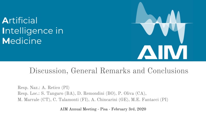
AIM, CSN5, 2019-2021 Artificial Intelligence in Medicine Discussion, General Remarks and Conclusions Resp. Naz.: A. Retico (PI) Resp. Loc.: S. Tangaro (BA), D. Remondini (BO), P. Oliva (CA), M. Marrale (CT), C. Talamonti (FI), A. Chincarini (GE), M.E. Fantacci (PI) AIM Annual Meeting - Pisa - February 3rd, 2020
2 AIM, CSN5, 2019-2021 Discussion - - Consuntivi 2019 Plans for 2020 - - SW repositories? Collaboration website? - Thematic Meeting in Bari in June/September - Collaboration with AIFM
3 AIM, CSN5, 2019-2021 Links among partners (the added value of AIM) In 2019 the following research collaborations have been strengthened (the kick-off meeting on Jan 30th 2019 was very useful to create links among researchers of different sites): FI-PI-CA-CT in AIM3.T1 BO-CT in AIM3.T3 AIM3.T4 and AIM3.T5 ??? How to enhance scientific output?
4 AIM, CSN5, 2019-2021 Other activities carried out in 2019 Monthly virtual meetings took place among group coordinators to monitor the work progress. To promote the exchange of knowledge of research problems and technical solution especially among young researchers, virtual seminars have been organized, referred to AIM tasks, in particular: AIM1.T1 - Confounding effects in Machine-Learning based studies of MRI data . 6 maggio alle 15.30 su eZuce. Elisa Ferrari (Pisa) AIM3.T4 - Predictive models for Systems Medicine . 20 maggio ore 15.00 su Vibe. Silvia Vitali e Tommaso Matteuzzi (Bologna) AIM3.T2 - Predictive models for CESM 10 giugno, ore 15.00 su Vibe, Annarita Fanizzi (Bari) Shall we continue with this line?
5 AIM, CSN5, 2019-2021 AIM s planned activity and milestones for 2020-21 Tasks Planned activity 2020 Milestones 2020 AIM1 AIM1.T1 PI: the datasample of about 2000 MRI of healthy subjects from the ABIDE consortium (14 different sites) is available for the test of the data harmonization strategy with GAN. The GAN-based MRI standardization procedure has to be implemented and its effects have to be quantified by a quality control analysis pipeline (e.g. ENIGMA QA pipeline). M1.1 (30-06-2020) Acquisition of suitable MRI data sample for testing (e.g. ABIDE, ADNI, ...), identification of test metrics and validation. BA: statistical validation of harmonization models for MRI BO: test the SVA algorithm used in PACS challenge onto other datasets, e.g. the ABIDE dataset mentioned above, in synergy with the other groups in AIM that are actually using other methods AIM1.T2 PI: characterization of 5 different mammographic systems and acquisition and evaluation of phantoms and clinical images. M1.2 (31-12-2020) Database consolidation and validation of the harmonization algorithm and publication of the results CA: implementation of the standardization algorithm also for for presentation images
6 AIM, CSN5, 2019-2021 AIM s planned activity and milestones for 2020-21 Tasks Planned activity 2020 Milestones 2020 AIM2 AIM2.T1 GE: On quantification, we intend to work towards greater accuracy in estimating the "ranking" error (i.e. in the measure of a value relative to a dataset normative), standardizing the results in the analysis to 2 quantifiers with respect to that with 3. M2.1 (31-12-2020) Method validation on EU multicentric dataset and all fluorinated tracers GE: We will then develop different brain clustering methods on the dataset we have collected and systematized during 2019. The parceling (once validated clinically) will then be used to build predictive and accumulation models in relation to clinical variables (including EEG). If we get the desired results, we could (the conditional is a must) be able to distinguish an inert accumulation (so-called incidental amyloidosis) from a pathological accumulation. If verified, yes would have an objective and extremely sophisticated examination for early diagnosis. Alternatively, if different accumulation patterns cannot be differentiated, we should at least be able to construct a causal model that allows a predictability on the risk of pathology. AIM2.T2 BO: Regarding ML and DL approaches to MR data, we have completed a fingerprint analysis via Deep Learning on simulated data, and we plan to move to data coming from real samples. M2.2: (31-12-2020) Application of Deep Learning tool to real MRF data.
7 AIM, CSN5, 2019-2021 AIM s planned activity and milestones for 2020-21 Tasks Planned activity 2020 Milestones 2020 AIM3 AIM3.T1 FI: Update and maintenance of the data sample of subjects with medulloblastoma (MRI, CT, treatment plans, patients outcome). M3.1 (31-12-2020) Software development for the selection of the most important features and first test on data PI + FI + CA: selection of relevant radiomics and dosiomics features from subjects data to predict radiation-related toxicity. Cross validation of results on available data cohorts. CT: The analysis will be carried out on the kV and MV (acquired before each treatment fraction) CT images. After a manual contouring of the lesion done by the radiation oncologists, features which characterize the tumor tissues will be extracted. Afterwards, a software will be developed to select the features that better correlate with the most favorable prognosis of patients with lung tumors that underwent radiation therapy with tomotherapy apparatus. This analysis will be carried out on both kV and MV CT images. The final aim is to be able to predict the prognosis of the patients from CT images. AIM3.T2 PI: The database of about 10000 mammograms comprising both healthy and pathologic cases will be collected and ready for the validation of the CNN classifier of breast density. Submission of results relevant for the clinical community on a peer-reviewed high impact journal. M3.2a (30-06-20) Validation of the CNN on the complete data sample of mammograms and publication of the results CA: implementation of the standardization algorithm on the heterogeneous database. Optimization and validation of the CNN on the harmonized dataset. M3.2b (31-12-2020) Further patient data acquisition and application of the analysis software on the data acquired in the first year and validation of an automatic classification method BA: further development of the automated software for breast cancer diagnosis on CESM data and validation on the extended data sample.
8 AIM, CSN5, 2019-2021 AIM s planned activity and milestones for 2020-21 Tasks Planned activity 2020 Milestones 2020 AIM3 AIM3.T3 CT: The analysis will be carried out on structural T1- and T2- weighted images and on diffusion weighted images. After segmentation of T1-weighted images to recognize the various regions of the cortex as well as the subcortical regions, the fibers from cortex to thalamus will be obtained through probabilistic tractography. This information allows for the thalamus parcellation and the following identification of the target of the treatment. In a retrospective analysis this target will be compared with the lesion produced during the ablation treatment. M3.3 (31-12-2020) Further patient data acquisition and application of the analysis software on the data acquired on the first year BO: A software will be developed to select from images features that better correlate with the clinical results for patients that underwent trans cranial-MR guided Focused Ultrasound Surgery (tc-MRgFUS) AIM3.T4 BO: in collaboration with IRCCS Meldola we will continue multiple analyses on NGS data from cancer patients (gene fusions, integration of genetics and metabolomics), some of which should be completed within the year M3.4 (31-12-2020) Application of the pipeline to real patient case studies for personalized targeting AIM3.T5 CT+BO: The analysis will be carried out on MRI images of prostate for patients with suspect lesion for tumors. For all patients the independent clinical results of biopsy is available. After a manual contouring of the lesion done from the radiologists, features which characterize the lesion tissues will be extracted. Afterwards, a software will be developed to select the features that better correlate with the type of lesion (benign or malignant one as identified at biopsy). The final aim is to be able to predict the type of lesion from MR images possibly without biopsy. M3.5 (31-12-2020) Application of the analysis software on the data acquired in the first year and validation of an automatic classification method. Acquisition of data from other patients.
9 AIM, CSN5, 2019-2021 thank you
